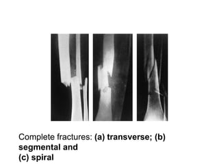
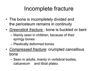
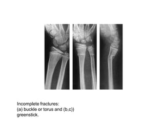
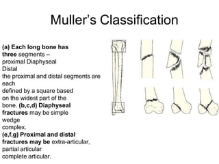
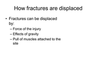
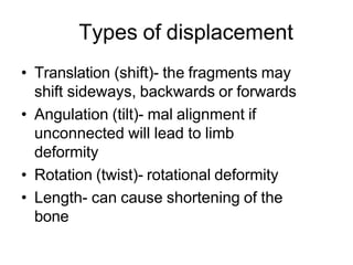
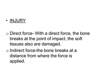
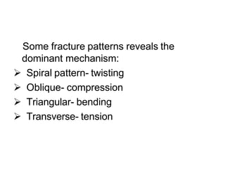
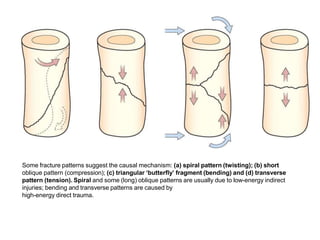
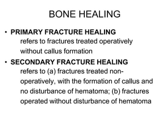
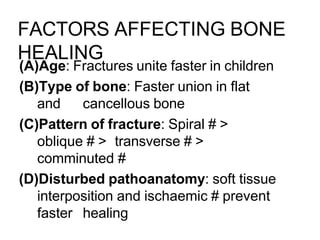
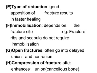
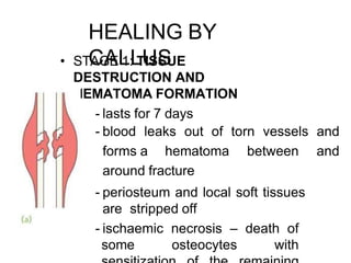
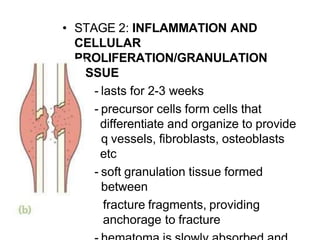
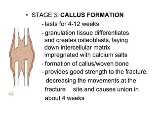
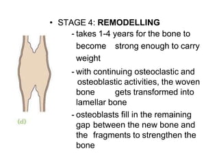
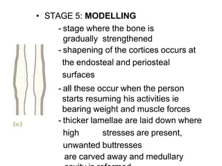
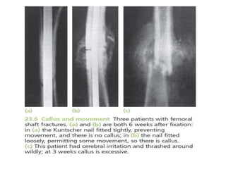
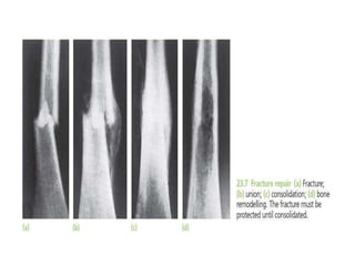
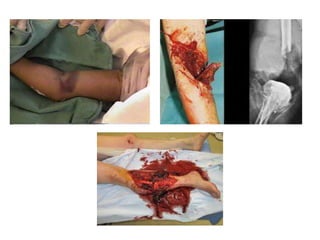
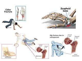
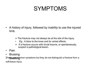
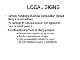
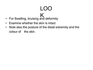
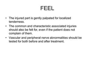
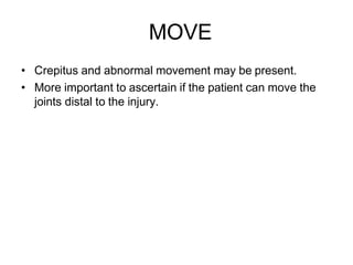
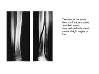
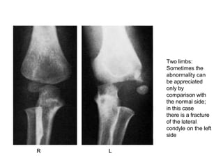
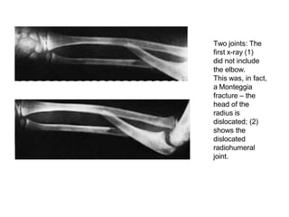
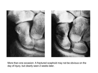
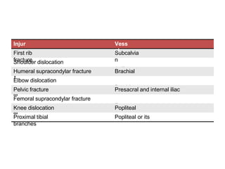
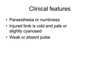
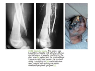
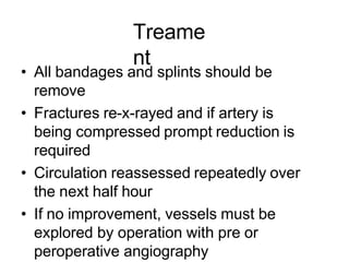
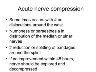
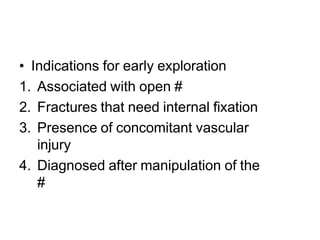
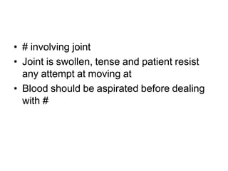
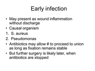
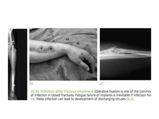
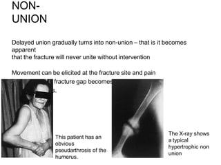
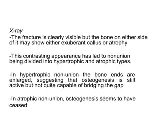
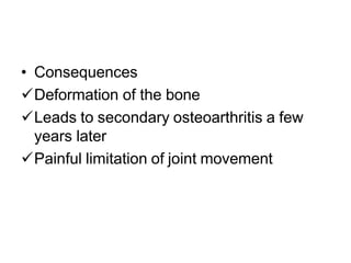
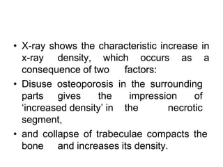
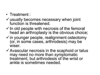
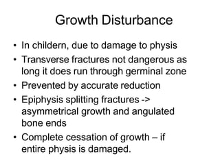
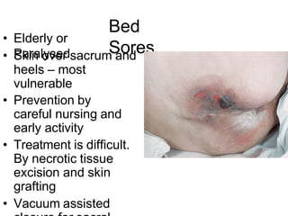
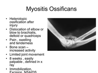
SlideShare a Scribd company logo
1 of 112
Fractur
e
Dr. Uzair Rashid
Introductio
n
• Break in the structural continuity of a bone
• If the overlying skin remains intact -
closed (simple fracture)
• If skin or one of the body cavities is
breached- open (compound fracture)
Types of Fracture
• Divided in
to
– Complete
– Incomplet
e
Complete
fracture
Bone is split into two or more fragments. The
fracture pattern on x-ray can help predict
behaviour after reduction
• in a transverse fracture the fragments usually
remain in place after reduction
• if it is oblique or spiral, they tend to shorten
and re- displace even if the bone is splinted.
• In an impacted fracture the fragments are
jammed tightly together and the fracture line is
indistinct.
• A comminuted fracture is one in which there are
more than two fragments
Complete fractures: (a) transverse; (b)
segmental and
(c) spiral
Incomplete fracture
• The bone is incompletely divided and
the periosteum remains in continuity
• Greenstick fracture : bone is buckled or bent
– Mainly seen in children, because of their
springy bones
– Plastically deformed bones
• Compressed fracture: crumpled cancellous
bone
– Seen in adults, mainly in vertebral bodies,
calcaneum and tibial plateu
Incomplete fractures:
(a) buckle or torus and (b,c))
greenstick.
Muller’s Classification
(a) Each long bone has
three segments –
proximal Diaphyseal
Distal
the proximal and distal segments are
each
defined by a square based
on the widest part of the
bone. (b,c,d) Diaphyseal
fractures may be simple
wedge
complex.
(e,f,g) Proximal and distal
fractures may be extra-articular,
partial articular
complete articular.
How fractures are displaced
• Fractures can be displaced
by:
– Force of the injury
– Effects of gravity
– Pull of muscles attached to the
site
Types of displacement
• Translation (shift)- the fragments may
shift sideways, backwards or forwards
• Angulation (tilt)- mal alignment if
unconnected will lead to limb
deformity
• Rotation (twist)- rotational deformity
• Length- can cause shortening of the
bone
Mechanism of injury
• Injury
• Repetitive stress
• Pathological
fractures
• INJURY
o Direct force- With a direct force, the bone
breaks at the point of impact; the soft
tissues also are damaged.
o Indirect force-the bone breaks at a
distance from where the force is
applied.
Some fracture patterns reveals the
dominant mechanism:
 Spiral pattern- twisting
 Oblique- compression
 Triangular- bending
 Transverse- tension
Some fracture patterns suggest the causal mechanism: (a) spiral pattern (twisting); (b) short
oblique pattern (compression); (c) triangular ‘butterfly’ fragment (bending) and (d) transverse
pattern (tension). Spiral and some (long) oblique patterns are usually due to low-energy indirect
injuries; bending and transverse patterns are caused by
high-energy direct trauma.
• FATIGUE OR STRESS FRACTURES-
 Occur in normal bone, subject to
repeated heavy loading, typically in
athletes, dancers or military personnel.
Drugs like steroids and methotrexate
• PATHOLOGICAL FRACTURES- Occurs
in a bone that is made weak by some
disease.
Causes-
 Inflammatory- Osteomyelitis
Neoplastic- giant cell tumour, Ewings
sarcoma, secondaries
 Miscellaneous bone conditions- simple
bone cyst, anuerysmal bone cyst
Heriditary- Osteogenesis
imperfecta, Osteopetrosis
 Other acquired generalised
diseases- Osteoporosis,
osteomalacia, rickets
BONE HEALING
• PRIMARY FRACTURE HEALING
refers to fractures treated operatively
without callus formation
• SECONDARY FRACTURE HEALING
refers to (a) fractures treated non-
operatively, with the formation of callus and
no disturbance of hematoma; (b) fractures
operated without disturbance of hematoma
FACTORS AFFECTING BONE
HEALING
(A)Age: Fractures unite faster in children
(B)Type of bone: Faster union in flat
and cancellous bone
(C)Pattern of fracture: Spiral # >
oblique # > transverse # >
comminuted #
(D)Disturbed pathoanatomy: soft tissue
interposition and ischaemic # prevent
faster healing
(E)Type of reduction: good
apposition of fracture results
in faster healing
(F)Immobilisation: depends on the
fracture site eg. Fracture
ribs and scapula do not require
immobilisation
(G)Open fractures: often go into delayed
union and non-union
(H)Compression of fracture site:
enhances union(cancellous bone)
HEALING BY
CALLUS
• STAGE 1: TISSUE
DESTRUCTION AND
HEMATOMA FORMATION
- lasts for 7 days
- blood leaks out of torn vessels and
forms a hematoma between and
around fracture
- periosteum and local soft tissues
are stripped off
- ischaemic necrosis – death of
some osteocytes with
• STAGE 2: INFLAMMATION AND
CELLULAR
PROLIFERATION/GRANULATION
TISSUE
- lasts for 2-3 weeks
- precursor cells form cells that
differentiate and organize to provide
q vessels, fibroblasts, osteoblasts
etc
- soft granulation tissue formed
between
fracture fragments, providing
anchorage to fracture
• STAGE 3: CALLUS FORMATION
- lasts for 4-12 weeks
- granulation tissue differentiates
and creates osteoblasts, laying
down intercellular matrix
impregnated with calcium salts
- formation of callus/woven bone
- provides good strength to the fracture,
decreasing the movements at the
fracture site and causes union in
about 4 weeks
• STAGE 4: REMODELLING
- takes 1-4 years for the bone to
become strong enough to carry
weight
- with continuing osteoclastic and
osteoblastic activities, the woven
bone gets transformed into
lamellar bone
- osteoblasts fill in the remaining
gap between the new bone and
the fragments to strengthen the
bone
• STAGE 5: MODELLING
- stage where the bone is
gradually strengthened
- shapening of the cortices occurs at
the endosteal and periosteal
surfaces
- all these occur when the person
starts resuming his activities ie
bearing weight and muscle forces
- thicker lamellae are laid down where
high stresses are present,
unwanted buttresses
are carved away and medullary
HEALING BY DIRECT
UNION
• Formation of callus requires stimulus
from movement
• In cases of impacted fracture in
cancellous bone or a fracture
immobilised by the use of metal plate,
callus will not be formed and hence the
fracture will heal by direct union
1. Osteoblastic new bone formation occurs
directly between the fracture fragments
2. New capillaries and osteoprogenitor cells
grow in from the edges and lay down new
bone on the exposed surface (gap healing)
3. Lamellar bone is produced in the narrow
crevices (<200μm) while woven bone is
produced in the wider gaps which is then
remodelled into lamellar bone
4. Penetration and bridging of the fracture
by osteoclasts and osteoblasts in
3-4 weeks.
5. Intimacy of the contact surfaces lead to
internal bridging without intermediate
stages (contact healing)
CLINICAL
PRESENTATION
OF FRACTURES
In the Casualty..
• Take a Brief History.
• General Particulars:
• AGE & SEX
 Children and the elderly
 Different mechanisms of injury : Traumatic , Pathological
 Post menopausal women : Osteoporosis and pathological fractures.
• HISTORY OF TRAUMA – Ascertaining the mechanism
of injury is important, helps understand symptoms and
aids examination.
SYMPTOMS
• A history of injury, followed by inability to use the injured
limb.
 The fracture may not always be at the site of the injury.
• Eg : A blow to the knee and its varied effects.
 If a fracture occurs with trivial trauma, or spontaneously,
suspect a pathological lesion.
• Pain
• Bruising
• Swelling
These are common symptoms but they do not distinguish a fracture from a
soft-tissue injury.
• Deformity – More suggestive of a fracture.
• Enquire about symptoms of associated
injuries:
 Pain and swelling elsewhere (it is a common mistake to get
distracted by the main injury, particularly if it is severe),
 Numbness or Loss of movement.
 Skin pallor or cyanosis.
 Blood in the urine.
 Abdominal pain.
 Difficulty with breathing.
 Transient loss of consciousness.
• Ask about previous injuries, or any other
musculoskeletal abnormality that might
cause confusion when the x-ray is seen.
Finally, a general medical history is
important, in preparation for anaesthesia
or operation.
GENERAL SIGNS
• First follow the ABCs: look for, and if necessary
attend to,
• Airway obstruction,
• Breathing problems,
• Circulatory problems
• Cervical spine injury.
• Secondary survey – Examine the main injury-
ascertain the type of fracture, classify, plan a
management protocol and look out for
complications.
• It will also be necessary to exclude other
previously unsuspected injuries.
LOCAL SIGNS
• Familiar headings of clinical examination should
always be considered,
• (or damage to arteries, nerves and ligaments
may be overlooked.)
• A systematic approach is always helpful:
 Examine the most obviously injured part.
 Test for artery and nerve damage.
 Look for associated injuries in the region.
 Look for associated injuries in distant parts.
LOO
K
• For Swelling, bruising and deformity
• Examine whether the skin is intact
• Note also the posture of the distal extremity and the
colour of the skin.
FEEL
• The injured part is gently palpated for localized
tenderness.
• The common and characteristic associated injuries
should also be felt for, even if the patient does not
complain of them.
• Vascular and peripheral nerve abnormalities should be
tested for both before and after treatment.
MOVE
• Crepitus and abnormal movement may be present.
• More important to ascertain if the patient can move the
joints distal to the injury.
X-RAY INVESTIGATION
• X-ray examination is mandatory.
• Rule of twos:
Two views – A fracture or a dislocation may not be
seen on a single x-ray film, and at least two
views (anteroposterior and lateral) must be taken.
Two limbs – In children, the appearance of
immature epiphyses may confuse the diagnosis
of a fracture; x- rays of the uninjured limb are
needed for comparison.
Two films of the same
tibia: the fracture may be
‘invisible’ in one
view and perfectly plain in
a view at right angles to
that.
Two limbs:
Sometimes the
abnormality can
be appreciated
only by
comparison with
the normal side;
in this case
there is a fracture
of the lateral
condyle on the left
side
R L
 Two joints – In the forearm or leg, one bone may be
fractured and angulated.Angulation, however, is
impossible unless the other bone is also broken, or a joint
dislocated. The joints above and below the fracture must
both be included on the x- ray films.
 Two injuries – Severe force often causes injuries at more
than one level. Eg: In fractures of the calcaneum or
femur it is important to also x-ray the pelvis and spine.
 Two occasions – Some fractures are notoriously
difficult to detect soon after injury, but another x-ray
examination a week or two later may show the lesion.
Eg: Undisplaced fractures of the distal end of the
clavicle, scaphoid, femoral neck and lateral malleolus,
and also stress fractures and physeal injuries.
Two joints: The
first x-ray (1)
did not include
the elbow.
This was, in fact,
a Monteggia
fracture – the
head of the
radius is
dislocated; (2)
shows the
dislocated
radiohumeral
joint.
More than one occasion: A fractured scaphoid may not be obvious on the
day of injury, but clearly seen 2 weeks later.
SPECIAL IMAGING
• Computed tomography (CT) may be helpful in lesions
of the spine or for complex joint fractures; help in
accurate visualization of fractures in ‘difficult’ sites
such as the calcaneum or acetabulum.
• Magnetic resonance imaging (MRI) may be the only
way of showing whether a fractured vertebra is
threatening to compress the spinal cord.
• Radioisotope scanning is helpful in diagnosing a
suspected stress fracture or other undisplaced
fractures.
FINAL DESCRIPTION
• Diagnosing a fracture is not enough; the
surgeon should describe it with its
properties:
• Is it open or closed?
• Which bone is broken, and where?
• Has it involved a joint surface?
• What is the shape of the break?
• Is it stable or unstable?
• Is it a high-energy or a low-energy injury?
In short, the examiner must learn to recognize what
has been aptly described as the ‘personality’ of the
fracture.
Complications of
Fracture
VISCERAL
INJURIES
• Often occur in fractures around the
trunk
• Penetration of lung by rib fractures
which causes pneumothorax
• Rupture of bladder or urethra in
pelvic fractures
• Require emergency treatment
VASCULAR
INJURY
Injur
y
Vess
el
First rib
fracture
Subcalvia
n
Shoulder dislocation
Axillar
y
Humeral supracondylar fracture Brachial
Elbow dislocation
Brachi
al
Pelvic fracture Presacral and internal iliac
Femoral supracondylar fracture
Femor
al
Knee dislocation Popliteal
Proximal tibial Popliteal or its
branches
Clinical features
• Paraesthesia or numbness
• Injured limb is cold and pale or
slightly cyanosed
• Weak or absent pulse
Treame
nt
• All bandages and splints should be
remove
• Fractures re-x-rayed and if artery is
being compressed prompt reduction is
required
• Circulation reassessed repeatedly over
the next half hour
• If no improvement, vessels must be
explored by operation with pre or
peroperative angiography
• Cut vessel can be sutured, or a segment
may be replaced by a vein graft, if it is
thrombosed, endarterectomy may restore
blood flow
• If vessel repair is done, stable fixation is
a must and fracture should be fixed
internally
NERVE
INJURY
Injury Nerve
Shoulder dislocation Axillary
Humeral shaft fracture Radial
Humeral supracondylar fracture Radial or median
Elbow medial condyle Ulnar
Moteggia fracture-dislocation Posterior-interosseous
Hip dislocation Sciatic
Knee dislocation peroneal
Closed nerve injuries
• Seldom severe and spontaneous
recovery occurs in 90% within 4
months
• Nerve should be explored if no recovery,
nerve conduction studies and EMG fail to
show evidence of recovery
Open nerve injuries
• Nerve should be explored at the time
of debridement and repair at the time
or
at wound closure
Acute nerve compression
• Sometimes occurs with # or
dislocations around the wrist
• Numbness or paraesthesia in
distribution of the median or ulnar
nerves
• # reduction or splitting of bandages
around the splint
• If no improvement within 48 hours,
nerve should be explored and
decompressed
• Indications for early exploration
1. Associated with open #
2. Fractures that need internal fixation
3. Presence of concomitant vascular
injury
4. Diagnosed after manipulation of the
#
HAEMARTHRO
SIS
• # involving joint
• Joint is swollen, tense and patient resist
any attempt at moving at
• Blood should be aspirated before dealing
with #
INFECTI
ON
Early infection
• May present as wound inflammation
without discharge
• Causal organism
1. S. aureus
2. Pseudomonas
• Antibiotics may allow # to proceed to union
as long as fixation remains stable
• But further surgery is likely later, when
antibiotics are stopped
Late
presentation
• May present with a sinus and xray
evidence of sequestra
• Implants and all avascular pieces of
bone should be removed
• External fixator can be used to bridge the
#
• If resulting defect too large for bone
grafting, patient should be referred to a
limb reconstruction centre
• When infection involves a joint, principles
of treatment are the same as with bone
infection, namely debridement and
drainage, drugs and splintage
• On resolution of infection, # is stabilize so
that joint movement can recommence
• If # cannot be stablized, joint should
be splinted in the optimum position
• Can lead to permanent stiffness
Compartment Syndrome
• Occurs with fracture of elbow, forearm
bones, proximal third of tibia, hands or
foot ;
• Crush injuries and circumferential burns
• Increase of pressure within the
osseofascial compartment
• Due to bleeding , oedema or inflammation
• Clinical features
– Pain : bursting sensation
– Altered sensibility
– Paresis / weakness in active muscle
contraction
– Testing the muscle by stretching them (
ischemic muscle is highly sensitive to stretch
and it causes pain )
• Confirmation of diagnosis
– Measuring the intracompartmental
pressures
• Introduced a split catheter into
the compartment
• Pressure measured close to level of the
fracture
• Differential pressure (difference between the
diastolic pressure and compartment
pressure ) is
<30mmHg– immediate decompression
• Management
– Remove any casts, bandages and dressings
Gas gangrene
• Produced by Clostridial infection
• Present in dirty wound with dead muscle
that are closed without adequate
debridement
• Toxins can cause necrosis of tissue
and promote the spread of the
disease
• Clinical features
– Within 24 hours of injury
– Intense pain
– swelling around the wound
– Brownish discharge
– Little or no pyrexia
– Increase pulse rate
– Characteristic smell
– Patient rapidly become toxemia and may
lapse into coma and death
• Prevention
– All dead tissue should be completely excised
– Doubt about tissue viability – wound should
be left open
• Treatment
– Early diagnosis
– Fluid replacement & IV antibiotics
– Hyperbaric O2 – limit the spread of gangrene
– Prompt decompression of the wound and
removal of dead tissue
– Amputation in advance cases
Fracture Blisters
• 2 types
– Clear fluid filled vesicles
– Blood stained
• Occur during limb swelling
• Due to elevation of the epidermal layer of
the skin from the dermis
• No advantage in puncturing the blister (
cause local infection )
• Surgical incision can be done after the
limb swelling decrease
Plaster and Pressure Sores
• Occur where the skin presses directly
onto bone
• Traction on a Thomas Splint ( wrong ring
size, excessive fixed traction and neglect)
• Prevention
– Padding the bony points
– Moulding the wet plaster so that pressure
is distributed to the soft tissue
around the bony points
• Treatment
– Patient feels the localised burning pain (
warning sign )
– Window must be immediately cut in the
plaster
– If unnoticed, skin necrosis will progress
LATE
COMPLICATIONS
DELAYED UNION
When a fracture takes more than usual
time to unite, it is said to have gone in
‘delayed union’.
Causes
Factors causing delayed union can be
summarized as: biological, biomechanical or
patient-related.
BIOLOGICAL
--Inadequate blood supply
--Severe soft tissue damage affects fracture
healing by:
(1)reducing the effectiveness of muscle splintage
(2)damaging the local blood supply and
(3)diminishing or eliminating the osteogenic
input from mesenchymal stem cells within
muscle
BIOMECHANICAL
--Imperfect splintage
--Over-rigid fixation
--Infection
PATIENT RELATED
In a less than ideal world, there are patients who
are:
• Immense
• Immoderate
• Immovable
• Impossible.
These factors must be accommodated
in an appropriate
fashion.
Clinical features
-Fracture tenderness persists
-Pain may be acute
-On x-ray- fracture line remains visible
- there is very little or incomplete
callus formation or periosteal
reaction
- However, the bone ends are not
sclerosed or atrophic (suggest that,
although the fracture has not
united, it eventually will)
Treatmen
t
Conservative principles are:
(1)to eliminate any possible cause of delayed union
(2)to promote healing by providing the most
appropriate environment
Immobilization (whether by cast or by internal
fixation) should be sufficient to prevent shear at the
fracture site,
but fracture loading is an important stimulus to union
and can be enhanced by:
(1)encouraging muscular exercise
(2)by weightbearing in the cast or brace
OPERATIVE
--If union is delayed for more than 6 months and
there is no sign of callus formation, internal fixation
and bone grafting are indicated.
NON-
UNION
Delayed union gradually turns into non-union – that is it becomes
apparent
that the fracture will never unite without intervention
Movement can be elicited at the fracture site and pain
diminishes; the fracture gap becomes a type of
pseudoarthrosis.
This patient has an
obvious
pseudarthrosis of the
humerus.
The X-ray shows
a typical
hypertrophic non
union
X-ray
-The fracture is clearly visible but the bone on either side
of it may show either exuberant callus or atrophy
-This contrasting appearance has led to nonunion
being divided into hypertrophic and atrophic types.
-In hypertrophic non-union the bone ends are
enlarged, suggesting that osteogenesis is still
active but not quite capable of bridging the gap
-In atrophic non-union, osteogenesis seems to have
ceased
Causes
Biological and patient related:
(1) poor soft tissues (from either the injury or
surgery)
(2)local infection
(3) associated drug abuse, anti-
inflammatory or cytotoxic
immunosuppressant medication
(4) non-compliance on the part of the patient.
TREATME
NT
CONSERVATIVE
Non-union is occasionally symptomless, needing
no treatment or, at most, a removable splint with
hypertrophic non-union, functional bracing may be
sufficient to induce union, but splintage often
needs to be prolonged.
Pulsed electromagnetic fields and low-
frequency, pulsed ultrasound can
also be used to stimulate union.
OPERATIVE
--hypertrophic non-union -- very rigid fixation
alone (internal or external) may lead to union.
--atrophic non-union, fixation alone is not
enough. Fibrous tissue in the fracture gap, as
well as the hard, sclerotic bone ends is excised
and bone grafts are packed around the
fracture. If there is
significant ‘die-back’, this will require more
extensive excision and the gap is then dealt
with by bone advancement using the Ilizarov
technique.
Malunion
• the fragments join in an unsatisfactory
position (unacceptable angulation, rotation
or shortening)
• Causes are :
failure to reduce a fracture adequately
failure to hold reduction while
healing proceeds
gradual collapse of comminuted
or osteoporotic bone.
• Common sites: fractures at the ends of a
bone
• E.g. supracondylar fracture of the
humerus, Colles fracture.
• Consequences:
Deformity
Shortening of the limb
Limitation of movements
• X-rays are essential to check the
position of the fracture while it is
uniting.
• Important during the first 3 weeks, when
the situation may change without
warning.
• Treatment
1. In adults,
• fractures should be reduced as near to
the anatomical position as possible.
• Angulation of more than 10–15 degrees in
a long bone or a noticeable rotational
deformity may need correction by
remanipulation, or by osteotomy and
fixation.
2. In children,
• angular deformities near the bone ends
(and especially if the deformity is in the
same plane as that of movement of the
nearby joint) will usually remodel with time;
• rotational deformities will not.
3. In the lower limb,
• shortening of more than 2.0 cm is
seldom acceptable to the patient
• a limb length equalizing procedure may
be indicated.
Avascular Necrosis
• Certain regions are notorious for their
propensity to develop ischaemia and bone
necrosis after injury
• Common sites:
the head of the femur (after fracture of
the femoral neck or dislocation of the
hip)
the proximal part of the scaphoid (after
fracture through its waist)
the lunate (following dislocation)
the body of the talus (after fracture of its
neck).
• Consequences
Deformation of the bone
Leads to secondary osteoarthritis a few
years later
Painful limitation of joint movement
• X-ray shows the characteristic increase in
x-ray density, which occurs as a
consequence of two factors:
• Disuse osteoporosis in the surrounding
parts gives the impression of
‘increased density’ in the necrotic
segment,
• and collapse of trabeculae compacts the
bone and increases its density.
• Treatment :
usually becomes necessary when joint
function is threatened.
In old people with necrosis of the femoral
head an arthroplasty is the obvious choice;
in younger people, realignment osteotomy
(or, in some cases, arthrodesis) may be
wiser.
Avascular necrosis in the scaphoid or talus
may need no more than symptomatic
treatment, but arthrodesis of the wrist or
ankle is sometimes needed.
Growth Disturbance
• In childern, due to damage to physis
• Transverse fractures not dangerous as
long it does run through germinal zone
• Prevented by accurate reduction
• Epiphysis splitting fractures ->
asymmetrical growth and angulated
bone ends
• Complete cessation of growth – if
entire physis is damaged.
Bed
Sores
• Elderly or
Paralysed
• Skin over sacrum and
heels – most
vulnerable
• Prevention by
careful nursing and
early activity
• Treatment is difficult.
By necrotic tissue
excision and skin
grafting
• Vacuum assisted
Myositis Ossificans
• Heterotopic
ossification after
injury
• Dislocation of elbow or
blow to brachialis,
deltoid or quadriceps
• Pain , swelling
and tenderness
• Bone scan –
increased activity
• Limited joint movement
• 8 weeks , easily
palpable , defined in x
ray
• Immobilization,
Tendon Lesions
• Tendinitis – Tibialis posterior tendon
after medial malleolar fractures
• Rupture of EPL tendon 6-12 weeks
after fracture of lower radius
• Corrected by transfer of extensor
indicis proprius tendon
Nerve Compression
• Lateral popliteal nerve – external
rotation
• Radial palsy – crutches
• Numbness, paraesthesia, loss of power
and wasting
• Ulnar, Median and Posterior tibial nerve
• Decompression
Muscle
Contracture
• Ischaemic contractures of affected
muscles
• Deformity and stiffness
• Forearm and hand, leg and foot
• Wasting and Clawing of fingers
• Due to injuries and tight bandage
• Calf ischemia – popliteal artery or its
divisions
• Tendon transfer
Joint Instability and Stiffness
• Instability – Ligamentous laxity,
muscle weakness and bone loss
• Recurrent dislocation
• Stiffness – knee, elbow ,shoulder ,small
joints of hand
• Hemarthrosis – synovial adhesions
• Treatment is by exercise to prevent
occurence
Sudeck’s
dystrophy
• Complex regional pain syndrome
• Algodystrophy
• Reflex sympathetic dystrophy
• Type 1 – RSD develops after
injurious or noxious event
• Type 2 – Causalgia develops after nerve
injury
• Pain(burning),swelling,redness,pale skin
and atrophy
• Early recognition
• Elevation and active exercises
• NSAIDS, regional anaesthesia
• Drugs like amitriptyline, carbamazepine
and gabapentin
• Physiotherapy
Osteoarthritis
• Damage to articular cartilage –post
traumatic
• Irregular joint surface – local stress
• Intra – articular osteotomies and
repositioning of fragment
• Malunion – secondary osteoarthritis

More Related Content

Similar to fractures their classification and treatment.pptx

Fracture healing
Fracture healingFracture healing
Fracture healingNafeeyabano
 
Principles of fractures
Principles of fracturesPrinciples of fractures
Principles of fracturesAhmad Sulong
 
Tibia (Shinbone) Shaft Fractures.pptx
Tibia (Shinbone) Shaft Fractures.pptxTibia (Shinbone) Shaft Fractures.pptx
Tibia (Shinbone) Shaft Fractures.pptxKrishna Krish Krish
 
Lecture 5 (bone fractures and repair)
Lecture 5 (bone fractures and repair)Lecture 5 (bone fractures and repair)
Lecture 5 (bone fractures and repair)Ayub Abdi
 
RIYA FRACTURE PPT.pptx
RIYA FRACTURE PPT.pptxRIYA FRACTURE PPT.pptx
RIYA FRACTURE PPT.pptxanyaloreto813
 
Week7musculoskeletallecture
Week7musculoskeletallectureWeek7musculoskeletallecture
Week7musculoskeletallecturemoduledesign
 
Fractures &amp; dislocations general principles
Fractures &amp; dislocations general principlesFractures &amp; dislocations general principles
Fractures &amp; dislocations general principlesDr KAMBLE
 
Fracture and it's Nursing Management
Fracture and it's Nursing Management Fracture and it's Nursing Management
Fracture and it's Nursing Management Hari Nagar
 
P M S 2 Fractures And Healing
P M S 2  Fractures And  HealingP M S 2  Fractures And  Healing
P M S 2 Fractures And HealingAriff Anuar
 
Fractures - general view
Fractures - general view Fractures - general view
Fractures - general view Shanta Peter
 
Fracture,types and its management-MSN...
Fracture,types and its management-MSN...Fracture,types and its management-MSN...
Fracture,types and its management-MSN...RNRM Shalini Rana
 

Similar to fractures their classification and treatment.pptx (20)

Fractures
FracturesFractures
Fractures
 
Fractures
FracturesFractures
Fractures
 
fracture.pptx
fracture.pptxfracture.pptx
fracture.pptx
 
Fracture healing
Fracture healingFracture healing
Fracture healing
 
Principles of fractures
Principles of fracturesPrinciples of fractures
Principles of fractures
 
Tibia (Shinbone) Shaft Fractures.pptx
Tibia (Shinbone) Shaft Fractures.pptxTibia (Shinbone) Shaft Fractures.pptx
Tibia (Shinbone) Shaft Fractures.pptx
 
FRACTURES.pptx
FRACTURES.pptxFRACTURES.pptx
FRACTURES.pptx
 
Lecture 5 (bone fractures and repair)
Lecture 5 (bone fractures and repair)Lecture 5 (bone fractures and repair)
Lecture 5 (bone fractures and repair)
 
Fracture
FractureFracture
Fracture
 
RIYA FRACTURE PPT.pptx
RIYA FRACTURE PPT.pptxRIYA FRACTURE PPT.pptx
RIYA FRACTURE PPT.pptx
 
Week7musculoskeletallecture
Week7musculoskeletallectureWeek7musculoskeletallecture
Week7musculoskeletallecture
 
FRACTURE new.pptx
FRACTURE new.pptxFRACTURE new.pptx
FRACTURE new.pptx
 
Fractures &amp; dislocations general principles
Fractures &amp; dislocations general principlesFractures &amp; dislocations general principles
Fractures &amp; dislocations general principles
 
Fracture and it's Nursing Management
Fracture and it's Nursing Management Fracture and it's Nursing Management
Fracture and it's Nursing Management
 
P M S 2 Fractures And Healing
P M S 2  Fractures And  HealingP M S 2  Fractures And  Healing
P M S 2 Fractures And Healing
 
Fractures - general view
Fractures - general view Fractures - general view
Fractures - general view
 
Fractures
FracturesFractures
Fractures
 
Fracture,types and its management-MSN...
Fracture,types and its management-MSN...Fracture,types and its management-MSN...
Fracture,types and its management-MSN...
 
Salter bab 15 ppt
Salter bab 15 pptSalter bab 15 ppt
Salter bab 15 ppt
 
Fracture (1)
Fracture  (1)Fracture  (1)
Fracture (1)
 

Recently uploaded

Food safety_Challenges food safety laboratories_.pdf
Food safety_Challenges food safety laboratories_.pdfFood safety_Challenges food safety laboratories_.pdf
Food safety_Challenges food safety laboratories_.pdfSherif Taha
 
Jual Obat Aborsi Hongkong ( Asli No.1 ) 085657271886 Obat Penggugur Kandungan...
Jual Obat Aborsi Hongkong ( Asli No.1 ) 085657271886 Obat Penggugur Kandungan...Jual Obat Aborsi Hongkong ( Asli No.1 ) 085657271886 Obat Penggugur Kandungan...
Jual Obat Aborsi Hongkong ( Asli No.1 ) 085657271886 Obat Penggugur Kandungan...ZurliaSoop
 
FSB Advising Checklist - Orientation 2024
FSB Advising Checklist - Orientation 2024FSB Advising Checklist - Orientation 2024
FSB Advising Checklist - Orientation 2024Elizabeth Walsh
 
COMMUNICATING NEGATIVE NEWS - APPROACHES .pptx
COMMUNICATING NEGATIVE NEWS - APPROACHES .pptxCOMMUNICATING NEGATIVE NEWS - APPROACHES .pptx
COMMUNICATING NEGATIVE NEWS - APPROACHES .pptxannathomasp01
 
2024-NATIONAL-LEARNING-CAMP-AND-OTHER.pptx
2024-NATIONAL-LEARNING-CAMP-AND-OTHER.pptx2024-NATIONAL-LEARNING-CAMP-AND-OTHER.pptx
2024-NATIONAL-LEARNING-CAMP-AND-OTHER.pptxMaritesTamaniVerdade
 
How to Give a Domain for a Field in Odoo 17
How to Give a Domain for a Field in Odoo 17How to Give a Domain for a Field in Odoo 17
How to Give a Domain for a Field in Odoo 17Celine George
 
Sociology 101 Demonstration of Learning Exhibit
Sociology 101 Demonstration of Learning ExhibitSociology 101 Demonstration of Learning Exhibit
Sociology 101 Demonstration of Learning Exhibitjbellavia9
 
Kodo Millet PPT made by Ghanshyam bairwa college of Agriculture kumher bhara...
Kodo Millet  PPT made by Ghanshyam bairwa college of Agriculture kumher bhara...Kodo Millet  PPT made by Ghanshyam bairwa college of Agriculture kumher bhara...
Kodo Millet PPT made by Ghanshyam bairwa college of Agriculture kumher bhara...pradhanghanshyam7136
 
NO1 Top Black Magic Specialist In Lahore Black magic In Pakistan Kala Ilam Ex...
NO1 Top Black Magic Specialist In Lahore Black magic In Pakistan Kala Ilam Ex...NO1 Top Black Magic Specialist In Lahore Black magic In Pakistan Kala Ilam Ex...
NO1 Top Black Magic Specialist In Lahore Black magic In Pakistan Kala Ilam Ex...Amil baba
 
Python Notes for mca i year students osmania university.docx
Python Notes for mca i year students osmania university.docxPython Notes for mca i year students osmania university.docx
Python Notes for mca i year students osmania university.docxRamakrishna Reddy Bijjam
 
Micro-Scholarship, What it is, How can it help me.pdf
Micro-Scholarship, What it is, How can it help me.pdfMicro-Scholarship, What it is, How can it help me.pdf
Micro-Scholarship, What it is, How can it help me.pdfPoh-Sun Goh
 
How to setup Pycharm environment for Odoo 17.pptx
How to setup Pycharm environment for Odoo 17.pptxHow to setup Pycharm environment for Odoo 17.pptx
How to setup Pycharm environment for Odoo 17.pptxCeline George
 
Wellbeing inclusion and digital dystopias.pptx
Wellbeing inclusion and digital dystopias.pptxWellbeing inclusion and digital dystopias.pptx
Wellbeing inclusion and digital dystopias.pptxJisc
 
Accessible Digital Futures project (20/03/2024)
Accessible Digital Futures project (20/03/2024)Accessible Digital Futures project (20/03/2024)
Accessible Digital Futures project (20/03/2024)Jisc
 
How to Add New Custom Addons Path in Odoo 17
How to Add New Custom Addons Path in Odoo 17How to Add New Custom Addons Path in Odoo 17
How to Add New Custom Addons Path in Odoo 17Celine George
 
UGC NET Paper 1 Mathematical Reasoning & Aptitude.pdf
UGC NET Paper 1 Mathematical Reasoning & Aptitude.pdfUGC NET Paper 1 Mathematical Reasoning & Aptitude.pdf
UGC NET Paper 1 Mathematical Reasoning & Aptitude.pdfNirmal Dwivedi
 
How to Create and Manage Wizard in Odoo 17
How to Create and Manage Wizard in Odoo 17How to Create and Manage Wizard in Odoo 17
How to Create and Manage Wizard in Odoo 17Celine George
 
On National Teacher Day, meet the 2024-25 Kenan Fellows
On National Teacher Day, meet the 2024-25 Kenan FellowsOn National Teacher Day, meet the 2024-25 Kenan Fellows
On National Teacher Day, meet the 2024-25 Kenan FellowsMebane Rash
 
ICT Role in 21st Century Education & its Challenges.pptx
ICT Role in 21st Century Education & its Challenges.pptxICT Role in 21st Century Education & its Challenges.pptx
ICT Role in 21st Century Education & its Challenges.pptxAreebaZafar22
 
HMCS Vancouver Pre-Deployment Brief - May 2024 (Web Version).pptx
HMCS Vancouver Pre-Deployment Brief - May 2024 (Web Version).pptxHMCS Vancouver Pre-Deployment Brief - May 2024 (Web Version).pptx
HMCS Vancouver Pre-Deployment Brief - May 2024 (Web Version).pptxmarlenawright1
 

Recently uploaded (20)

Food safety_Challenges food safety laboratories_.pdf
Food safety_Challenges food safety laboratories_.pdfFood safety_Challenges food safety laboratories_.pdf
Food safety_Challenges food safety laboratories_.pdf
 
Jual Obat Aborsi Hongkong ( Asli No.1 ) 085657271886 Obat Penggugur Kandungan...
Jual Obat Aborsi Hongkong ( Asli No.1 ) 085657271886 Obat Penggugur Kandungan...Jual Obat Aborsi Hongkong ( Asli No.1 ) 085657271886 Obat Penggugur Kandungan...
Jual Obat Aborsi Hongkong ( Asli No.1 ) 085657271886 Obat Penggugur Kandungan...
 
FSB Advising Checklist - Orientation 2024
FSB Advising Checklist - Orientation 2024FSB Advising Checklist - Orientation 2024
FSB Advising Checklist - Orientation 2024
 
COMMUNICATING NEGATIVE NEWS - APPROACHES .pptx
COMMUNICATING NEGATIVE NEWS - APPROACHES .pptxCOMMUNICATING NEGATIVE NEWS - APPROACHES .pptx
COMMUNICATING NEGATIVE NEWS - APPROACHES .pptx
 
2024-NATIONAL-LEARNING-CAMP-AND-OTHER.pptx
2024-NATIONAL-LEARNING-CAMP-AND-OTHER.pptx2024-NATIONAL-LEARNING-CAMP-AND-OTHER.pptx
2024-NATIONAL-LEARNING-CAMP-AND-OTHER.pptx
 
How to Give a Domain for a Field in Odoo 17
How to Give a Domain for a Field in Odoo 17How to Give a Domain for a Field in Odoo 17
How to Give a Domain for a Field in Odoo 17
 
Sociology 101 Demonstration of Learning Exhibit
Sociology 101 Demonstration of Learning ExhibitSociology 101 Demonstration of Learning Exhibit
Sociology 101 Demonstration of Learning Exhibit
 
Kodo Millet PPT made by Ghanshyam bairwa college of Agriculture kumher bhara...
Kodo Millet  PPT made by Ghanshyam bairwa college of Agriculture kumher bhara...Kodo Millet  PPT made by Ghanshyam bairwa college of Agriculture kumher bhara...
Kodo Millet PPT made by Ghanshyam bairwa college of Agriculture kumher bhara...
 
NO1 Top Black Magic Specialist In Lahore Black magic In Pakistan Kala Ilam Ex...
NO1 Top Black Magic Specialist In Lahore Black magic In Pakistan Kala Ilam Ex...NO1 Top Black Magic Specialist In Lahore Black magic In Pakistan Kala Ilam Ex...
NO1 Top Black Magic Specialist In Lahore Black magic In Pakistan Kala Ilam Ex...
 
Python Notes for mca i year students osmania university.docx
Python Notes for mca i year students osmania university.docxPython Notes for mca i year students osmania university.docx
Python Notes for mca i year students osmania university.docx
 
Micro-Scholarship, What it is, How can it help me.pdf
Micro-Scholarship, What it is, How can it help me.pdfMicro-Scholarship, What it is, How can it help me.pdf
Micro-Scholarship, What it is, How can it help me.pdf
 
How to setup Pycharm environment for Odoo 17.pptx
How to setup Pycharm environment for Odoo 17.pptxHow to setup Pycharm environment for Odoo 17.pptx
How to setup Pycharm environment for Odoo 17.pptx
 
Wellbeing inclusion and digital dystopias.pptx
Wellbeing inclusion and digital dystopias.pptxWellbeing inclusion and digital dystopias.pptx
Wellbeing inclusion and digital dystopias.pptx
 
Accessible Digital Futures project (20/03/2024)
Accessible Digital Futures project (20/03/2024)Accessible Digital Futures project (20/03/2024)
Accessible Digital Futures project (20/03/2024)
 
How to Add New Custom Addons Path in Odoo 17
How to Add New Custom Addons Path in Odoo 17How to Add New Custom Addons Path in Odoo 17
How to Add New Custom Addons Path in Odoo 17
 
UGC NET Paper 1 Mathematical Reasoning & Aptitude.pdf
UGC NET Paper 1 Mathematical Reasoning & Aptitude.pdfUGC NET Paper 1 Mathematical Reasoning & Aptitude.pdf
UGC NET Paper 1 Mathematical Reasoning & Aptitude.pdf
 
How to Create and Manage Wizard in Odoo 17
How to Create and Manage Wizard in Odoo 17How to Create and Manage Wizard in Odoo 17
How to Create and Manage Wizard in Odoo 17
 
On National Teacher Day, meet the 2024-25 Kenan Fellows
On National Teacher Day, meet the 2024-25 Kenan FellowsOn National Teacher Day, meet the 2024-25 Kenan Fellows
On National Teacher Day, meet the 2024-25 Kenan Fellows
 
ICT Role in 21st Century Education & its Challenges.pptx
ICT Role in 21st Century Education & its Challenges.pptxICT Role in 21st Century Education & its Challenges.pptx
ICT Role in 21st Century Education & its Challenges.pptx
 
HMCS Vancouver Pre-Deployment Brief - May 2024 (Web Version).pptx
HMCS Vancouver Pre-Deployment Brief - May 2024 (Web Version).pptxHMCS Vancouver Pre-Deployment Brief - May 2024 (Web Version).pptx
HMCS Vancouver Pre-Deployment Brief - May 2024 (Web Version).pptx
 

fractures their classification and treatment.pptx

  • 2. Introductio n • Break in the structural continuity of a bone • If the overlying skin remains intact - closed (simple fracture) • If skin or one of the body cavities is breached- open (compound fracture)
  • 3. Types of Fracture • Divided in to – Complete – Incomplet e
  • 4. Complete fracture Bone is split into two or more fragments. The fracture pattern on x-ray can help predict behaviour after reduction • in a transverse fracture the fragments usually remain in place after reduction • if it is oblique or spiral, they tend to shorten and re- displace even if the bone is splinted. • In an impacted fracture the fragments are jammed tightly together and the fracture line is indistinct. • A comminuted fracture is one in which there are more than two fragments
  • 5. Complete fractures: (a) transverse; (b) segmental and (c) spiral
  • 6. Incomplete fracture • The bone is incompletely divided and the periosteum remains in continuity • Greenstick fracture : bone is buckled or bent – Mainly seen in children, because of their springy bones – Plastically deformed bones • Compressed fracture: crumpled cancellous bone – Seen in adults, mainly in vertebral bodies, calcaneum and tibial plateu
  • 7. Incomplete fractures: (a) buckle or torus and (b,c)) greenstick.
  • 8. Muller’s Classification (a) Each long bone has three segments – proximal Diaphyseal Distal the proximal and distal segments are each defined by a square based on the widest part of the bone. (b,c,d) Diaphyseal fractures may be simple wedge complex. (e,f,g) Proximal and distal fractures may be extra-articular, partial articular complete articular.
  • 9. How fractures are displaced • Fractures can be displaced by: – Force of the injury – Effects of gravity – Pull of muscles attached to the site
  • 10. Types of displacement • Translation (shift)- the fragments may shift sideways, backwards or forwards • Angulation (tilt)- mal alignment if unconnected will lead to limb deformity • Rotation (twist)- rotational deformity • Length- can cause shortening of the bone
  • 11. Mechanism of injury • Injury • Repetitive stress • Pathological fractures
  • 12. • INJURY o Direct force- With a direct force, the bone breaks at the point of impact; the soft tissues also are damaged. o Indirect force-the bone breaks at a distance from where the force is applied.
  • 13. Some fracture patterns reveals the dominant mechanism:  Spiral pattern- twisting  Oblique- compression  Triangular- bending  Transverse- tension
  • 14. Some fracture patterns suggest the causal mechanism: (a) spiral pattern (twisting); (b) short oblique pattern (compression); (c) triangular ‘butterfly’ fragment (bending) and (d) transverse pattern (tension). Spiral and some (long) oblique patterns are usually due to low-energy indirect injuries; bending and transverse patterns are caused by high-energy direct trauma.
  • 15. • FATIGUE OR STRESS FRACTURES-  Occur in normal bone, subject to repeated heavy loading, typically in athletes, dancers or military personnel. Drugs like steroids and methotrexate
  • 16. • PATHOLOGICAL FRACTURES- Occurs in a bone that is made weak by some disease. Causes-  Inflammatory- Osteomyelitis Neoplastic- giant cell tumour, Ewings sarcoma, secondaries
  • 17.  Miscellaneous bone conditions- simple bone cyst, anuerysmal bone cyst Heriditary- Osteogenesis imperfecta, Osteopetrosis  Other acquired generalised diseases- Osteoporosis, osteomalacia, rickets
  • 18. BONE HEALING • PRIMARY FRACTURE HEALING refers to fractures treated operatively without callus formation • SECONDARY FRACTURE HEALING refers to (a) fractures treated non- operatively, with the formation of callus and no disturbance of hematoma; (b) fractures operated without disturbance of hematoma
  • 19. FACTORS AFFECTING BONE HEALING (A)Age: Fractures unite faster in children (B)Type of bone: Faster union in flat and cancellous bone (C)Pattern of fracture: Spiral # > oblique # > transverse # > comminuted # (D)Disturbed pathoanatomy: soft tissue interposition and ischaemic # prevent faster healing
  • 20. (E)Type of reduction: good apposition of fracture results in faster healing (F)Immobilisation: depends on the fracture site eg. Fracture ribs and scapula do not require immobilisation (G)Open fractures: often go into delayed union and non-union (H)Compression of fracture site: enhances union(cancellous bone)
  • 21. HEALING BY CALLUS • STAGE 1: TISSUE DESTRUCTION AND HEMATOMA FORMATION - lasts for 7 days - blood leaks out of torn vessels and forms a hematoma between and around fracture - periosteum and local soft tissues are stripped off - ischaemic necrosis – death of some osteocytes with
  • 22. • STAGE 2: INFLAMMATION AND CELLULAR PROLIFERATION/GRANULATION TISSUE - lasts for 2-3 weeks - precursor cells form cells that differentiate and organize to provide q vessels, fibroblasts, osteoblasts etc - soft granulation tissue formed between fracture fragments, providing anchorage to fracture
  • 23. • STAGE 3: CALLUS FORMATION - lasts for 4-12 weeks - granulation tissue differentiates and creates osteoblasts, laying down intercellular matrix impregnated with calcium salts - formation of callus/woven bone - provides good strength to the fracture, decreasing the movements at the fracture site and causes union in about 4 weeks
  • 24. • STAGE 4: REMODELLING - takes 1-4 years for the bone to become strong enough to carry weight - with continuing osteoclastic and osteoblastic activities, the woven bone gets transformed into lamellar bone - osteoblasts fill in the remaining gap between the new bone and the fragments to strengthen the bone
  • 25. • STAGE 5: MODELLING - stage where the bone is gradually strengthened - shapening of the cortices occurs at the endosteal and periosteal surfaces - all these occur when the person starts resuming his activities ie bearing weight and muscle forces - thicker lamellae are laid down where high stresses are present, unwanted buttresses are carved away and medullary
  • 26.
  • 27.
  • 28. HEALING BY DIRECT UNION • Formation of callus requires stimulus from movement • In cases of impacted fracture in cancellous bone or a fracture immobilised by the use of metal plate, callus will not be formed and hence the fracture will heal by direct union
  • 29. 1. Osteoblastic new bone formation occurs directly between the fracture fragments 2. New capillaries and osteoprogenitor cells grow in from the edges and lay down new bone on the exposed surface (gap healing) 3. Lamellar bone is produced in the narrow crevices (<200μm) while woven bone is produced in the wider gaps which is then remodelled into lamellar bone
  • 30. 4. Penetration and bridging of the fracture by osteoclasts and osteoblasts in 3-4 weeks. 5. Intimacy of the contact surfaces lead to internal bridging without intermediate stages (contact healing)
  • 32. In the Casualty.. • Take a Brief History. • General Particulars: • AGE & SEX  Children and the elderly  Different mechanisms of injury : Traumatic , Pathological  Post menopausal women : Osteoporosis and pathological fractures. • HISTORY OF TRAUMA – Ascertaining the mechanism of injury is important, helps understand symptoms and aids examination.
  • 33.
  • 34.
  • 35. SYMPTOMS • A history of injury, followed by inability to use the injured limb.  The fracture may not always be at the site of the injury. • Eg : A blow to the knee and its varied effects.  If a fracture occurs with trivial trauma, or spontaneously, suspect a pathological lesion. • Pain • Bruising • Swelling These are common symptoms but they do not distinguish a fracture from a soft-tissue injury.
  • 36.
  • 37. • Deformity – More suggestive of a fracture. • Enquire about symptoms of associated injuries:  Pain and swelling elsewhere (it is a common mistake to get distracted by the main injury, particularly if it is severe),  Numbness or Loss of movement.  Skin pallor or cyanosis.  Blood in the urine.  Abdominal pain.  Difficulty with breathing.  Transient loss of consciousness. • Ask about previous injuries, or any other musculoskeletal abnormality that might cause confusion when the x-ray is seen. Finally, a general medical history is important, in preparation for anaesthesia or operation.
  • 38. GENERAL SIGNS • First follow the ABCs: look for, and if necessary attend to, • Airway obstruction, • Breathing problems, • Circulatory problems • Cervical spine injury. • Secondary survey – Examine the main injury- ascertain the type of fracture, classify, plan a management protocol and look out for complications. • It will also be necessary to exclude other previously unsuspected injuries.
  • 39. LOCAL SIGNS • Familiar headings of clinical examination should always be considered, • (or damage to arteries, nerves and ligaments may be overlooked.) • A systematic approach is always helpful:  Examine the most obviously injured part.  Test for artery and nerve damage.  Look for associated injuries in the region.  Look for associated injuries in distant parts.
  • 40. LOO K • For Swelling, bruising and deformity • Examine whether the skin is intact • Note also the posture of the distal extremity and the colour of the skin.
  • 41. FEEL • The injured part is gently palpated for localized tenderness. • The common and characteristic associated injuries should also be felt for, even if the patient does not complain of them. • Vascular and peripheral nerve abnormalities should be tested for both before and after treatment.
  • 42. MOVE • Crepitus and abnormal movement may be present. • More important to ascertain if the patient can move the joints distal to the injury.
  • 43. X-RAY INVESTIGATION • X-ray examination is mandatory. • Rule of twos: Two views – A fracture or a dislocation may not be seen on a single x-ray film, and at least two views (anteroposterior and lateral) must be taken. Two limbs – In children, the appearance of immature epiphyses may confuse the diagnosis of a fracture; x- rays of the uninjured limb are needed for comparison.
  • 44. Two films of the same tibia: the fracture may be ‘invisible’ in one view and perfectly plain in a view at right angles to that.
  • 45. Two limbs: Sometimes the abnormality can be appreciated only by comparison with the normal side; in this case there is a fracture of the lateral condyle on the left side R L
  • 46.  Two joints – In the forearm or leg, one bone may be fractured and angulated.Angulation, however, is impossible unless the other bone is also broken, or a joint dislocated. The joints above and below the fracture must both be included on the x- ray films.  Two injuries – Severe force often causes injuries at more than one level. Eg: In fractures of the calcaneum or femur it is important to also x-ray the pelvis and spine.  Two occasions – Some fractures are notoriously difficult to detect soon after injury, but another x-ray examination a week or two later may show the lesion. Eg: Undisplaced fractures of the distal end of the clavicle, scaphoid, femoral neck and lateral malleolus, and also stress fractures and physeal injuries.
  • 47. Two joints: The first x-ray (1) did not include the elbow. This was, in fact, a Monteggia fracture – the head of the radius is dislocated; (2) shows the dislocated radiohumeral joint.
  • 48. More than one occasion: A fractured scaphoid may not be obvious on the day of injury, but clearly seen 2 weeks later.
  • 49. SPECIAL IMAGING • Computed tomography (CT) may be helpful in lesions of the spine or for complex joint fractures; help in accurate visualization of fractures in ‘difficult’ sites such as the calcaneum or acetabulum. • Magnetic resonance imaging (MRI) may be the only way of showing whether a fractured vertebra is threatening to compress the spinal cord. • Radioisotope scanning is helpful in diagnosing a suspected stress fracture or other undisplaced fractures.
  • 50. FINAL DESCRIPTION • Diagnosing a fracture is not enough; the surgeon should describe it with its properties: • Is it open or closed? • Which bone is broken, and where? • Has it involved a joint surface? • What is the shape of the break? • Is it stable or unstable? • Is it a high-energy or a low-energy injury? In short, the examiner must learn to recognize what has been aptly described as the ‘personality’ of the fracture.
  • 53. • Often occur in fractures around the trunk • Penetration of lung by rib fractures which causes pneumothorax • Rupture of bladder or urethra in pelvic fractures • Require emergency treatment
  • 55. Injur y Vess el First rib fracture Subcalvia n Shoulder dislocation Axillar y Humeral supracondylar fracture Brachial Elbow dislocation Brachi al Pelvic fracture Presacral and internal iliac Femoral supracondylar fracture Femor al Knee dislocation Popliteal Proximal tibial Popliteal or its branches
  • 56. Clinical features • Paraesthesia or numbness • Injured limb is cold and pale or slightly cyanosed • Weak or absent pulse
  • 57.
  • 58. Treame nt • All bandages and splints should be remove • Fractures re-x-rayed and if artery is being compressed prompt reduction is required • Circulation reassessed repeatedly over the next half hour • If no improvement, vessels must be explored by operation with pre or peroperative angiography
  • 59. • Cut vessel can be sutured, or a segment may be replaced by a vein graft, if it is thrombosed, endarterectomy may restore blood flow • If vessel repair is done, stable fixation is a must and fracture should be fixed internally
  • 61. Injury Nerve Shoulder dislocation Axillary Humeral shaft fracture Radial Humeral supracondylar fracture Radial or median Elbow medial condyle Ulnar Moteggia fracture-dislocation Posterior-interosseous Hip dislocation Sciatic Knee dislocation peroneal
  • 62. Closed nerve injuries • Seldom severe and spontaneous recovery occurs in 90% within 4 months • Nerve should be explored if no recovery, nerve conduction studies and EMG fail to show evidence of recovery
  • 63. Open nerve injuries • Nerve should be explored at the time of debridement and repair at the time or at wound closure
  • 64. Acute nerve compression • Sometimes occurs with # or dislocations around the wrist • Numbness or paraesthesia in distribution of the median or ulnar nerves • # reduction or splitting of bandages around the splint • If no improvement within 48 hours, nerve should be explored and decompressed
  • 65. • Indications for early exploration 1. Associated with open # 2. Fractures that need internal fixation 3. Presence of concomitant vascular injury 4. Diagnosed after manipulation of the #
  • 67. • # involving joint • Joint is swollen, tense and patient resist any attempt at moving at • Blood should be aspirated before dealing with #
  • 69. Early infection • May present as wound inflammation without discharge • Causal organism 1. S. aureus 2. Pseudomonas • Antibiotics may allow # to proceed to union as long as fixation remains stable • But further surgery is likely later, when antibiotics are stopped
  • 70. Late presentation • May present with a sinus and xray evidence of sequestra • Implants and all avascular pieces of bone should be removed • External fixator can be used to bridge the # • If resulting defect too large for bone grafting, patient should be referred to a limb reconstruction centre
  • 71. • When infection involves a joint, principles of treatment are the same as with bone infection, namely debridement and drainage, drugs and splintage • On resolution of infection, # is stabilize so that joint movement can recommence • If # cannot be stablized, joint should be splinted in the optimum position • Can lead to permanent stiffness
  • 72.
  • 73. Compartment Syndrome • Occurs with fracture of elbow, forearm bones, proximal third of tibia, hands or foot ; • Crush injuries and circumferential burns • Increase of pressure within the osseofascial compartment • Due to bleeding , oedema or inflammation
  • 74. • Clinical features – Pain : bursting sensation – Altered sensibility – Paresis / weakness in active muscle contraction – Testing the muscle by stretching them ( ischemic muscle is highly sensitive to stretch and it causes pain )
  • 75. • Confirmation of diagnosis – Measuring the intracompartmental pressures • Introduced a split catheter into the compartment • Pressure measured close to level of the fracture • Differential pressure (difference between the diastolic pressure and compartment pressure ) is <30mmHg– immediate decompression • Management – Remove any casts, bandages and dressings
  • 76. Gas gangrene • Produced by Clostridial infection • Present in dirty wound with dead muscle that are closed without adequate debridement • Toxins can cause necrosis of tissue and promote the spread of the disease
  • 77. • Clinical features – Within 24 hours of injury – Intense pain – swelling around the wound – Brownish discharge – Little or no pyrexia – Increase pulse rate – Characteristic smell – Patient rapidly become toxemia and may lapse into coma and death
  • 78. • Prevention – All dead tissue should be completely excised – Doubt about tissue viability – wound should be left open • Treatment – Early diagnosis – Fluid replacement & IV antibiotics – Hyperbaric O2 – limit the spread of gangrene – Prompt decompression of the wound and removal of dead tissue – Amputation in advance cases
  • 79. Fracture Blisters • 2 types – Clear fluid filled vesicles – Blood stained • Occur during limb swelling • Due to elevation of the epidermal layer of the skin from the dermis • No advantage in puncturing the blister ( cause local infection ) • Surgical incision can be done after the limb swelling decrease
  • 80. Plaster and Pressure Sores • Occur where the skin presses directly onto bone • Traction on a Thomas Splint ( wrong ring size, excessive fixed traction and neglect) • Prevention – Padding the bony points – Moulding the wet plaster so that pressure is distributed to the soft tissue around the bony points
  • 81. • Treatment – Patient feels the localised burning pain ( warning sign ) – Window must be immediately cut in the plaster – If unnoticed, skin necrosis will progress
  • 82. LATE COMPLICATIONS DELAYED UNION When a fracture takes more than usual time to unite, it is said to have gone in ‘delayed union’.
  • 83. Causes Factors causing delayed union can be summarized as: biological, biomechanical or patient-related. BIOLOGICAL --Inadequate blood supply --Severe soft tissue damage affects fracture healing by: (1)reducing the effectiveness of muscle splintage (2)damaging the local blood supply and (3)diminishing or eliminating the osteogenic input from mesenchymal stem cells within muscle
  • 84. BIOMECHANICAL --Imperfect splintage --Over-rigid fixation --Infection PATIENT RELATED In a less than ideal world, there are patients who are: • Immense • Immoderate • Immovable • Impossible. These factors must be accommodated in an appropriate fashion.
  • 85. Clinical features -Fracture tenderness persists -Pain may be acute -On x-ray- fracture line remains visible - there is very little or incomplete callus formation or periosteal reaction - However, the bone ends are not sclerosed or atrophic (suggest that, although the fracture has not united, it eventually will)
  • 86. Treatmen t Conservative principles are: (1)to eliminate any possible cause of delayed union (2)to promote healing by providing the most appropriate environment Immobilization (whether by cast or by internal fixation) should be sufficient to prevent shear at the fracture site, but fracture loading is an important stimulus to union and can be enhanced by: (1)encouraging muscular exercise (2)by weightbearing in the cast or brace
  • 87. OPERATIVE --If union is delayed for more than 6 months and there is no sign of callus formation, internal fixation and bone grafting are indicated.
  • 88. NON- UNION Delayed union gradually turns into non-union – that is it becomes apparent that the fracture will never unite without intervention Movement can be elicited at the fracture site and pain diminishes; the fracture gap becomes a type of pseudoarthrosis. This patient has an obvious pseudarthrosis of the humerus. The X-ray shows a typical hypertrophic non union
  • 89. X-ray -The fracture is clearly visible but the bone on either side of it may show either exuberant callus or atrophy -This contrasting appearance has led to nonunion being divided into hypertrophic and atrophic types. -In hypertrophic non-union the bone ends are enlarged, suggesting that osteogenesis is still active but not quite capable of bridging the gap -In atrophic non-union, osteogenesis seems to have ceased
  • 90. Causes Biological and patient related: (1) poor soft tissues (from either the injury or surgery) (2)local infection (3) associated drug abuse, anti- inflammatory or cytotoxic immunosuppressant medication (4) non-compliance on the part of the patient.
  • 91. TREATME NT CONSERVATIVE Non-union is occasionally symptomless, needing no treatment or, at most, a removable splint with hypertrophic non-union, functional bracing may be sufficient to induce union, but splintage often needs to be prolonged. Pulsed electromagnetic fields and low- frequency, pulsed ultrasound can also be used to stimulate union.
  • 92. OPERATIVE --hypertrophic non-union -- very rigid fixation alone (internal or external) may lead to union. --atrophic non-union, fixation alone is not enough. Fibrous tissue in the fracture gap, as well as the hard, sclerotic bone ends is excised and bone grafts are packed around the fracture. If there is significant ‘die-back’, this will require more extensive excision and the gap is then dealt with by bone advancement using the Ilizarov technique.
  • 93. Malunion • the fragments join in an unsatisfactory position (unacceptable angulation, rotation or shortening) • Causes are : failure to reduce a fracture adequately failure to hold reduction while healing proceeds gradual collapse of comminuted or osteoporotic bone.
  • 94. • Common sites: fractures at the ends of a bone • E.g. supracondylar fracture of the humerus, Colles fracture. • Consequences: Deformity Shortening of the limb Limitation of movements
  • 95. • X-rays are essential to check the position of the fracture while it is uniting. • Important during the first 3 weeks, when the situation may change without warning.
  • 96. • Treatment 1. In adults, • fractures should be reduced as near to the anatomical position as possible. • Angulation of more than 10–15 degrees in a long bone or a noticeable rotational deformity may need correction by remanipulation, or by osteotomy and fixation.
  • 97. 2. In children, • angular deformities near the bone ends (and especially if the deformity is in the same plane as that of movement of the nearby joint) will usually remodel with time; • rotational deformities will not.
  • 98. 3. In the lower limb, • shortening of more than 2.0 cm is seldom acceptable to the patient • a limb length equalizing procedure may be indicated.
  • 99. Avascular Necrosis • Certain regions are notorious for their propensity to develop ischaemia and bone necrosis after injury • Common sites: the head of the femur (after fracture of the femoral neck or dislocation of the hip) the proximal part of the scaphoid (after fracture through its waist) the lunate (following dislocation) the body of the talus (after fracture of its neck).
  • 100. • Consequences Deformation of the bone Leads to secondary osteoarthritis a few years later Painful limitation of joint movement
  • 101. • X-ray shows the characteristic increase in x-ray density, which occurs as a consequence of two factors: • Disuse osteoporosis in the surrounding parts gives the impression of ‘increased density’ in the necrotic segment, • and collapse of trabeculae compacts the bone and increases its density.
  • 102. • Treatment : usually becomes necessary when joint function is threatened. In old people with necrosis of the femoral head an arthroplasty is the obvious choice; in younger people, realignment osteotomy (or, in some cases, arthrodesis) may be wiser. Avascular necrosis in the scaphoid or talus may need no more than symptomatic treatment, but arthrodesis of the wrist or ankle is sometimes needed.
  • 103. Growth Disturbance • In childern, due to damage to physis • Transverse fractures not dangerous as long it does run through germinal zone • Prevented by accurate reduction • Epiphysis splitting fractures -> asymmetrical growth and angulated bone ends • Complete cessation of growth – if entire physis is damaged.
  • 104. Bed Sores • Elderly or Paralysed • Skin over sacrum and heels – most vulnerable • Prevention by careful nursing and early activity • Treatment is difficult. By necrotic tissue excision and skin grafting • Vacuum assisted
  • 105. Myositis Ossificans • Heterotopic ossification after injury • Dislocation of elbow or blow to brachialis, deltoid or quadriceps • Pain , swelling and tenderness • Bone scan – increased activity • Limited joint movement • 8 weeks , easily palpable , defined in x ray • Immobilization,
  • 106. Tendon Lesions • Tendinitis – Tibialis posterior tendon after medial malleolar fractures • Rupture of EPL tendon 6-12 weeks after fracture of lower radius • Corrected by transfer of extensor indicis proprius tendon
  • 107. Nerve Compression • Lateral popliteal nerve – external rotation • Radial palsy – crutches • Numbness, paraesthesia, loss of power and wasting • Ulnar, Median and Posterior tibial nerve • Decompression
  • 108. Muscle Contracture • Ischaemic contractures of affected muscles • Deformity and stiffness • Forearm and hand, leg and foot • Wasting and Clawing of fingers • Due to injuries and tight bandage • Calf ischemia – popliteal artery or its divisions • Tendon transfer
  • 109. Joint Instability and Stiffness • Instability – Ligamentous laxity, muscle weakness and bone loss • Recurrent dislocation • Stiffness – knee, elbow ,shoulder ,small joints of hand • Hemarthrosis – synovial adhesions • Treatment is by exercise to prevent occurence
  • 110. Sudeck’s dystrophy • Complex regional pain syndrome • Algodystrophy • Reflex sympathetic dystrophy • Type 1 – RSD develops after injurious or noxious event • Type 2 – Causalgia develops after nerve injury • Pain(burning),swelling,redness,pale skin and atrophy
  • 111. • Early recognition • Elevation and active exercises • NSAIDS, regional anaesthesia • Drugs like amitriptyline, carbamazepine and gabapentin • Physiotherapy
  • 112. Osteoarthritis • Damage to articular cartilage –post traumatic • Irregular joint surface – local stress • Intra – articular osteotomies and repositioning of fragment • Malunion – secondary osteoarthritis