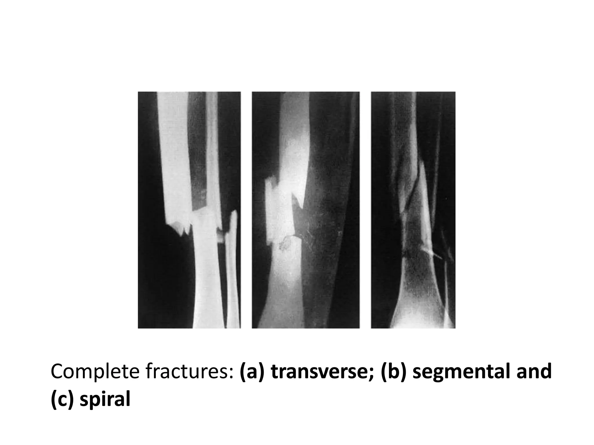
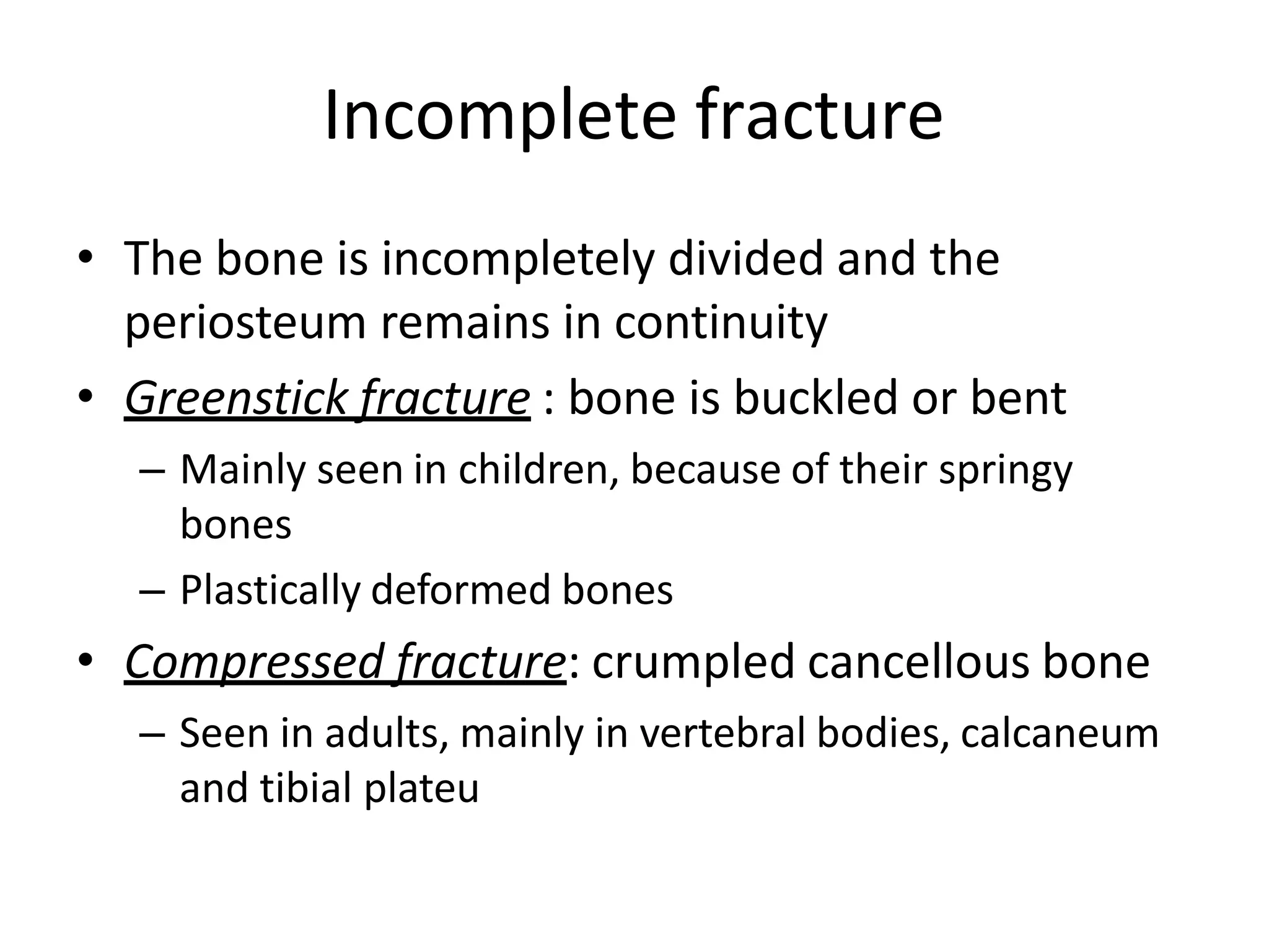
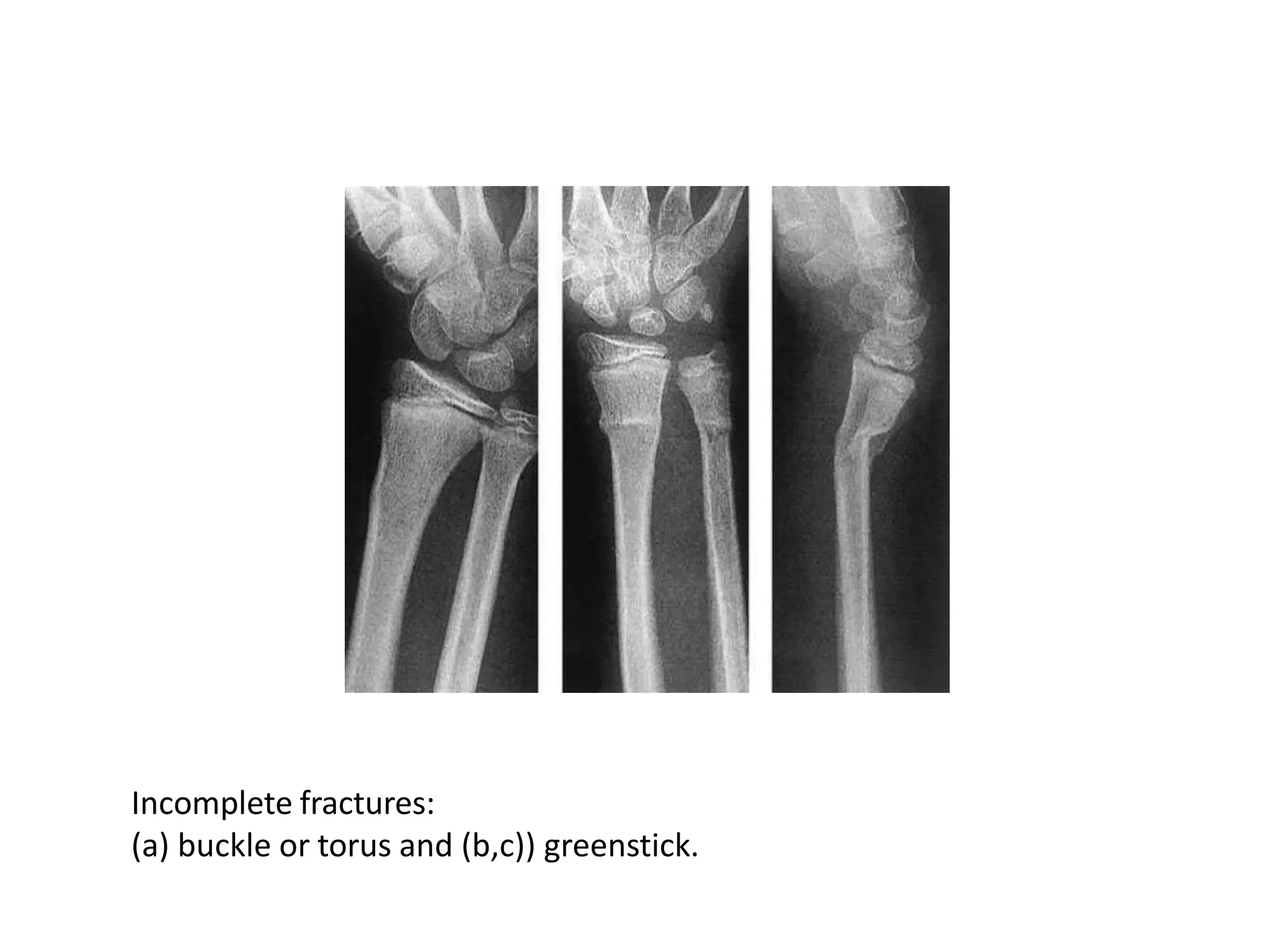
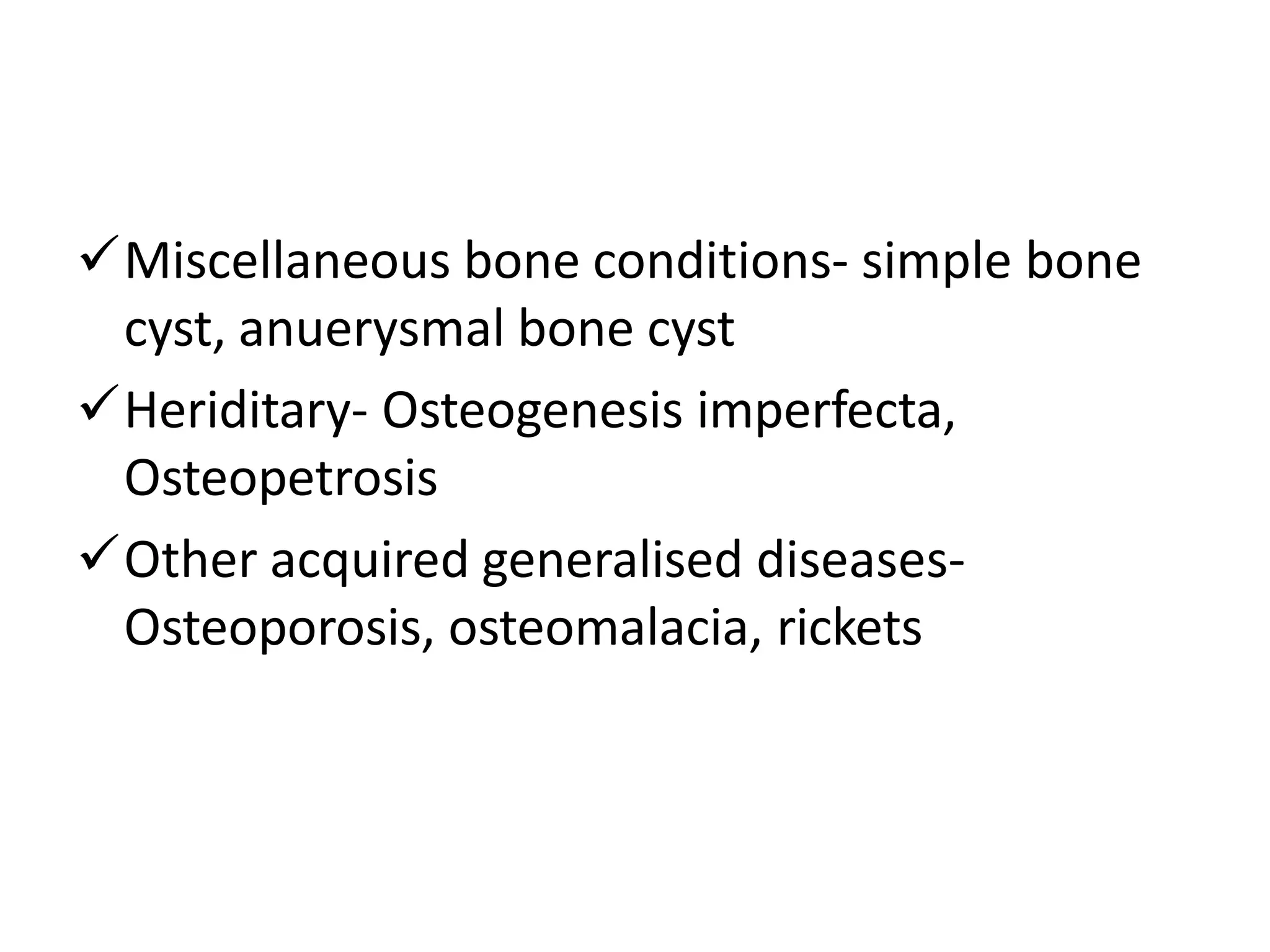
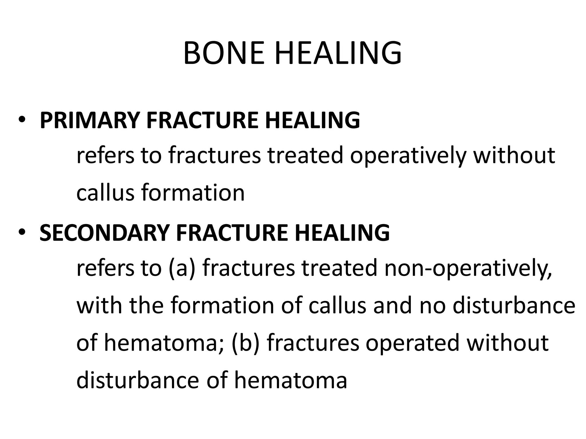
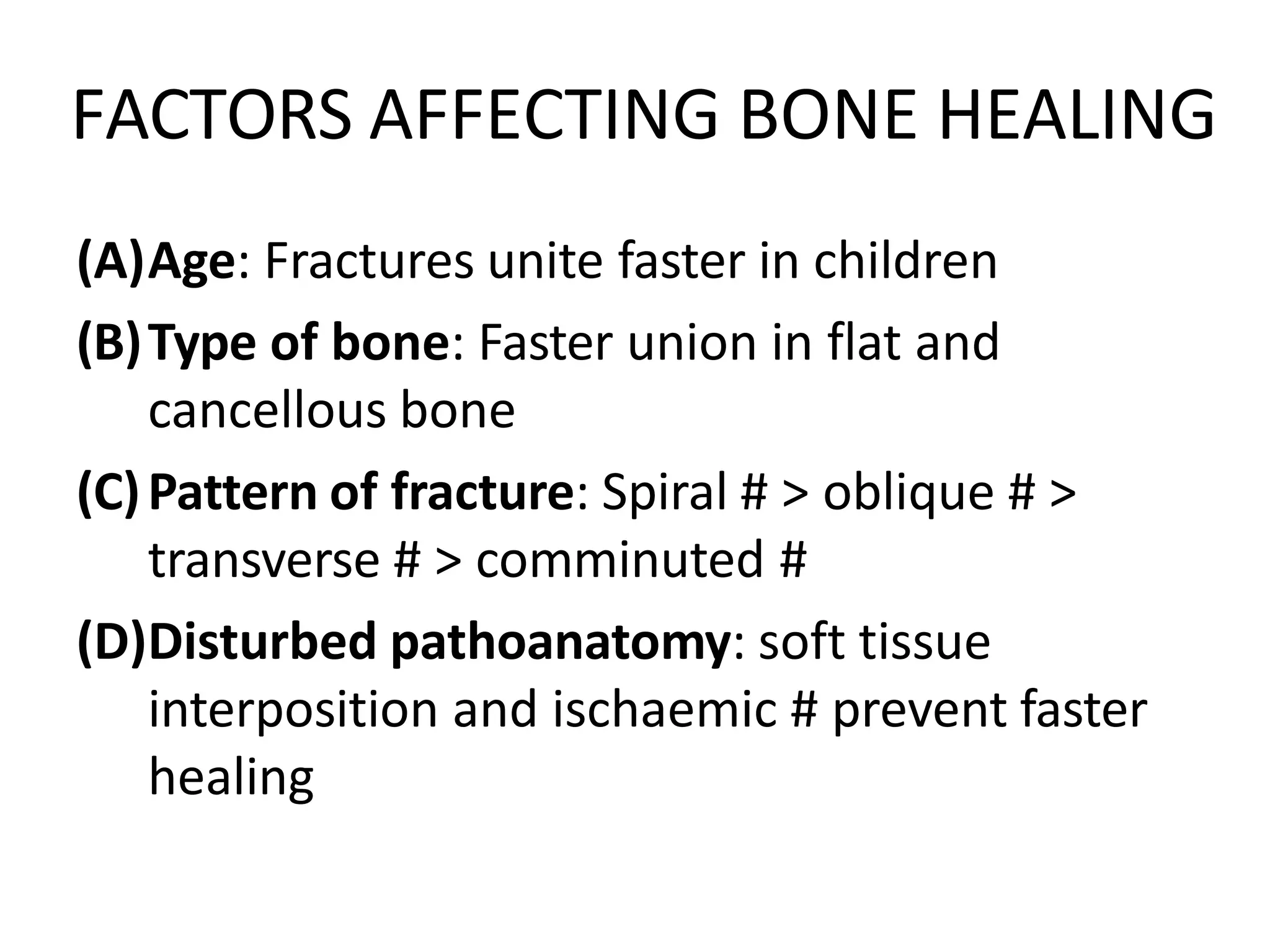
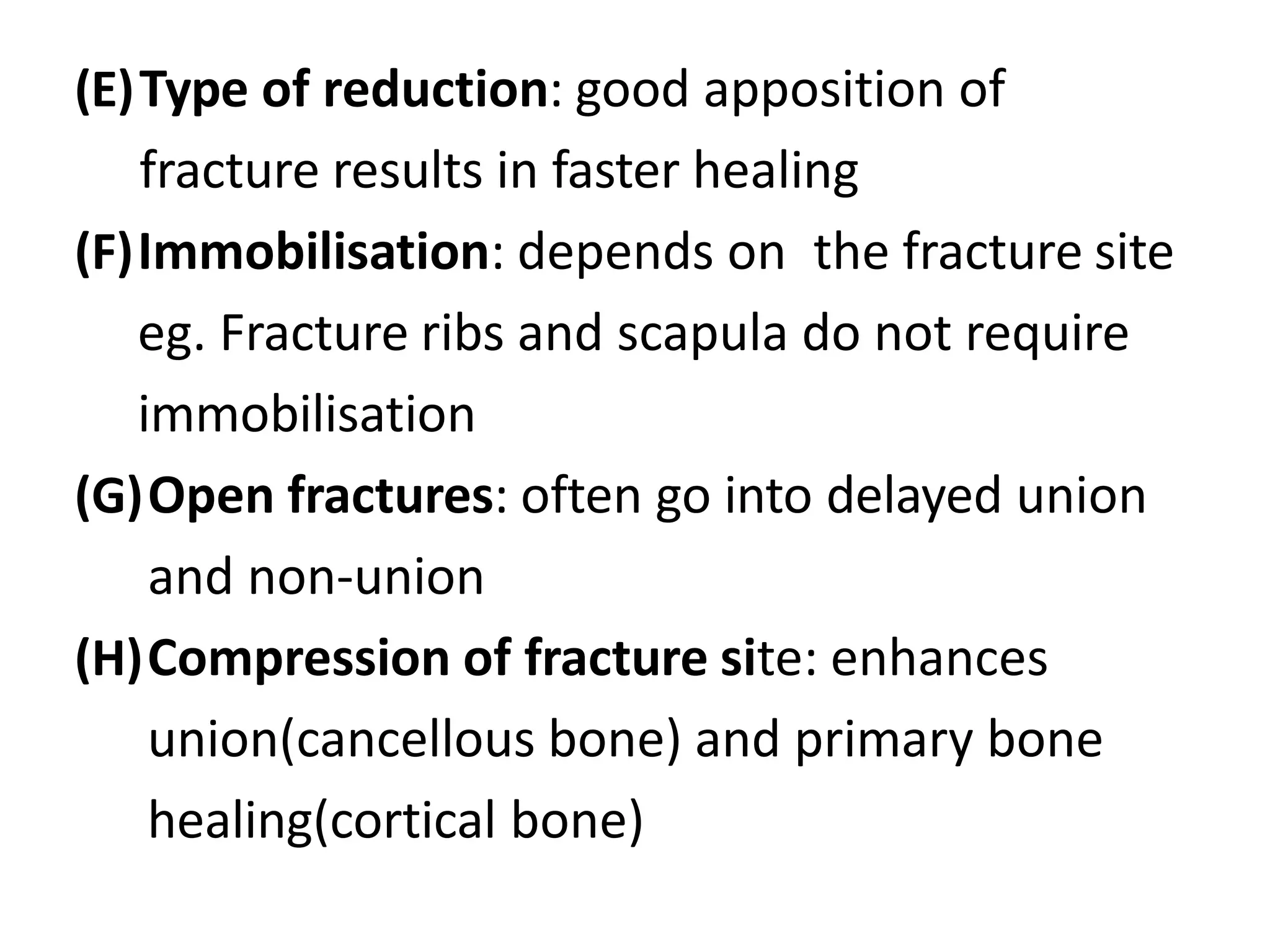
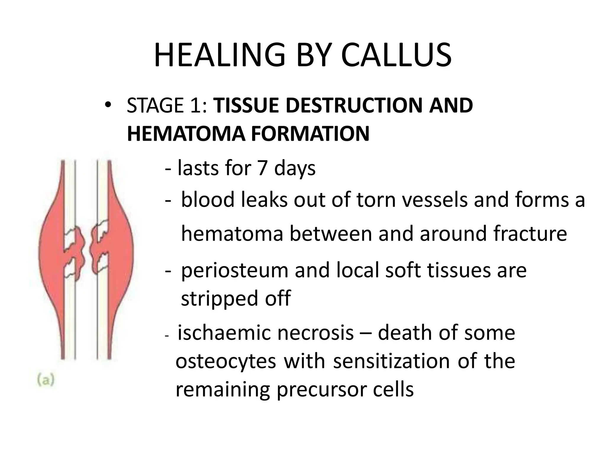
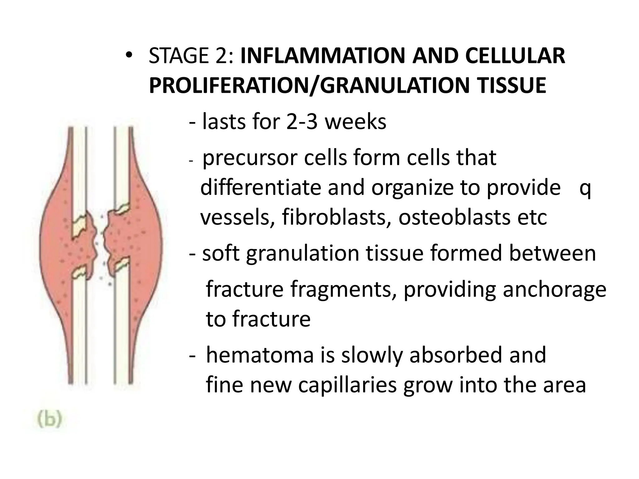
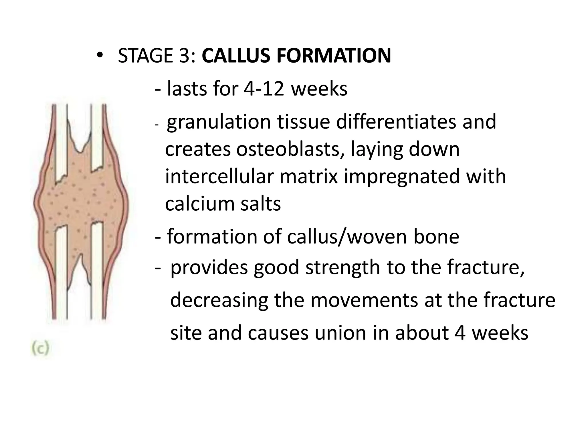
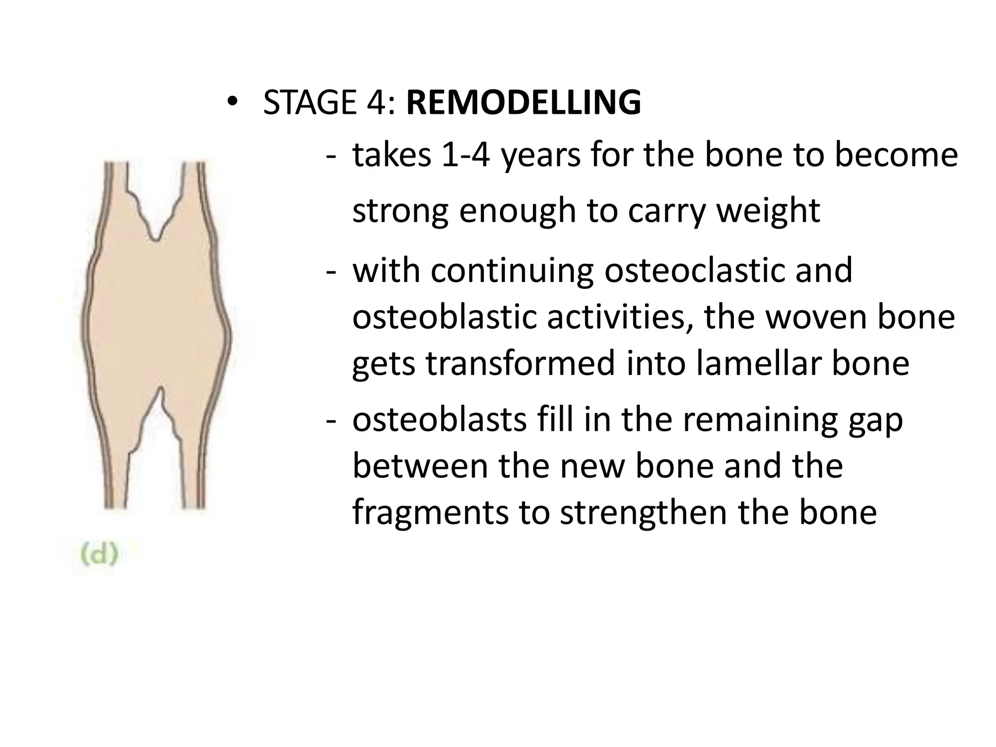
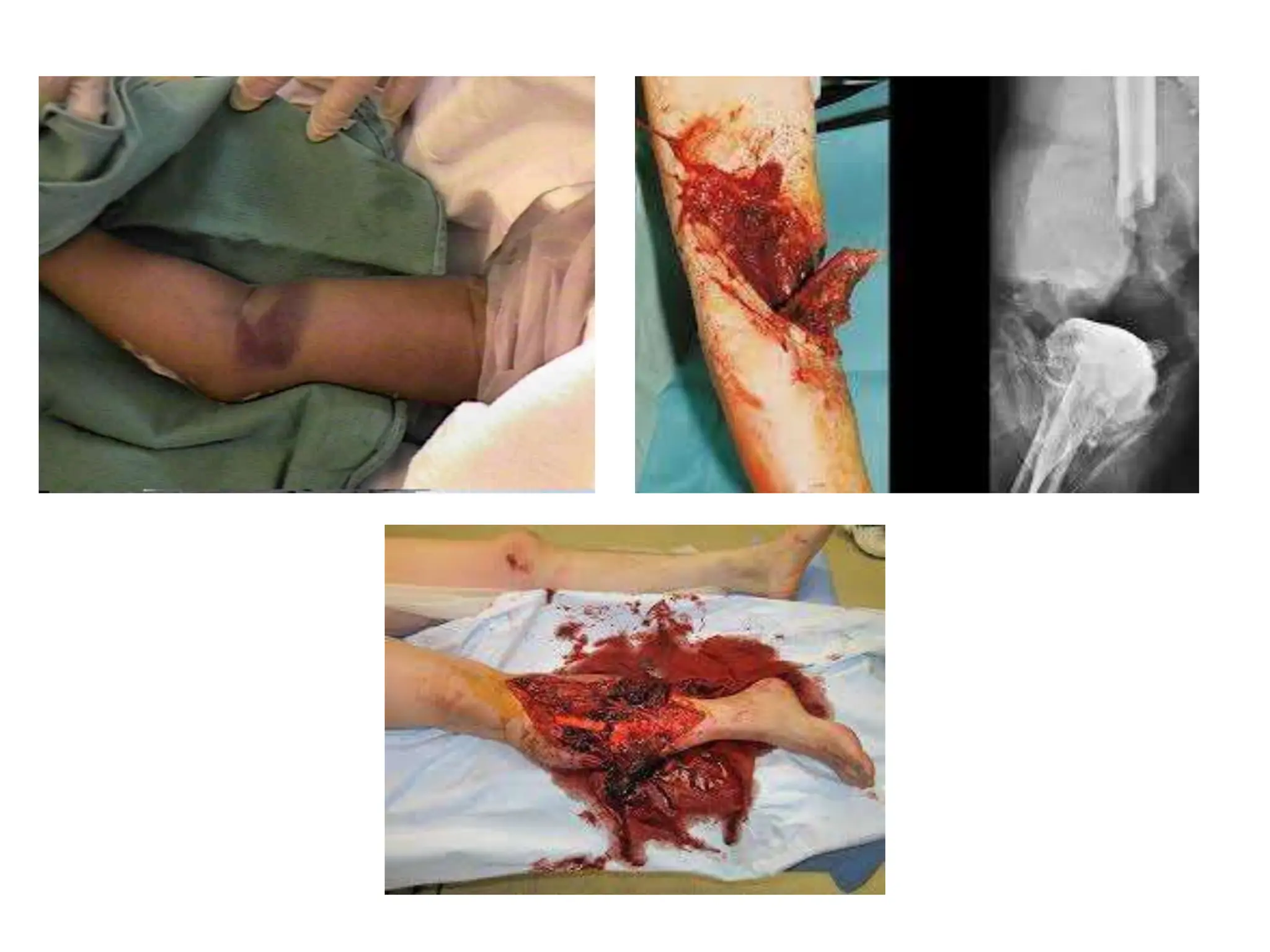
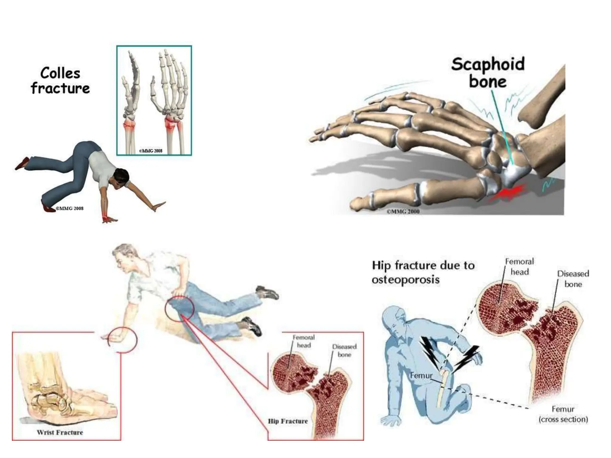
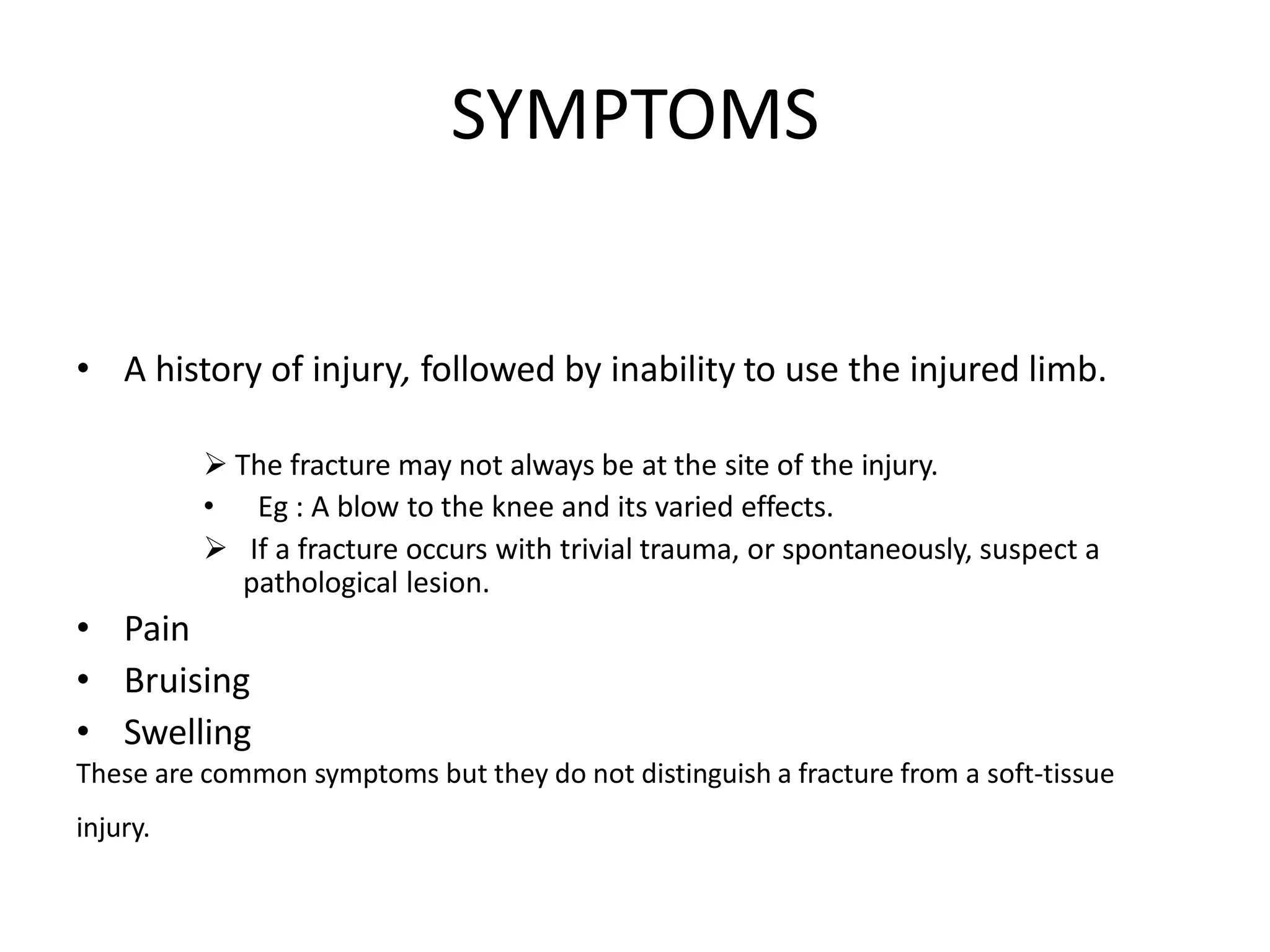
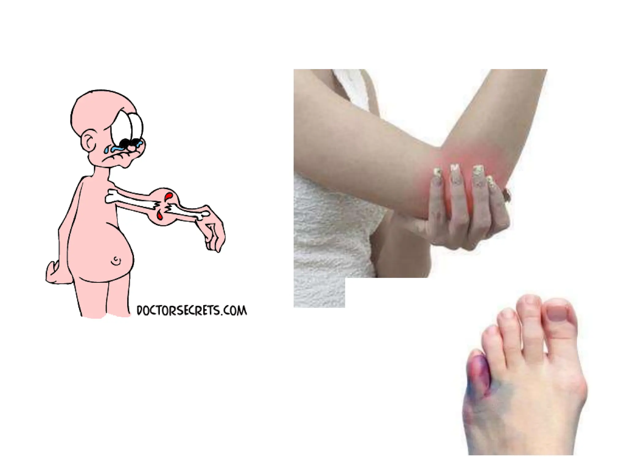
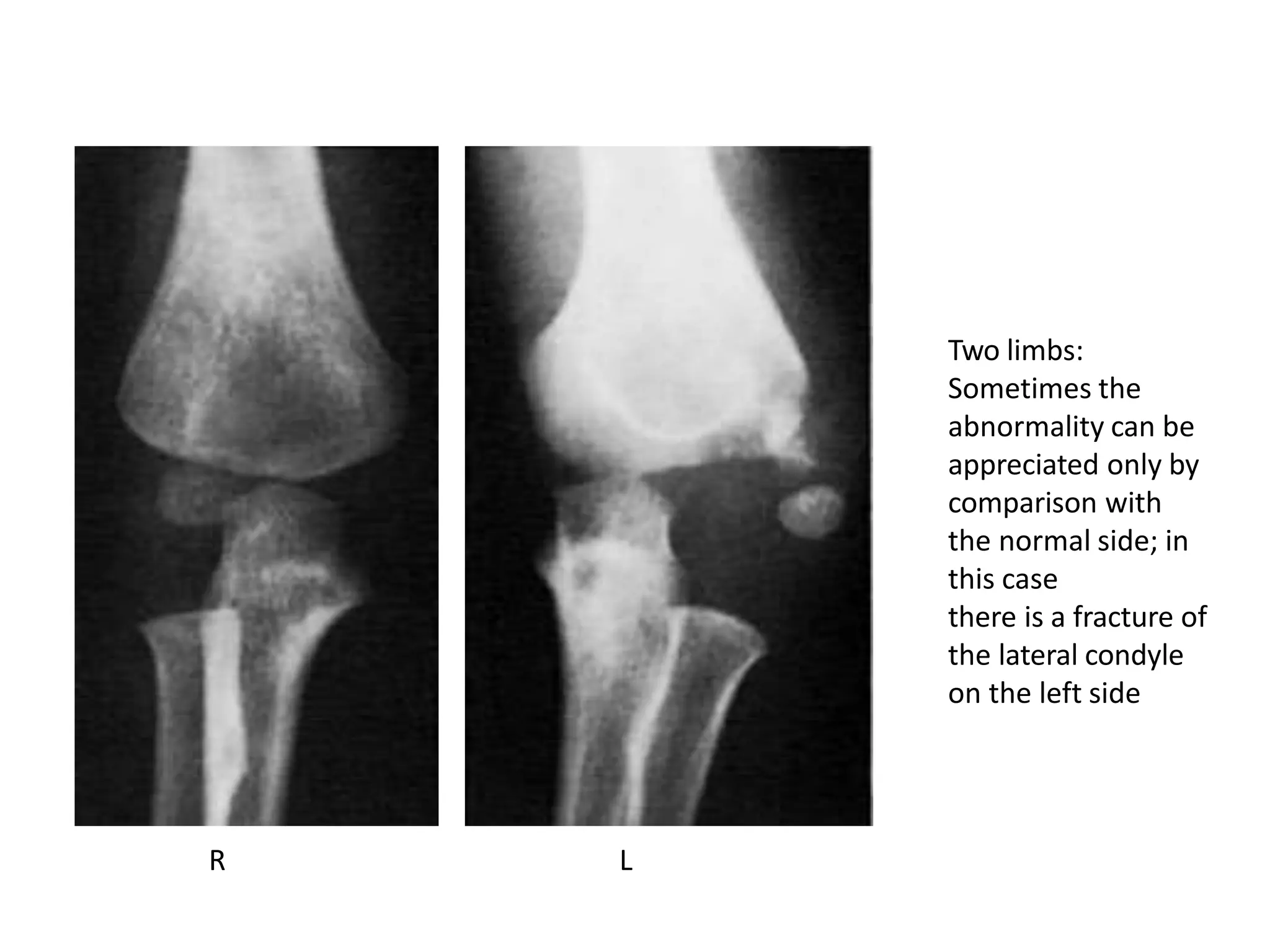
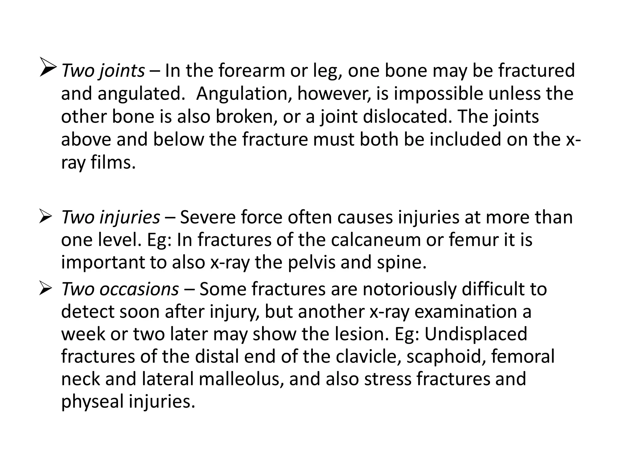
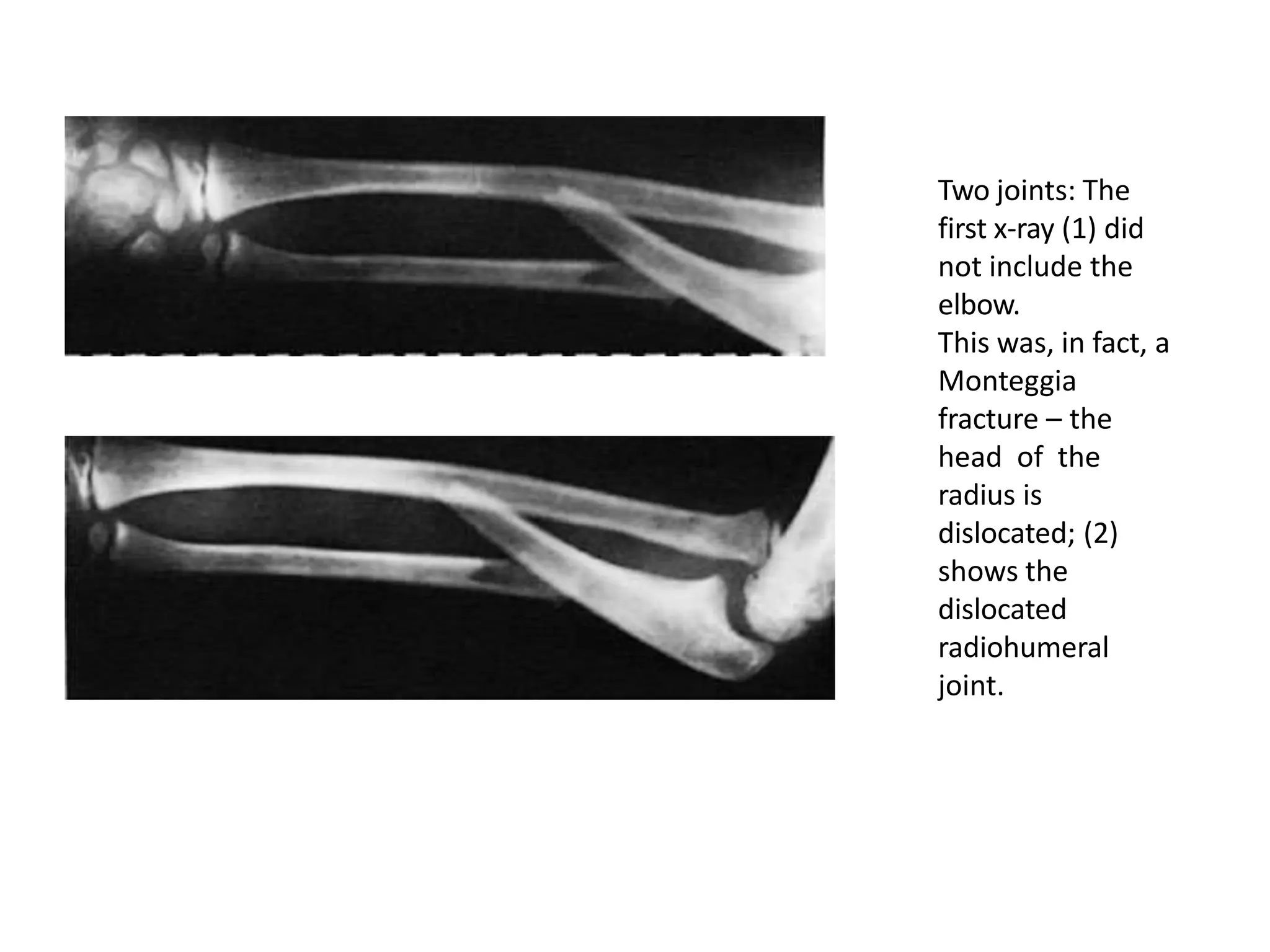
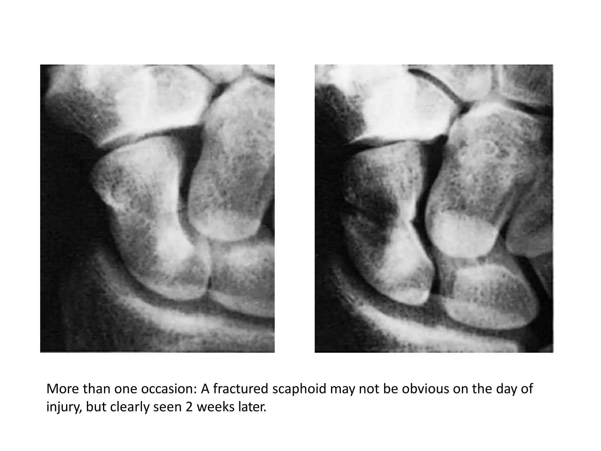
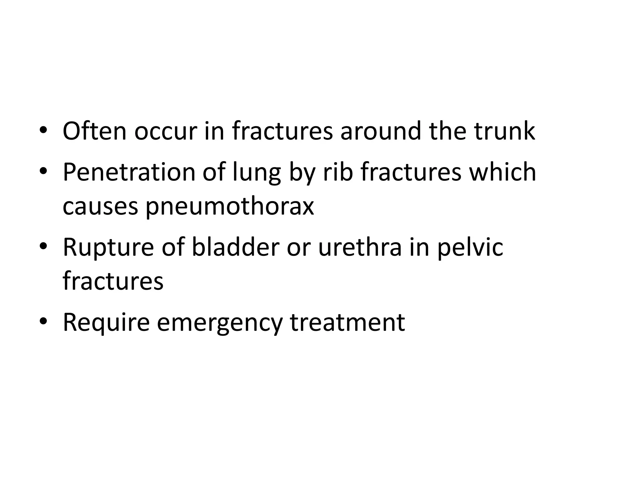
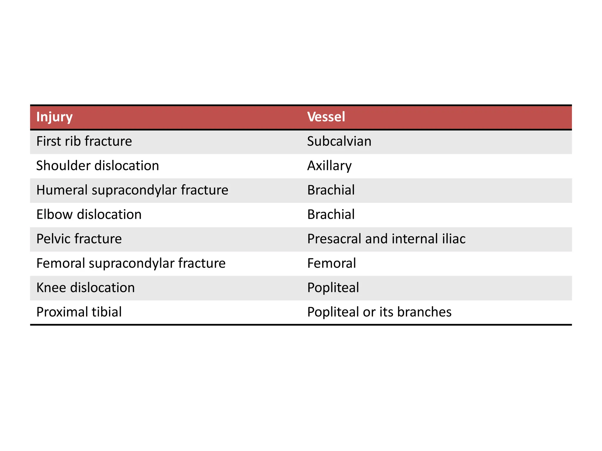
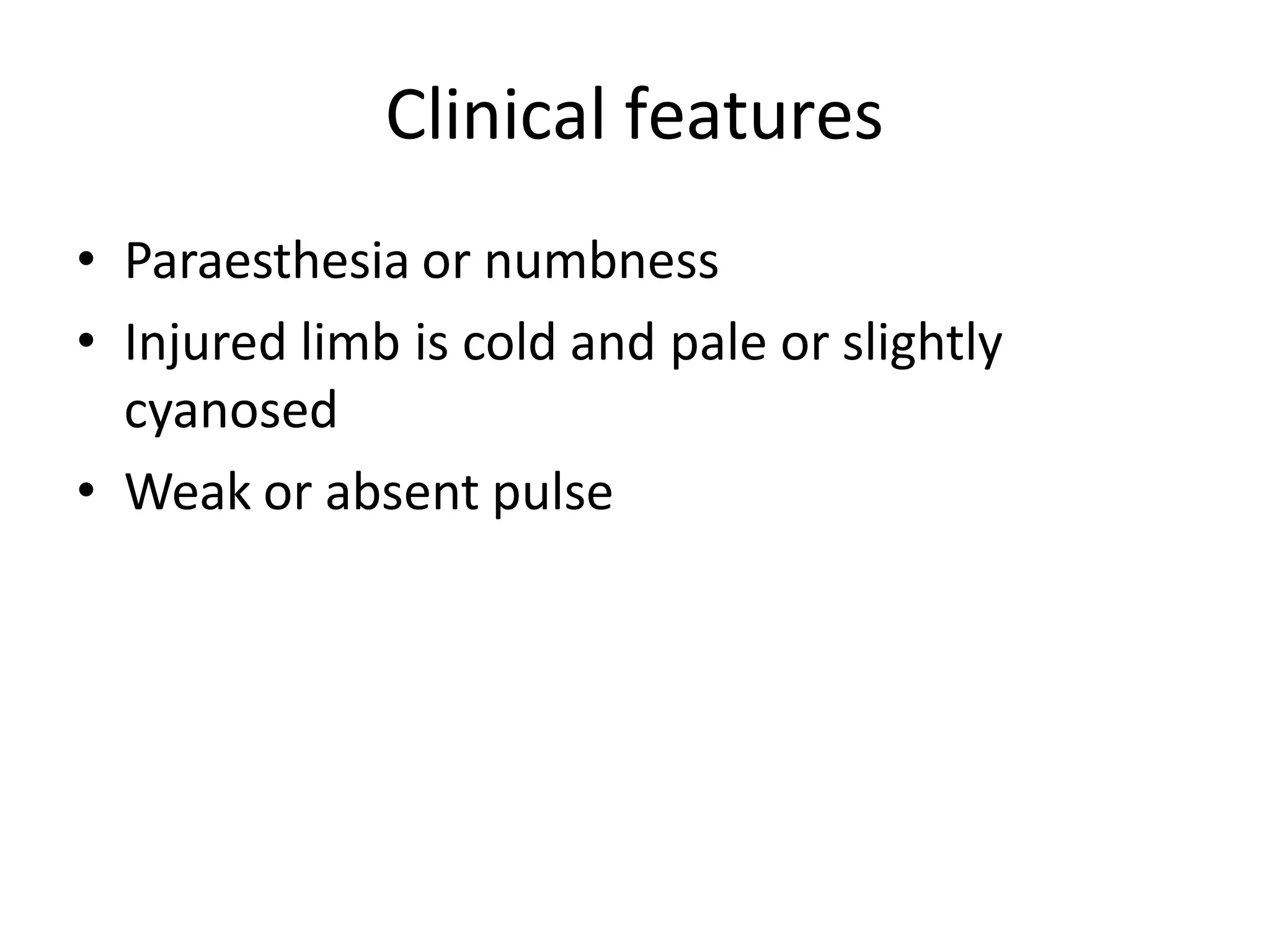
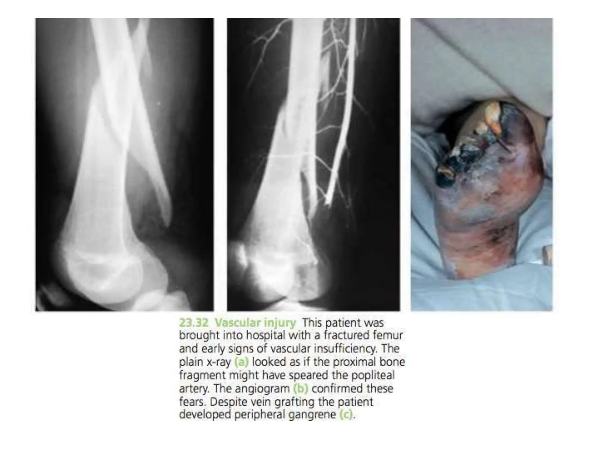
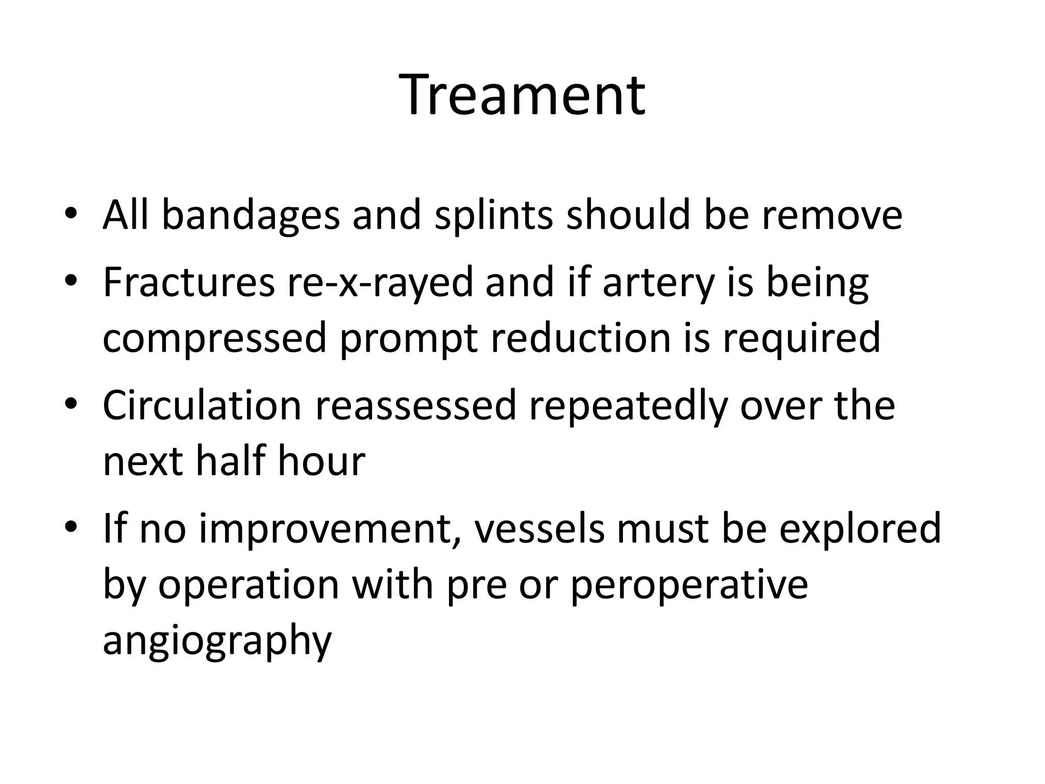
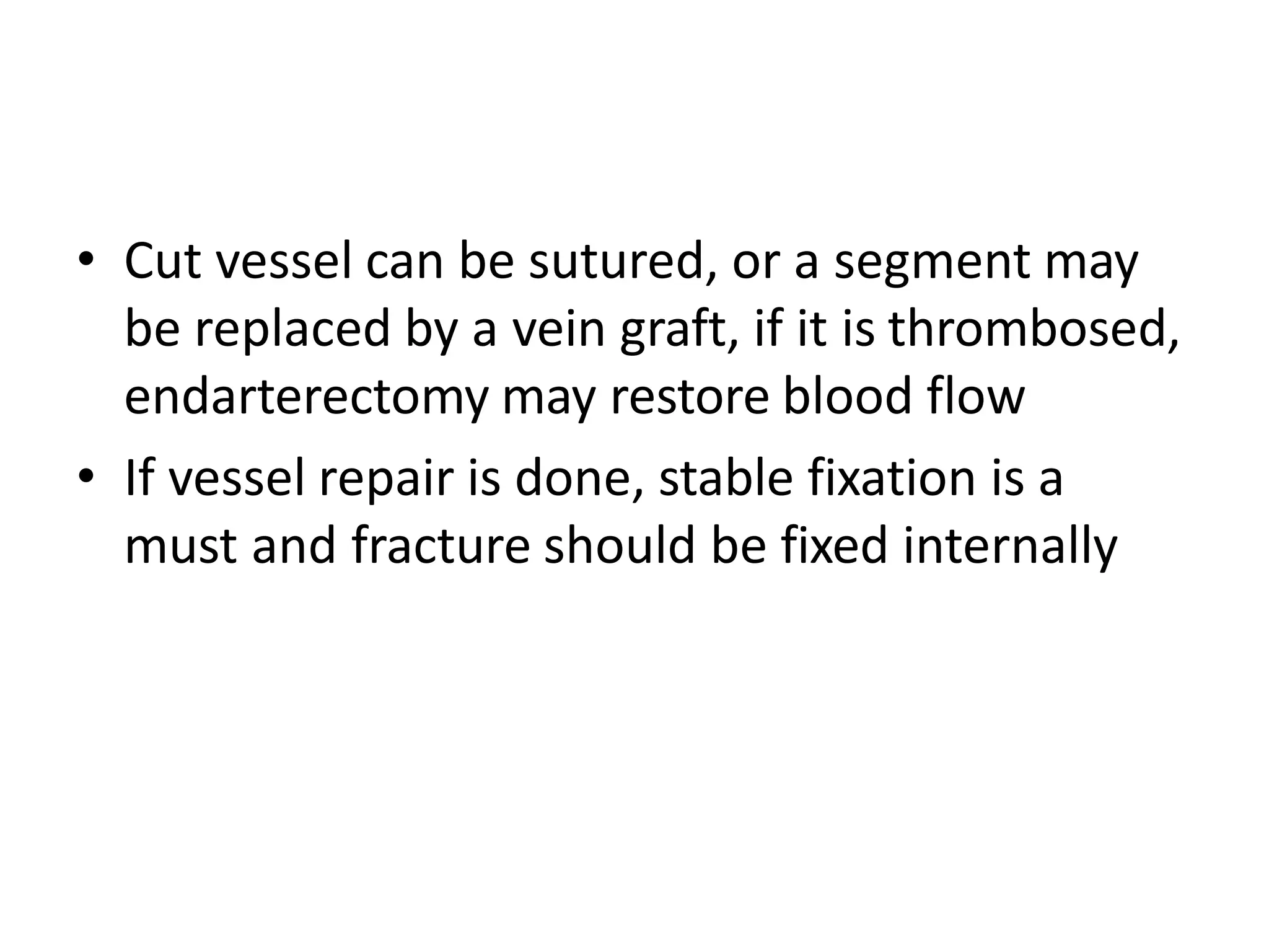
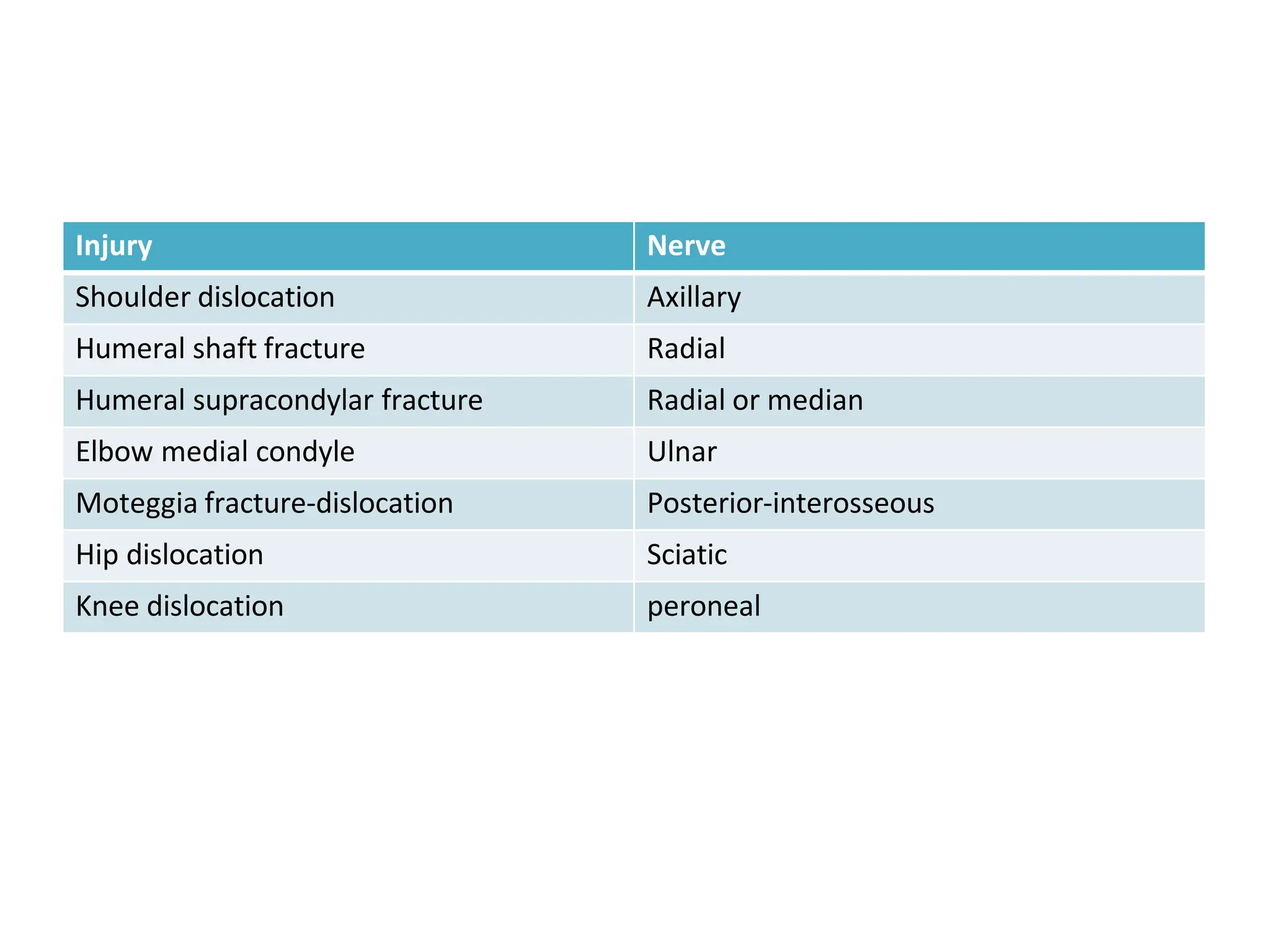
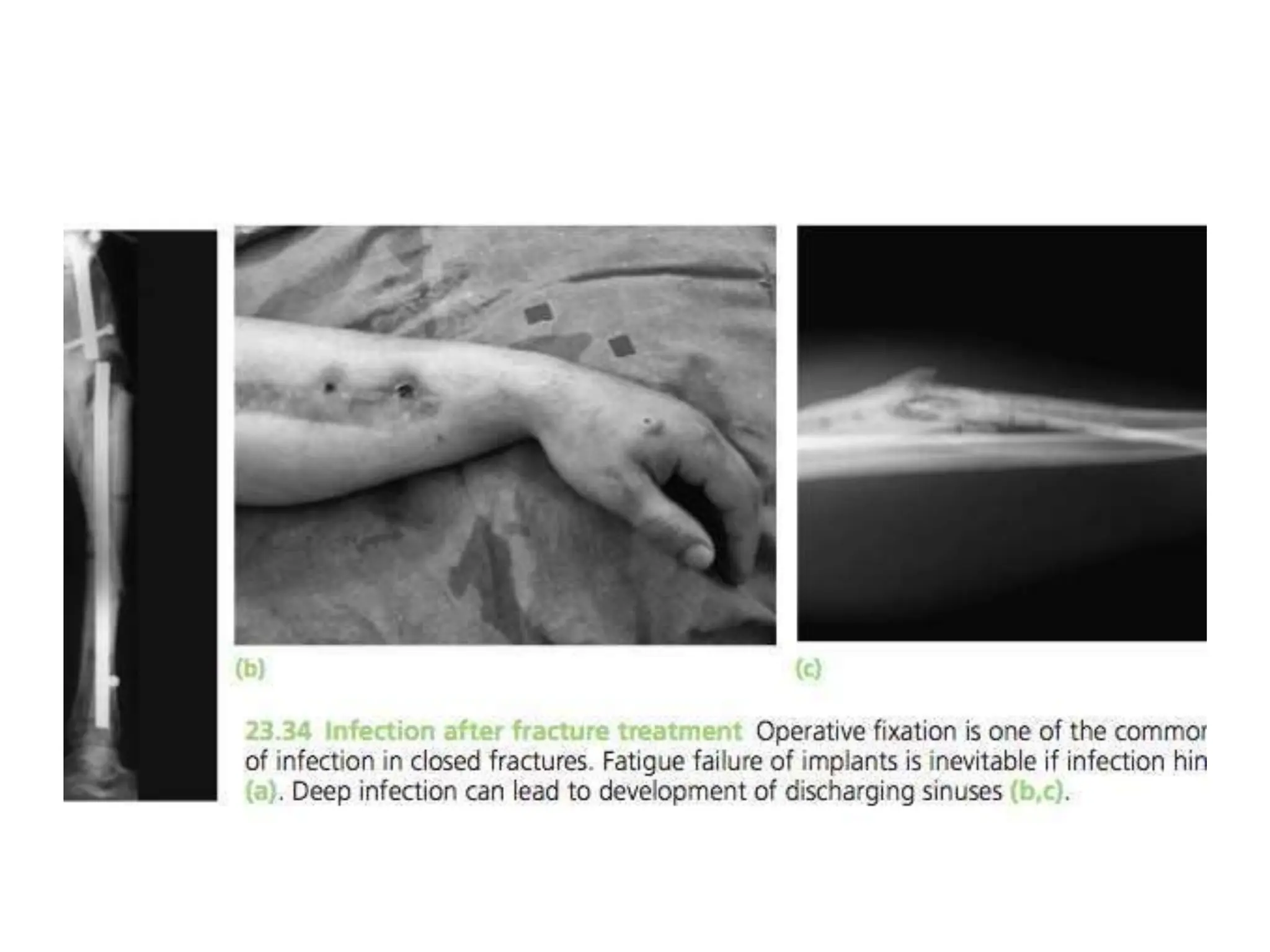
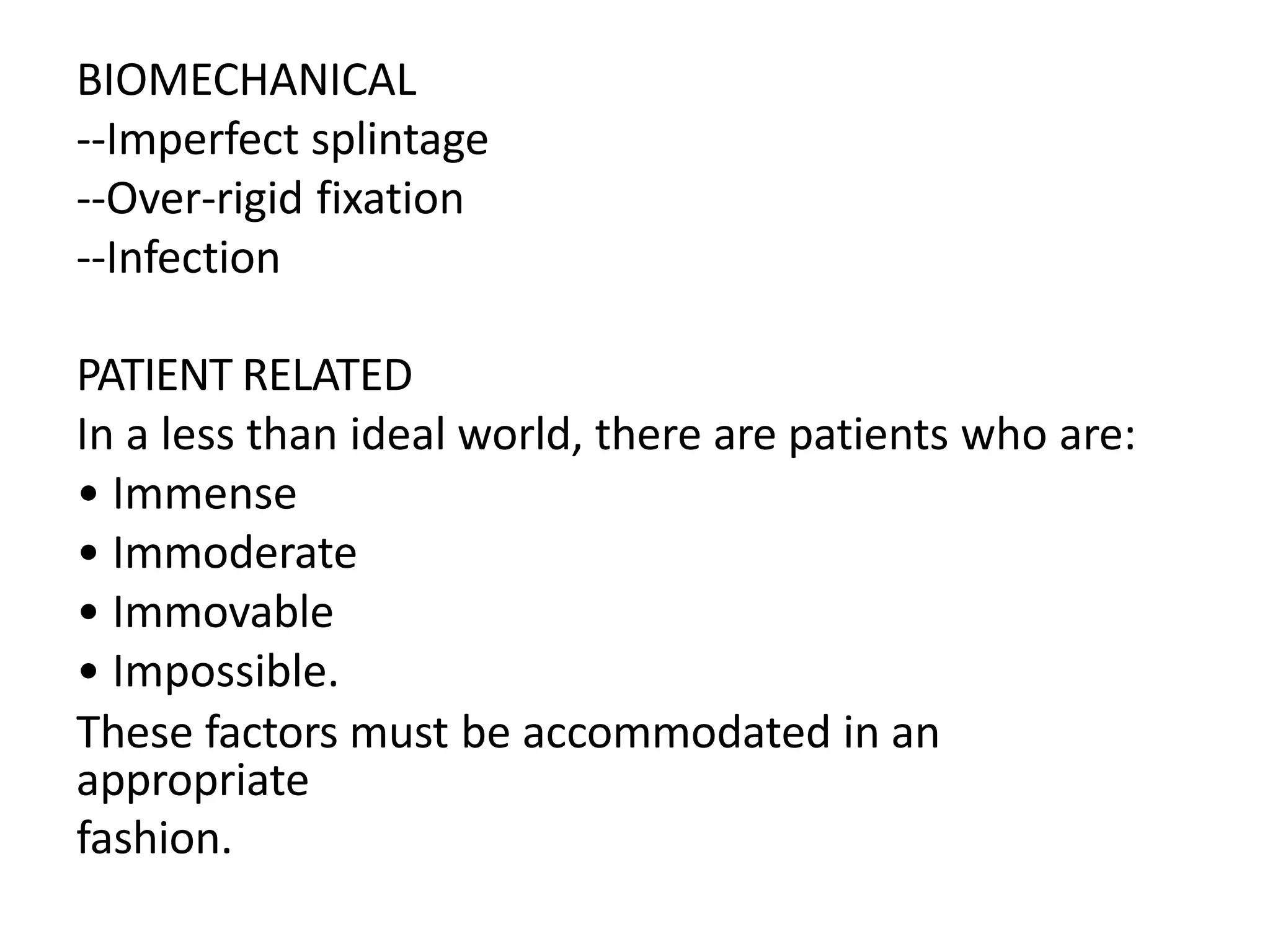
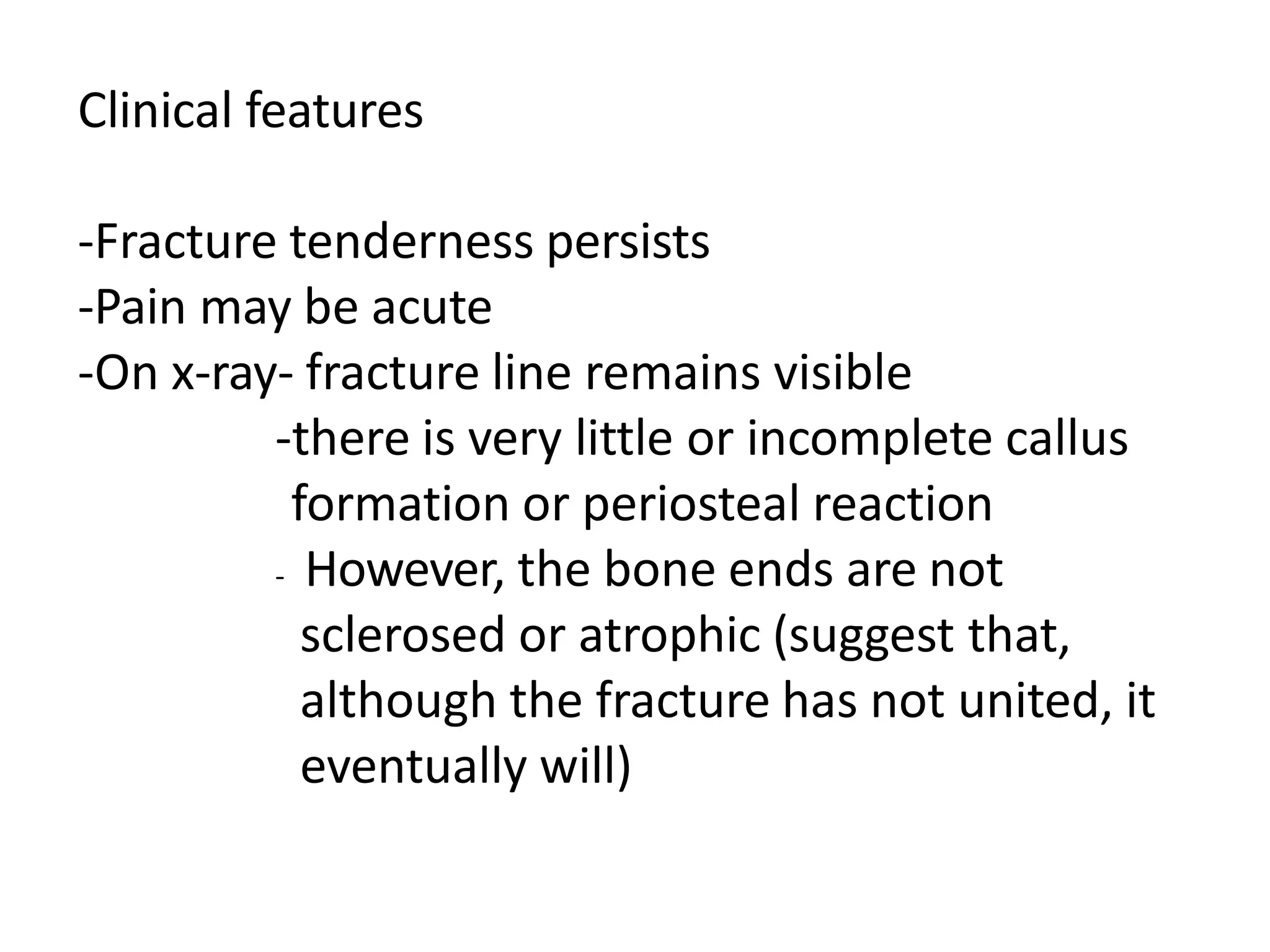
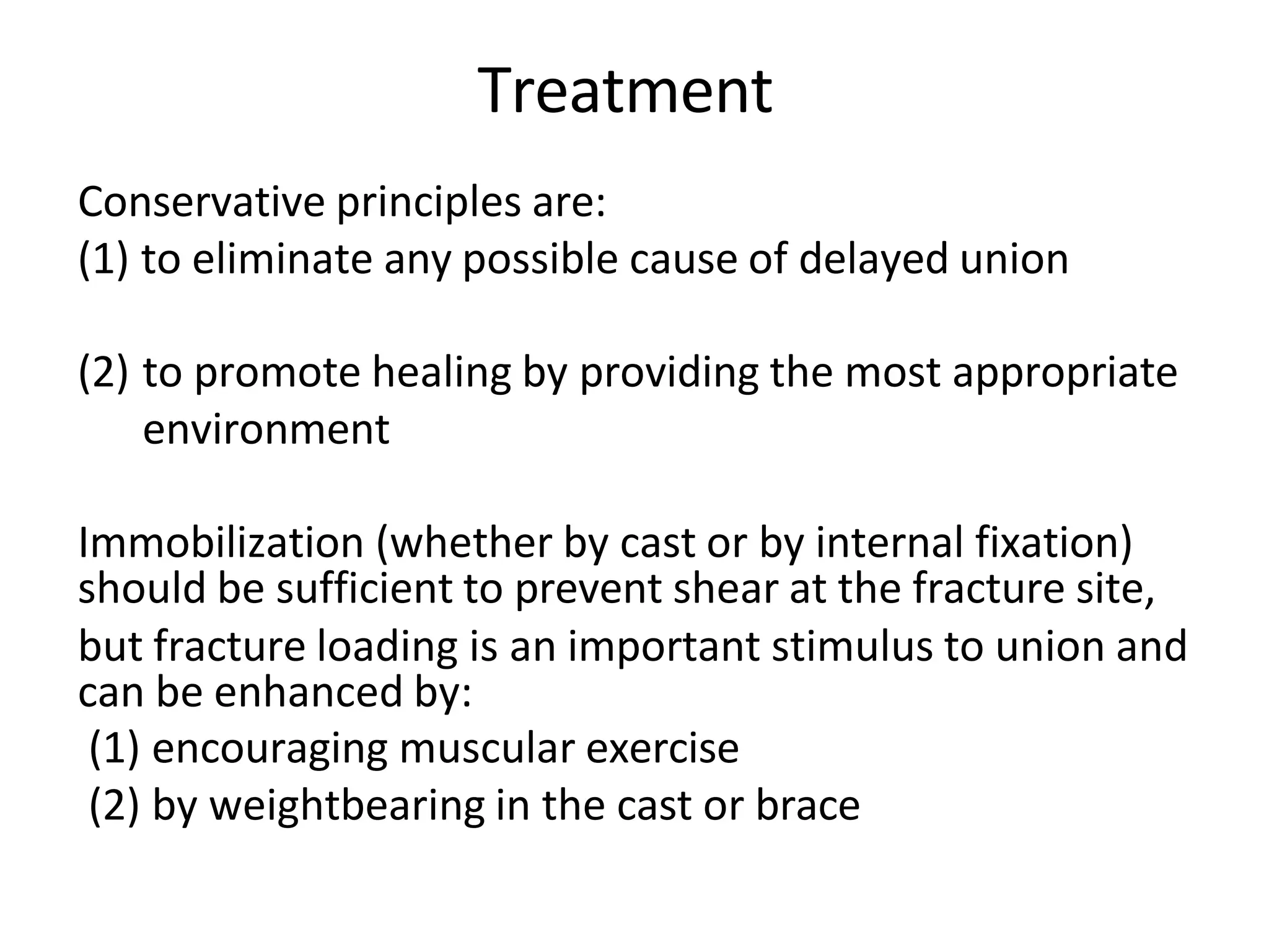
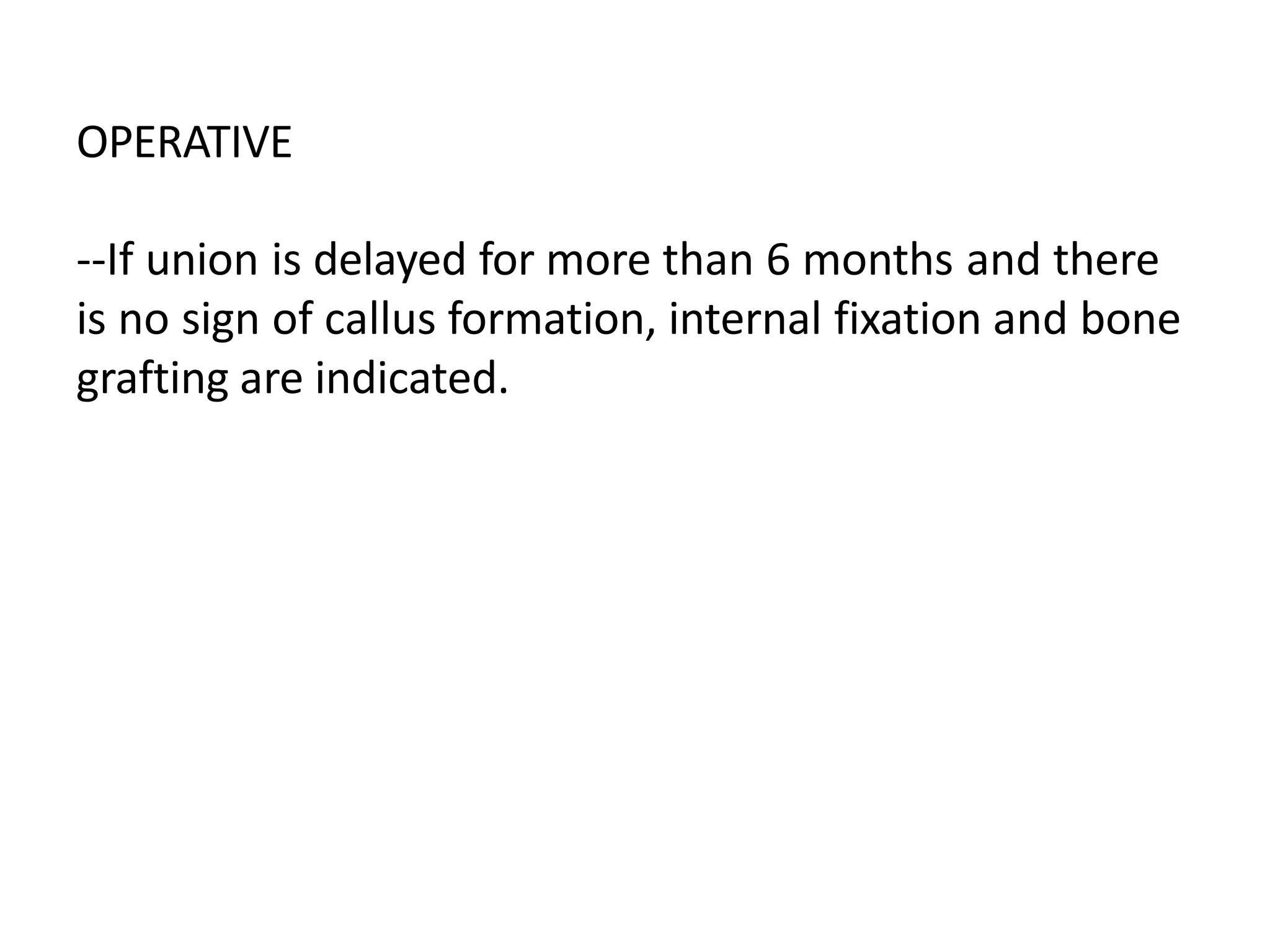
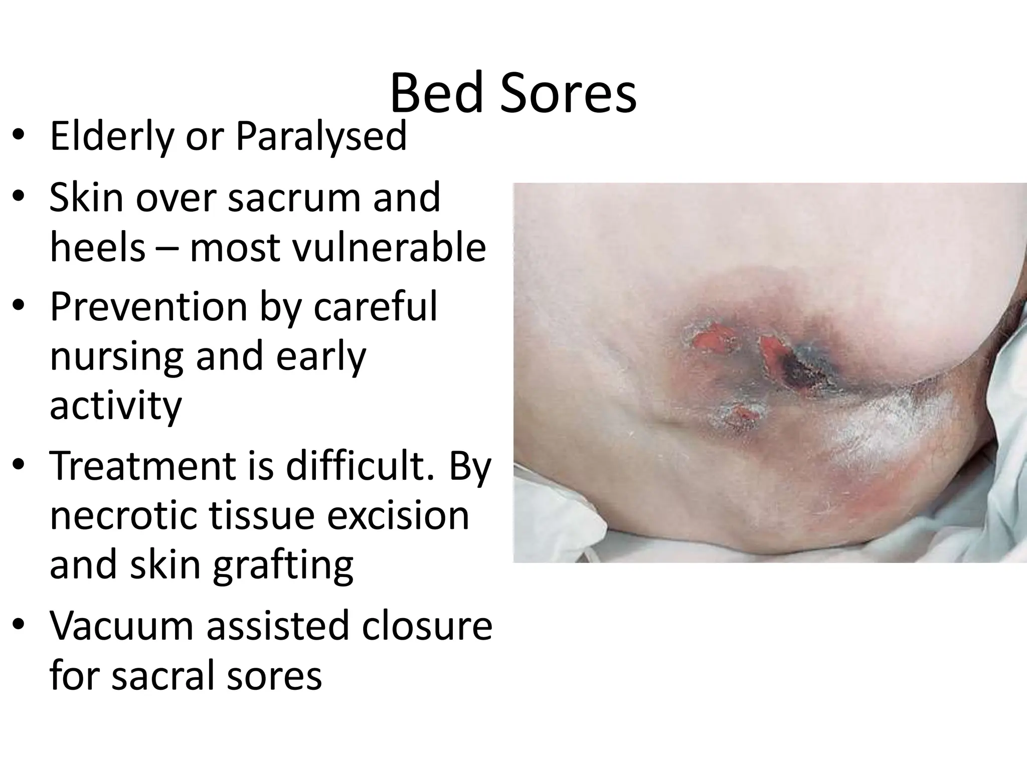
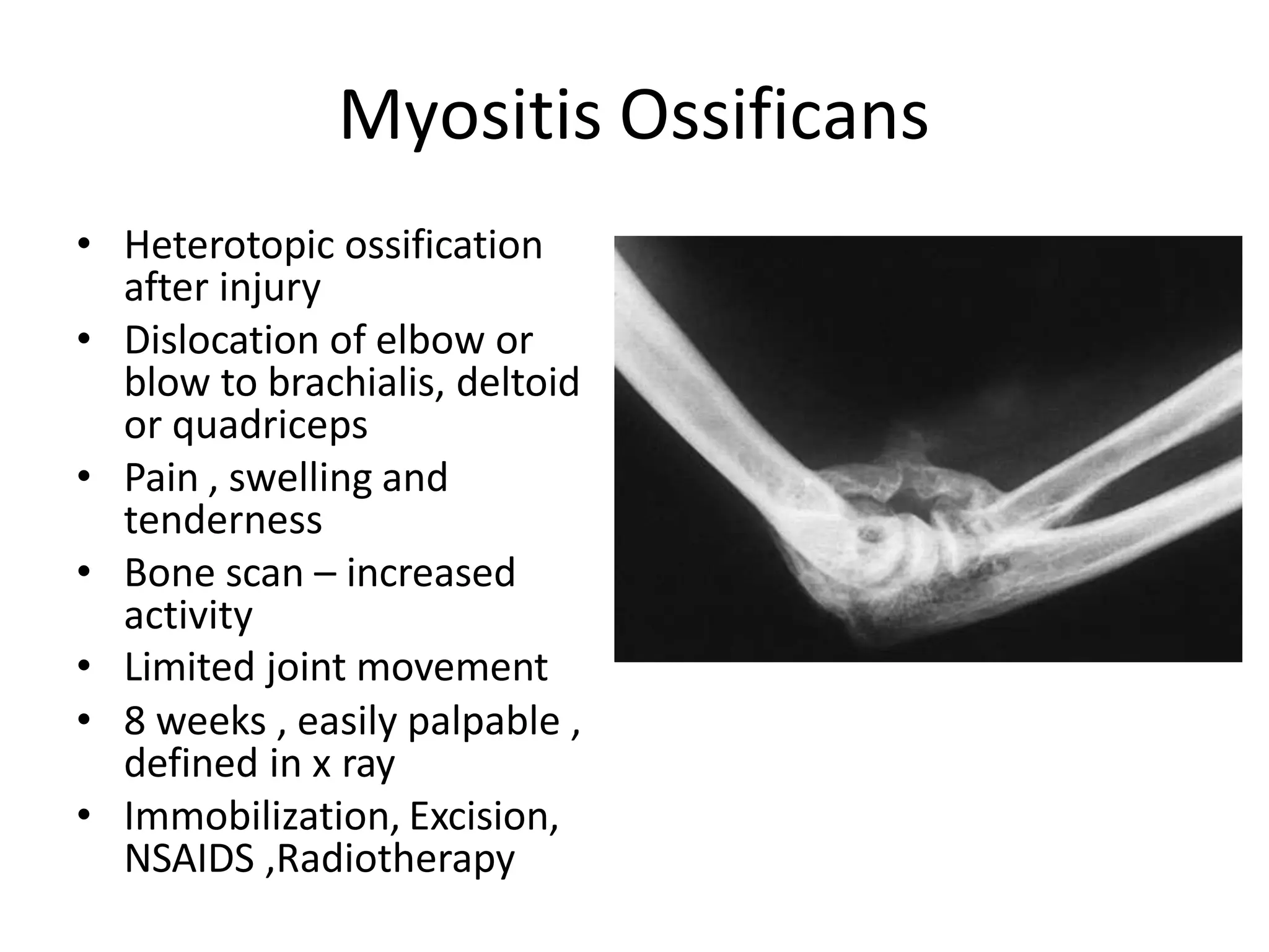
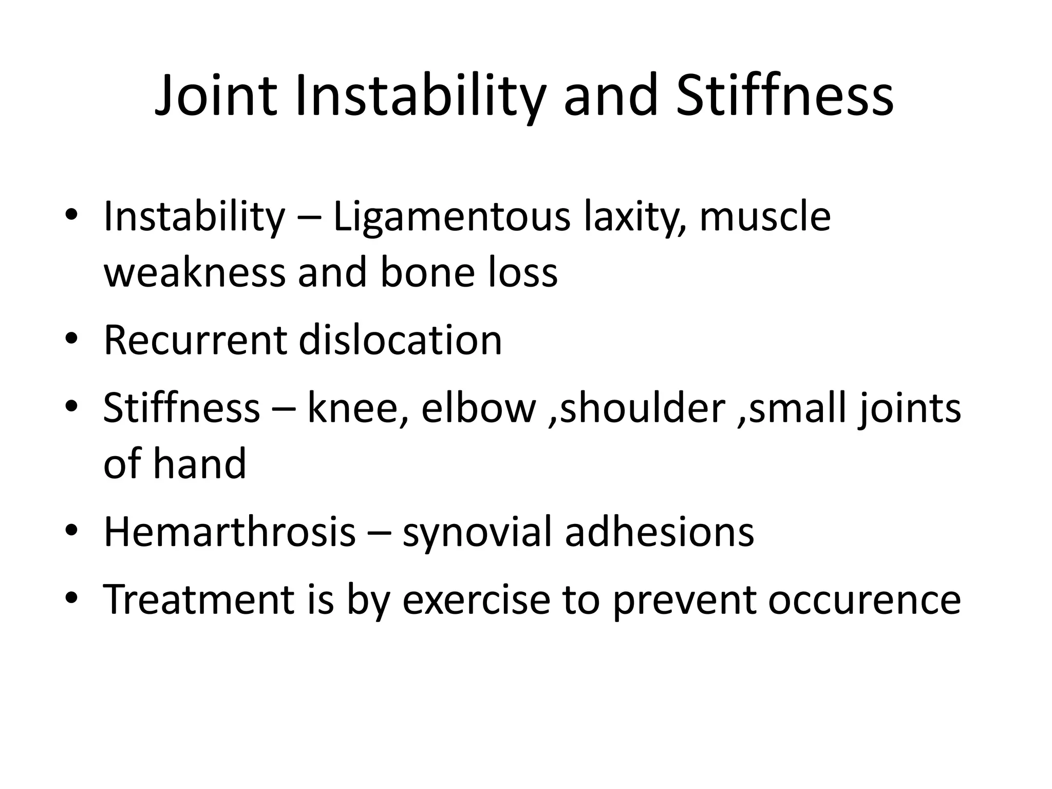
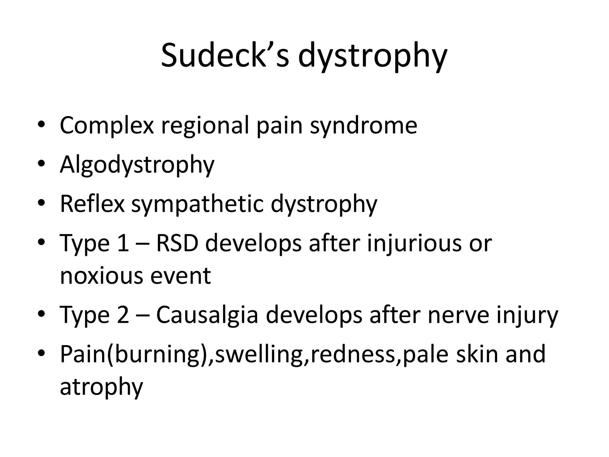
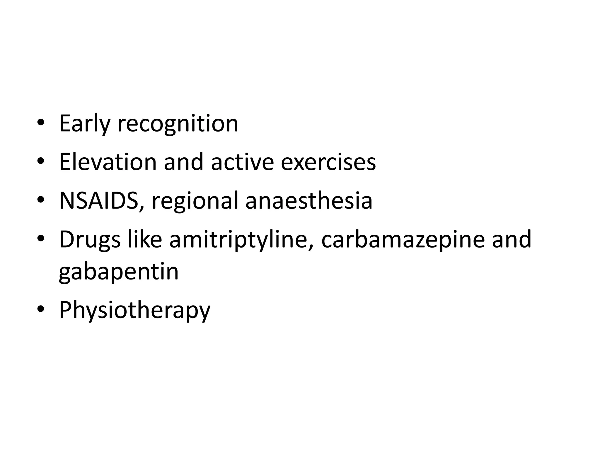
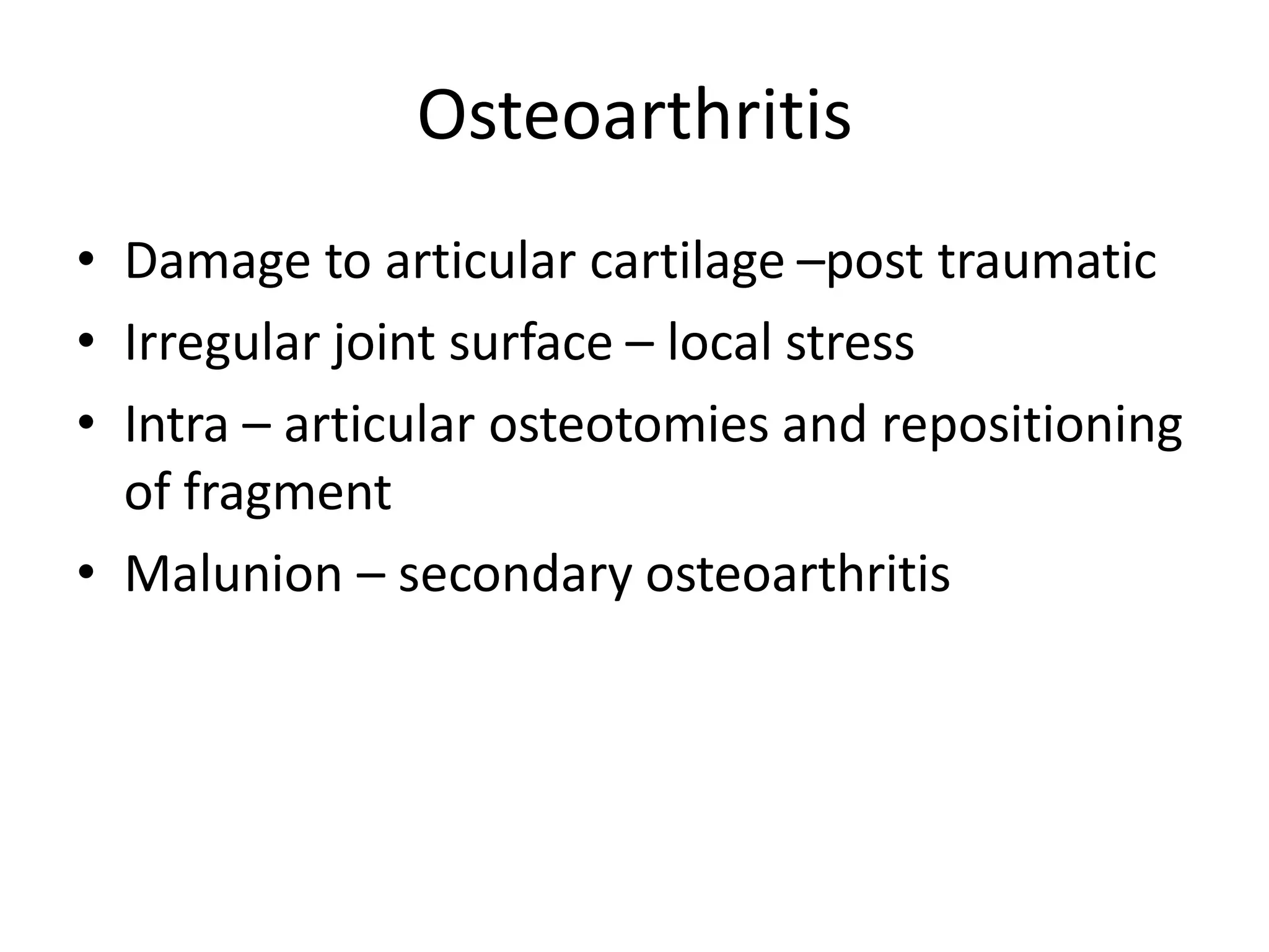
SlideShare a Scribd company logo
1 of 112
FRACTURE-TYPES, HEALING,
COMPLICATIONS, TREATMENT
Introduction
• Break in the structural continuity of a bone
• If the overlying skin remains intact - closed
(simple fracture)
• If skin or one of the body cavities is breached-
open (compound fracture)
Types of Fracture
• Divided in to
– Complete
– Incomplete
Complete fracture
Bone is split into two or more fragments. The
fracture pattern on x-ray can help predict behaviour
after reduction
• in a transverse fracture the fragments usually remain in
place after reduction
• if it is oblique or spiral, they tend to shorten and re-
displace even if the bone is splinted.
• In an impacted fracture the fragments are jammed
tightly together and the fracture line is indistinct.
• A comminuted fracture is one in which there are more
than two fragments
Complete fractures: (a) transverse; (b) segmental and
(c) spiral
Incomplete fracture
• The bone is incompletely divided and the
periosteum remains in continuity
• Greenstick fracture : bone is buckled or bent
– Mainly seen in children, because of their springy
bones
– Plastically deformed bones
• Compressed fracture: crumpled cancellous bone
– Seen in adults, mainly in vertebral bodies, calcaneum
and tibial plateu
Incomplete fractures:
(a) buckle or torus and (b,c)) greenstick.
Muller’s Classification
(a) Each long bone has three
segments – proximal
Diaphyseal
Distal
the proximal and distal segments are each
defined by a square based on
the widest part of the bone.
(b,c,d) Diaphyseal fractures
may be simple
wedge
complex.
(e,f,g) Proximal and distal fractures may
be extra-articular,
partial articular
complete articular.
How fractures are displaced
• Fractures can be displaced by:
– Force of the injury
– Effects of gravity
– Pull of muscles attached to the site
Types of displacement
• Translation (shift)- the fragments may shift
sideways, backwards or forwards
• Angulation (tilt)- mal alignment if
unconnected will lead to limb deformity
• Rotation (twist)- rotational deformity
• Length- can cause shortening of the bone
Mechanism of injury
• Injury
• Repetitive stress
• Pathological fractures
• INJURY
o Direct force- With a direct force, the bone
breaks at the point of impact; the soft tissues
also are damaged.
o Indirect force-the bone breaks at a distance
from where the force is applied.
Some fracture patterns reveals the dominant
mechanism:
 Spiral pattern- twisting
 Oblique- compression
 Triangular- bending
 Transverse- tension
Some fracture patterns suggest the causal mechanism: (a) spiral pattern (twisting); (b) short
oblique pattern (compression); (c) triangular ‘butterfly’ fragment (bending) and (d) transverse pattern
(tension). Spiral and some (long) oblique patterns are usually due to low-energy indirect injuries; bending and
transverse patterns are caused by
high-energy direct trauma.
• FATIGUE OR STRESS FRACTURES-
Occur in normal bone, subject to repeated
heavy loading, typically in athletes, dancers or
military personnel.
Drugs like steroids and methotrexate
• PATHOLOGICAL FRACTURES- Occurs in a bone
that is made weak by some disease.
Causes-
 Inflammatory- Osteomyelitis
Neoplastic- giant cell tumour, Ewings sarcoma,
secondaries
Miscellaneous bone conditions- simple bone
cyst, anuerysmal bone cyst
Heriditary- Osteogenesis imperfecta,
Osteopetrosis
Other acquired generalised diseases-
Osteoporosis, osteomalacia, rickets
BONE HEALING
• PRIMARY FRACTURE HEALING
refers to fractures treated operatively without
callus formation
• SECONDARY FRACTURE HEALING
refers to (a) fractures treated non-operatively,
with the formation of callus and no disturbance
of hematoma; (b) fractures operated without
disturbance of hematoma
FACTORS AFFECTING BONE HEALING
(A)Age: Fractures unite faster in children
(B)Type of bone: Faster union in flat and
cancellous bone
(C)Pattern of fracture: Spiral # > oblique # >
transverse # > comminuted #
(D)Disturbed pathoanatomy: soft tissue
interposition and ischaemic # prevent faster
healing
(E)Type of reduction: good apposition of
fracture results in faster healing
(F)Immobilisation: depends on the fracture site
eg. Fracture ribs and scapula do not require
immobilisation
(G)Open fractures: often go into delayed union
and non-union
(H)Compression of fracture site: enhances
union(cancellous bone) and primary bone
healing(cortical bone)
HEALING BY CALLUS
• STAGE 1: TISSUE DESTRUCTION AND
HEMATOMA FORMATION
- lasts for 7 days
- blood leaks out of torn vessels and forms a
hematoma between and around fracture
- periosteum and local soft tissues are
stripped off
- ischaemic necrosis – death of some
osteocytes with sensitization of the
remaining precursor cells
• STAGE 2: INFLAMMATION AND CELLULAR
PROLIFERATION/GRANULATION TISSUE
- lasts for 2-3 weeks
- precursor cells form cells that
differentiate and organize to provide q
vessels, fibroblasts, osteoblasts etc
- soft granulation tissue formed between
fracture fragments, providing anchorage
to fracture
- hematoma is slowly absorbed and
fine new capillaries grow into the area
• STAGE 3: CALLUS FORMATION
- lasts for 4-12 weeks
- granulation tissue differentiates and
creates osteoblasts, laying down
intercellular matrix impregnated with
calcium salts
- formation of callus/woven bone
- provides good strength to the fracture,
decreasing the movements at the fracture
site and causes union in about 4 weeks
• STAGE 4: REMODELLING
- takes 1-4 years for the bone to become
strong enough to carry weight
- with continuing osteoclastic and
osteoblastic activities, the woven bone
gets transformed into lamellar bone
- osteoblasts fill in the remaining gap
between the new bone and the
fragments to strengthen the bone
• STAGE 5: MODELLING
- stage where the bone is gradually
strengthened
- shapening of the cortices occurs at the
endosteal and periosteal surfaces
- all these occur when the person starts
resuming his activities ie bearing weight
and muscle forces
- thicker lamellae are laid down where high
stresses are present, unwanted buttresses
are carved away and medullary cavity is
reformed
HEALING BY DIRECT UNION
• Formation of callus requires stimulus from
movement
• In cases of impacted fracture in cancellous
bone or a fracture immobilised by the use of
metal plate, callus will not be formed and
hence the fracture will heal by direct union
1. Osteoblastic new bone formation occurs directly
between the fracture fragments
2. New capillaries and osteoprogenitor cells grow in
from the edges and lay down new bone on the
exposed surface (gap healing)
3. Lamellar bone is produced in the narrow crevices
(<200Îźm) while woven bone is produced in the
wider gaps which is then remodelled into lamellar
bone
4. Penetration and bridging of the fracture by
osteoclasts and osteoblasts in 3-4 weeks.
5. Intimacy of the contact surfaces lead to
internal bridging without intermediate stages
(contact healing)
CLINICAL PRESENTATION OF
FRACTURES
In the Casualty..
• Take a Brief History.
• General Particulars:
• AGE & SEX
 Children and the elderly
 Different mechanisms of injury : Traumatic , Pathological
 Post menopausal women : Osteoporosis and pathological fractures.
• HISTORY OF TRAUMA – Ascertaining the mechanism of injury
is important, helps understand symptoms and aids
examination.
SYMPTOMS
• A history of injury, followed by inability to use the injured limb.
 The fracture may not always be at the site of the injury.
• Eg : A blow to the knee and its varied effects.
 If a fracture occurs with trivial trauma, or spontaneously, suspect a
pathological lesion.
• Pain
• Bruising
• Swelling
These are common symptoms but they do not distinguish a fracture from a soft-tissue
injury.
• Deformity – More suggestive of a fracture.
• Enquire about symptoms of associated injuries:
 Pain and swelling elsewhere (it is a common mistake to get distracted by
the main injury, particularly if it is severe),
 Numbness or Loss of movement.
 Skin pallor or cyanosis.
 Blood in the urine.
 Abdominal pain.
 Difficulty with breathing.
 Transient loss of consciousness.
• Ask about previous injuries, or any other
musculoskeletal abnormality that might cause
confusion when the x-ray is seen. Finally, a
general medical history is important, in
preparation for anaesthesia or operation.
GENERAL SIGNS
• First follow the ABCs: look for, and if necessary attend
to,
• Airway obstruction,
• Breathing problems,
• Circulatory problems
• Cervical spine injury.
• Secondary survey – Examine the main injury- ascertain
the type of fracture, classify, plan a management
protocol and look out for complications.
• It will also be necessary to exclude other previously
unsuspected injuries.
LOCAL SIGNS
• Familiar headings of clinical examination should always be
considered,
• (or damage to arteries, nerves and ligaments may be
overlooked.)
• A systematic approach is always helpful:
 Examine the most obviously injured part.
 Test for artery and nerve damage.
 Look for associated injuries in the region.
 Look for associated injuries in distant parts.
LOOK
• For Swelling, bruising and deformity
• Examine whether the skin is intact
• Note also the posture of the distal extremity and the colour of
the skin.
FEEL
• The injured part is gently palpated for localized tenderness.
• The common and characteristic associated injuries should also
be felt for, even if the patient does not complain of them.
• Vascular and peripheral nerve abnormalities should be tested
for both before and after treatment.
MOVE
• Crepitus and abnormal movement may be present.
• More important to ascertain if the patient can move the joints
distal to the injury.
X-RAY INVESTIGATION
• X-ray examination is mandatory.
• Rule of twos:
Two views – A fracture or a dislocation may not be seen
on a single x-ray film, and at least two views
(anteroposterior and lateral) must be taken.
Two limbs – In children, the appearance of immature
epiphyses may confuse the diagnosis of a fracture; x-
rays of the uninjured limb are needed for comparison.
Two films of the same tibia:
the fracture may be
‘invisible’ in one
view and perfectly plain in a
view at right angles to that.
Two limbs:
Sometimes the
abnormality can be
appreciated only by
comparison with
the normal side; in
this case
there is a fracture of
the lateral condyle
on the left side
R L
Two joints – In the forearm or leg, one bone may be fractured
and angulated. Angulation, however, is impossible unless the
other bone is also broken, or a joint dislocated. The joints
above and below the fracture must both be included on the x-
ray films.
 Two injuries – Severe force often causes injuries at more than
one level. Eg: In fractures of the calcaneum or femur it is
important to also x-ray the pelvis and spine.
 Two occasions – Some fractures are notoriously difficult to
detect soon after injury, but another x-ray examination a
week or two later may show the lesion. Eg: Undisplaced
fractures of the distal end of the clavicle, scaphoid, femoral
neck and lateral malleolus, and also stress fractures and
physeal injuries.
Two joints: The
first x-ray (1) did
not include the
elbow.
This was, in fact, a
Monteggia
fracture – the
head of the
radius is
dislocated; (2)
shows the
dislocated
radiohumeral
joint.
More than one occasion: A fractured scaphoid may not be obvious on the day of
injury, but clearly seen 2 weeks later.
SPECIAL IMAGING
• Computed tomography (CT) may be helpful in lesions of the
spine or for complex joint fractures; help in accurate
visualization of fractures in ‘difficult’ sites such as the
calcaneum or acetabulum.
• Magnetic resonance imaging (MRI) may be the only way of
showing whether a fractured vertebra is threatening to
compress the spinal cord.
• Radioisotope scanning is helpful in diagnosing a suspected
stress fracture or other undisplaced fractures.
FINAL DESCRIPTION
• Diagnosing a fracture is not enough; the surgeon
should describe it with its properties:
• Is it open or closed?
• Which bone is broken, and where?
• Has it involved a joint surface?
• What is the shape of the break?
• Is it stable or unstable?
• Is it a high-energy or a low-energy injury?
In short, the examiner must learn to recognize what has
been aptly described as the ‘personality’ of the fracture.
Complications of Fracture
VISCERAL INJURIES
• Often occur in fractures around the trunk
• Penetration of lung by rib fractures which
causes pneumothorax
• Rupture of bladder or urethra in pelvic
fractures
• Require emergency treatment
VASCULAR INJURY
Injury Vessel
First rib fracture Subcalvian
Shoulder dislocation Axillary
Humeral supracondylar fracture Brachial
Elbow dislocation Brachial
Pelvic fracture Presacral and internal iliac
Femoral supracondylar fracture Femoral
Knee dislocation Popliteal
Proximal tibial Popliteal or its branches
Clinical features
• Paraesthesia or numbness
• Injured limb is cold and pale or slightly
cyanosed
• Weak or absent pulse
Treament
• All bandages and splints should be remove
• Fractures re-x-rayed and if artery is being
compressed prompt reduction is required
• Circulation reassessed repeatedly over the
next half hour
• If no improvement, vessels must be explored
by operation with pre or peroperative
angiography
• Cut vessel can be sutured, or a segment may
be replaced by a vein graft, if it is thrombosed,
endarterectomy may restore blood flow
• If vessel repair is done, stable fixation is a
must and fracture should be fixed internally
NERVE INJURY
Injury Nerve
Shoulder dislocation Axillary
Humeral shaft fracture Radial
Humeral supracondylar fracture Radial or median
Elbow medial condyle Ulnar
Moteggia fracture-dislocation Posterior-interosseous
Hip dislocation Sciatic
Knee dislocation peroneal
Closed nerve injuries
• Seldom severe and spontaneous recovery
occurs in 90% within 4 months
• Nerve should be explored if no recovery, nerve
conduction studies and EMG fail to show
evidence of recovery
Open nerve injuries
• Nerve should be explored at the time of
debridement and repair at the time or at
wound closure
Acute nerve compression
• Sometimes occurs with # or dislocations
around the wrist
• Numbness or paraesthesia in distribution of
the median or ulnar nerves
• # reduction or splitting of bandages around
the splint
• If no improvement within 48 hours, nerve
should be explored and decompressed
• Indications for early exploration
1. Associated with open #
2. Fractures that need internal fixation
3. Presence of concomitant vascular injury
4. Diagnosed after manipulation of the #
HAEMARTHROSIS
• # involving joint
• Joint is swollen, tense and patient resist any
attempt at moving at
• Blood should be aspirated before dealing with
#
INFECTION
Early infection
• May present as wound inflammation without
discharge
• Causal organism
1. S. aureus
2. Pseudomonas
• Antibiotics may allow # to proceed to union as
long as fixation remains stable
• But further surgery is likely later, when antibiotics
are stopped
Late presentation
• May present with a sinus and xray evidence of
sequestra
• Implants and all avascular pieces of bone
should be removed
• External fixator can be used to bridge the #
• If resulting defect too large for bone grafting,
patient should be referred to a limb
reconstruction centre
• When infection involves a joint, principles of
treatment are the same as with bone
infection, namely debridement and drainage,
drugs and splintage
• On resolution of infection, # is stabilize so that
joint movement can recommence
• If # cannot be stablized, joint should be
splinted in the optimum position
• Can lead to permanent stiffness
Compartment Syndrome
• Occurs with fracture of elbow, forearm bones,
proximal third of tibia, hands or foot ;
• Crush injuries and circumferential burns
• Increase of pressure within the osseofascial
compartment
• Due to bleeding , oedema or inflammation
• Clinical features
– Pain : bursting sensation
– Altered sensibility
– Paresis / weakness in active muscle contraction
– Testing the muscle by stretching them ( ischemic
muscle is highly sensitive to stretch and it causes
pain )
• Confirmation of diagnosis
– Measuring the intracompartmental pressures
• Introduced a split catheter into the
compartment
• Pressure measured close to level of the fracture
• Differential pressure (difference between the
diastolic pressure and compartment pressure ) is
<30mmHg – immediate decompression
• Management
– Remove any casts, bandages and dressings
– Differential pressure < 30mmHg – immediate
fasciotomy
Gas gangrene
• Produced by Clostridial infection
• Present in dirty wound with dead muscle that
are closed without adequate debridement
• Toxins can cause necrosis of tissue and
promote the spread of the disease
• Clinical features
– Within 24 hours of injury
– Intense pain
– swelling around the wound
– Brownish discharge
– Little or no pyrexia
– Increase pulse rate
– Characteristic smell
– Patient rapidly become toxemia and may lapse
into coma and death
• Prevention
– All dead tissue should be completely excised
– Doubt about tissue viability – wound should be
left open
• Treatment
– Early diagnosis
– Fluid replacement & IV antibiotics
– Hyperbaric O2 – limit the spread of gangrene
– Prompt decompression of the wound and removal
of dead tissue
– Amputation in advance cases
Fracture Blisters
• 2 types
– Clear fluid filled vesicles
– Blood stained
• Occur during limb swelling
• Due to elevation of the epidermal layer of the
skin from the dermis
• No advantage in puncturing the blister ( cause
local infection )
• Surgical incision can be done after the limb
swelling decrease
Plaster and Pressure Sores
• Occur where the skin presses directly onto
bone
• Traction on a Thomas Splint ( wrong ring size,
excessive fixed traction and neglect)
• Prevention
– Padding the bony points
– Moulding the wet plaster so that pressure is
distributed to the soft tissue around the bony
points
• Treatment
– Patient feels the localised burning pain ( warning
sign )
– Window must be immediately cut in the plaster
– If unnoticed, skin necrosis will progress
LATE COMPLICATIONS
DELAYED UNION
When a fracture takes more than usual time to
unite, it is said to have gone in ‘delayed union’.
Causes
Factors causing delayed union can be summarized as:
biological, biomechanical or patient-related.
BIOLOGICAL
--Inadequate blood supply
--Severe soft tissue damage affects fracture healing by:
(1) reducing the effectiveness of muscle splintage
(2) damaging the local blood supply and
(3)diminishing or eliminating the osteogenic input
from mesenchymal stem cells within muscle
--Periosteal stripping
BIOMECHANICAL
--Imperfect splintage
--Over-rigid fixation
--Infection
PATIENT RELATED
In a less than ideal world, there are patients who are:
• Immense
• Immoderate
• Immovable
• Impossible.
These factors must be accommodated in an
appropriate
fashion.
Clinical features
-Fracture tenderness persists
-Pain may be acute
-On x-ray- fracture line remains visible
-there is very little or incomplete callus
formation or periosteal reaction
- However, the bone ends are not
sclerosed or atrophic (suggest that,
although the fracture has not united, it
eventually will)
Treatment
Conservative principles are:
(1) to eliminate any possible cause of delayed union
(2) to promote healing by providing the most appropriate
environment
Immobilization (whether by cast or by internal fixation)
should be sufficient to prevent shear at the fracture site,
but fracture loading is an important stimulus to union and
can be enhanced by:
(1) encouraging muscular exercise
(2) by weightbearing in the cast or brace
OPERATIVE
--If union is delayed for more than 6 months and there
is no sign of callus formation, internal fixation and bone
grafting are indicated.
NON-UNION
Delayed union gradually turns into non-union – that is it becomes apparent
that the fracture will never unite without intervention
Movement can be elicited at the fracture site and pain
diminishes; the fracture gap becomes a type of pseudoarthrosis.
This patient has an
obvious pseudarthrosis
of the humerus.
The X-ray shows a
typical hypertrophic
non union
X-ray
-The fracture is clearly visible but the bone on either side of it
may show either exuberant callus or atrophy
-This contrasting appearance has led to nonunion being
divided into hypertrophic and atrophic types.
-In hypertrophic non-union the bone ends are enlarged,
suggesting that osteogenesis is still active but not quite
capable of bridging the gap
-In atrophic non-union, osteogenesis seems to have ceased
Causes
Biological and patient related:
(1) poor soft tissues (from either the injury or surgery)
(2)local infection
(3)associated drug abuse, anti-inflammatory or
cytotoxic immunosuppressant medication
(4) non-compliance on the part of the patient.
TREATMENT
CONSERVATIVE
Non-union is occasionally symptomless, needing no
treatment or, at most, a removable splint with
hypertrophic non-union, functional bracing may be
sufficient to induce union, but splintage often needs to
be prolonged.
Pulsed electromagnetic fields and low-frequency,
pulsed ultrasound can
also be used to stimulate union.
OPERATIVE
--hypertrophic non-union -- very rigid fixation alone
(internal or external) may lead to union.
--atrophic non-union, fixation alone is not enough.
Fibrous tissue in the fracture gap, as well as the
hard, sclerotic bone ends is excised and bone grafts
are packed around the fracture. If there is
significant ‘die-back’, this will require more
extensive excision and the gap is then dealt with by
bone advancement using the Ilizarov technique.
Malunion
• the fragments join in an unsatisfactory
position (unacceptable angulation, rotation or
shortening)
• Causes are :
failure to reduce a fracture adequately
failure to hold reduction while healing
proceeds
gradual collapse of comminuted or
osteoporotic bone.
• Common sites: fractures at the ends of a bone
• E.g. supracondylar fracture of the humerus,
Colles fracture.
• Consequences:
Deformity
Shortening of the limb
Limitation of movements
• X-rays are essential to check the position of
the fracture while it is uniting.
• Important during the first 3 weeks, when the
situation may change without warning.
• Treatment
1. In adults,
• fractures should be reduced as near to the
anatomical position as possible.
• Angulation of more than 10–15 degrees in a
long bone or a noticeable rotational deformity
may need correction by remanipulation, or by
osteotomy and fixation.
2. In children,
• angular deformities near the bone ends (and
especially if the deformity is in the same plane
as that of movement of the nearby joint) will
usually remodel with time;
• rotational deformities will not.
3. In the lower limb,
• shortening of more than 2.0 cm is seldom
acceptable to the patient
• a limb length equalizing procedure may be
indicated.
Avascular Necrosis
• Certain regions are notorious for their propensity
to develop ischaemia and bone necrosis after
injury
• Common sites:
the head of the femur (after fracture of the
femoral neck or dislocation of the hip)
the proximal part of the scaphoid (after fracture
through its waist)
the lunate (following dislocation)
the body of the talus (after fracture of its neck).
• Consequences
Deformation of the bone
Leads to secondary osteoarthritis a few years
later
Painful limitation of joint movement
• X-ray shows the characteristic increase in x-ray
density, which occurs as a consequence of two
factors:
• Disuse osteoporosis in the surrounding parts
gives the impression of ‘increased density’ in
the necrotic segment,
• and collapse of trabeculae compacts the bone
and increases its density.
• Treatment :
usually becomes necessary when joint function is
threatened.
In old people with necrosis of the femoral head
an arthroplasty is the obvious choice;
in younger people, realignment osteotomy (or, in
some cases, arthrodesis) may be wiser.
Avascular necrosis in the scaphoid or talus may
need no more than symptomatic treatment, but
arthrodesis of the wrist or ankle is sometimes
needed.
Growth Disturbance
• In childern, due to damage to physis
• Transverse fractures not dangerous as long it
does run through germinal zone
• Prevented by accurate reduction
• Epiphysis splitting fractures -> asymmetrical
growth and angulated bone ends
• Complete cessation of growth – if entire
physis is damaged.
Bed Sores
• Elderly or Paralysed
• Skin over sacrum and
heels – most vulnerable
• Prevention by careful
nursing and early
activity
• Treatment is difficult. By
necrotic tissue excision
and skin grafting
• Vacuum assisted closure
for sacral sores
Myositis Ossificans
• Heterotopic ossification
after injury
• Dislocation of elbow or
blow to brachialis, deltoid
or quadriceps
• Pain , swelling and
tenderness
• Bone scan – increased
activity
• Limited joint movement
• 8 weeks , easily palpable ,
defined in x ray
• Immobilization, Excision,
NSAIDS ,Radiotherapy
Tendon Lesions
• Tendinitis – Tibialis posterior tendon after
medial malleolar fractures
• Rupture of EPL tendon 6-12 weeks after
fracture of lower radius
• Corrected by transfer of extensor indicis
proprius tendon
Nerve Compression
• Lateral popliteal nerve – external rotation
• Radial palsy – crutches
• Numbness, paraesthesia, loss of power and
wasting
• Ulnar, Median and Posterior tibial nerve
• Decompression
Muscle Contracture
• Ischaemic contractures of affected muscles
• Deformity and stiffness
• Forearm and hand, leg and foot
• Wasting and Clawing of fingers
• Due to injuries and tight bandage
• Calf ischemia – popliteal artery or its divisions
• Tendon transfer
Joint Instability and Stiffness
• Instability – Ligamentous laxity, muscle
weakness and bone loss
• Recurrent dislocation
• Stiffness – knee, elbow ,shoulder ,small joints
of hand
• Hemarthrosis – synovial adhesions
• Treatment is by exercise to prevent occurence
Sudeck’s dystrophy
• Complex regional pain syndrome
• Algodystrophy
• Reflex sympathetic dystrophy
• Type 1 – RSD develops after injurious or
noxious event
• Type 2 – Causalgia develops after nerve injury
• Pain(burning),swelling,redness,pale skin and
atrophy
• Early recognition
• Elevation and active exercises
• NSAIDS, regional anaesthesia
• Drugs like amitriptyline, carbamazepine and
gabapentin
• Physiotherapy
Osteoarthritis
• Damage to articular cartilage –post traumatic
• Irregular joint surface – local stress
• Intra – articular osteotomies and repositioning
of fragment
• Malunion – secondary osteoarthritis

More Related Content

Similar to Fracture- Clinic presentation, types and complications

Fracture healing
Fracture healingFracture healing
Fracture healingNafeeyabano
 
Principles of fractures
Principles of fracturesPrinciples of fractures
Principles of fracturesAhmad Sulong
 
Fractures
FracturesFractures
FracturesSimba Syed
 
Fracture,types and its management-MSN...
Fracture,types and its management-MSN...Fracture,types and its management-MSN...
Fracture,types and its management-MSN...RNRM Shalini Rana
 
Week7musculoskeletallecture
Week7musculoskeletallectureWeek7musculoskeletallecture
Week7musculoskeletallecturemoduledesign
 
RIYA FRACTURE PPT.pptx
RIYA FRACTURE PPT.pptxRIYA FRACTURE PPT.pptx
RIYA FRACTURE PPT.pptxanyaloreto813
 
Fracture (1)
Fracture  (1)Fracture  (1)
Fracture (1)Anvin Thomas
 
Fracture and it's Nursing Management
Fracture and it's Nursing Management Fracture and it's Nursing Management
Fracture and it's Nursing Management Hari Nagar
 
Fracture
FractureFracture
FractureButsh HO
 
GENERAL ASPECTS OF FRACTURE.pptx
GENERAL ASPECTS OF FRACTURE.pptxGENERAL ASPECTS OF FRACTURE.pptx
GENERAL ASPECTS OF FRACTURE.pptxRakesh Singha
 
Bone healing
Bone healingBone healing
Bone healingBhaskarRaJ14
 
Fracture types - Plaster Of Paris tecniques and Complications
Fracture  types - Plaster  Of  Paris  tecniques  and  ComplicationsFracture  types - Plaster  Of  Paris  tecniques  and  Complications
Fracture types - Plaster Of Paris tecniques and ComplicationsVenkatesh Ghantasala
 
Lecture 5 (bone fractures and repair)
Lecture 5 (bone fractures and repair)Lecture 5 (bone fractures and repair)
Lecture 5 (bone fractures and repair)Ayub Abdi
 

Similar to Fracture- Clinic presentation, types and complications (20)

Fracture healing
Fracture healingFracture healing
Fracture healing
 
Fractures
FracturesFractures
Fractures
 
Principles of fractures
Principles of fracturesPrinciples of fractures
Principles of fractures
 
Salter bab 15 ppt
Salter bab 15 pptSalter bab 15 ppt
Salter bab 15 ppt
 
Fractures
FracturesFractures
Fractures
 
Fractures
FracturesFractures
Fractures
 
Fracture,types and its management-MSN...
Fracture,types and its management-MSN...Fracture,types and its management-MSN...
Fracture,types and its management-MSN...
 
Week7musculoskeletallecture
Week7musculoskeletallectureWeek7musculoskeletallecture
Week7musculoskeletallecture
 
RIYA FRACTURE PPT.pptx
RIYA FRACTURE PPT.pptxRIYA FRACTURE PPT.pptx
RIYA FRACTURE PPT.pptx
 
FRACTURES.pptx
FRACTURES.pptxFRACTURES.pptx
FRACTURES.pptx
 
Fracture (1)
Fracture  (1)Fracture  (1)
Fracture (1)
 
fracture.pptx
fracture.pptxfracture.pptx
fracture.pptx
 
Fracture and it's Nursing Management
Fracture and it's Nursing Management Fracture and it's Nursing Management
Fracture and it's Nursing Management
 
Fracture
FractureFracture
Fracture
 
GENERAL ASPECTS OF FRACTURE.pptx
GENERAL ASPECTS OF FRACTURE.pptxGENERAL ASPECTS OF FRACTURE.pptx
GENERAL ASPECTS OF FRACTURE.pptx
 
Bone healing
Bone healingBone healing
Bone healing
 
Fracture types - Plaster Of Paris tecniques and Complications
Fracture  types - Plaster  Of  Paris  tecniques  and  ComplicationsFracture  types - Plaster  Of  Paris  tecniques  and  Complications
Fracture types - Plaster Of Paris tecniques and Complications
 
FRACTURE new.pptx
FRACTURE new.pptxFRACTURE new.pptx
FRACTURE new.pptx
 
Lecture 5 (bone fractures and repair)
Lecture 5 (bone fractures and repair)Lecture 5 (bone fractures and repair)
Lecture 5 (bone fractures and repair)
 
Fracture
FractureFracture
Fracture
 

More from Praveen Yadav

SHOULDER DISLOCATION - Types, Clinical Features
SHOULDER DISLOCATION - Types, Clinical FeaturesSHOULDER DISLOCATION - Types, Clinical Features
SHOULDER DISLOCATION - Types, Clinical FeaturesPraveen Yadav
 
OSTEOMALACIA Basics , pathophysiology and treatment
OSTEOMALACIA Basics , pathophysiology and treatmentOSTEOMALACIA Basics , pathophysiology and treatment
OSTEOMALACIA Basics , pathophysiology and treatmentPraveen Yadav
 
rickets- Basic, clinical presentation, treatment
rickets- Basic, clinical presentation, treatmentrickets- Basic, clinical presentation, treatment
rickets- Basic, clinical presentation, treatmentPraveen Yadav
 
Rickets basics , pathology, diagnosis and management
Rickets basics , pathology, diagnosis and managementRickets basics , pathology, diagnosis and management
Rickets basics , pathology, diagnosis and managementPraveen Yadav
 
jointdisorders-inflammatory, metabolic and mechanical
jointdisorders-inflammatory, metabolic and mechanicaljointdisorders-inflammatory, metabolic and mechanical
jointdisorders-inflammatory, metabolic and mechanicalPraveen Yadav
 
osteomyelitis-Types, clinic features and treatment.pptx
osteomyelitis-Types, clinic features and treatment.pptxosteomyelitis-Types, clinic features and treatment.pptx
osteomyelitis-Types, clinic features and treatment.pptxPraveen Yadav
 
fracturemanagement-w-100423032635-phpapp01 (1).pptx
fracturemanagement-w-100423032635-phpapp01 (1).pptxfracturemanagement-w-100423032635-phpapp01 (1).pptx
fracturemanagement-w-100423032635-phpapp01 (1).pptxPraveen Yadav
 
Tendon and ligament injuries - Basics.pptx
Tendon and ligament injuries - Basics.pptxTendon and ligament injuries - Basics.pptx
Tendon and ligament injuries - Basics.pptxPraveen Yadav
 
principlesofphysiotherapyinspecialreferencetoorthopaedics
principlesofphysiotherapyinspecialreferencetoorthopaedicsprinciplesofphysiotherapyinspecialreferencetoorthopaedics
principlesofphysiotherapyinspecialreferencetoorthopaedicsPraveen Yadav
 
Tuberculosis of the skeletal system - surgical needs
Tuberculosis of the skeletal system - surgical needsTuberculosis of the skeletal system - surgical needs
Tuberculosis of the skeletal system - surgical needsPraveen Yadav
 
Copy of Basic_Communication_Skills_Shabbar_Suterwala_Leaders_Workshop.ppt
Copy of Basic_Communication_Skills_Shabbar_Suterwala_Leaders_Workshop.pptCopy of Basic_Communication_Skills_Shabbar_Suterwala_Leaders_Workshop.ppt
Copy of Basic_Communication_Skills_Shabbar_Suterwala_Leaders_Workshop.pptPraveen Yadav
 

More from Praveen Yadav (11)

SHOULDER DISLOCATION - Types, Clinical Features
SHOULDER DISLOCATION - Types, Clinical FeaturesSHOULDER DISLOCATION - Types, Clinical Features
SHOULDER DISLOCATION - Types, Clinical Features
 
OSTEOMALACIA Basics , pathophysiology and treatment
OSTEOMALACIA Basics , pathophysiology and treatmentOSTEOMALACIA Basics , pathophysiology and treatment
OSTEOMALACIA Basics , pathophysiology and treatment
 
rickets- Basic, clinical presentation, treatment
rickets- Basic, clinical presentation, treatmentrickets- Basic, clinical presentation, treatment
rickets- Basic, clinical presentation, treatment
 
Rickets basics , pathology, diagnosis and management
Rickets basics , pathology, diagnosis and managementRickets basics , pathology, diagnosis and management
Rickets basics , pathology, diagnosis and management
 
jointdisorders-inflammatory, metabolic and mechanical
jointdisorders-inflammatory, metabolic and mechanicaljointdisorders-inflammatory, metabolic and mechanical
jointdisorders-inflammatory, metabolic and mechanical
 
osteomyelitis-Types, clinic features and treatment.pptx
osteomyelitis-Types, clinic features and treatment.pptxosteomyelitis-Types, clinic features and treatment.pptx
osteomyelitis-Types, clinic features and treatment.pptx
 
fracturemanagement-w-100423032635-phpapp01 (1).pptx
fracturemanagement-w-100423032635-phpapp01 (1).pptxfracturemanagement-w-100423032635-phpapp01 (1).pptx
fracturemanagement-w-100423032635-phpapp01 (1).pptx
 
Tendon and ligament injuries - Basics.pptx
Tendon and ligament injuries - Basics.pptxTendon and ligament injuries - Basics.pptx
Tendon and ligament injuries - Basics.pptx
 
principlesofphysiotherapyinspecialreferencetoorthopaedics
principlesofphysiotherapyinspecialreferencetoorthopaedicsprinciplesofphysiotherapyinspecialreferencetoorthopaedics
principlesofphysiotherapyinspecialreferencetoorthopaedics
 
Tuberculosis of the skeletal system - surgical needs
Tuberculosis of the skeletal system - surgical needsTuberculosis of the skeletal system - surgical needs
Tuberculosis of the skeletal system - surgical needs
 
Copy of Basic_Communication_Skills_Shabbar_Suterwala_Leaders_Workshop.ppt
Copy of Basic_Communication_Skills_Shabbar_Suterwala_Leaders_Workshop.pptCopy of Basic_Communication_Skills_Shabbar_Suterwala_Leaders_Workshop.ppt
Copy of Basic_Communication_Skills_Shabbar_Suterwala_Leaders_Workshop.ppt
 

Recently uploaded

Bangalore Call Girls Hebbal Kempapura Number 7001035870 Meetin With Bangalor...
Bangalore Call Girls Hebbal Kempapura Number 7001035870  Meetin With Bangalor...Bangalore Call Girls Hebbal Kempapura Number 7001035870  Meetin With Bangalor...
Bangalore Call Girls Hebbal Kempapura Number 7001035870 Meetin With Bangalor...narwatsonia7
 
VIP Call Girls Pune Vrinda 9907093804 Short 1500 Night 6000 Best call girls S...
VIP Call Girls Pune Vrinda 9907093804 Short 1500 Night 6000 Best call girls S...VIP Call Girls Pune Vrinda 9907093804 Short 1500 Night 6000 Best call girls S...
VIP Call Girls Pune Vrinda 9907093804 Short 1500 Night 6000 Best call girls S...Miss joya
 
VIP Mumbai Call Girls Hiranandani Gardens Just Call 9920874524 with A/C Room ...
VIP Mumbai Call Girls Hiranandani Gardens Just Call 9920874524 with A/C Room ...VIP Mumbai Call Girls Hiranandani Gardens Just Call 9920874524 with A/C Room ...
VIP Mumbai Call Girls Hiranandani Gardens Just Call 9920874524 with A/C Room ...Garima Khatri
 
Call Girls Service Bellary Road Just Call 7001305949 Enjoy College Girls Service
Call Girls Service Bellary Road Just Call 7001305949 Enjoy College Girls ServiceCall Girls Service Bellary Road Just Call 7001305949 Enjoy College Girls Service
Call Girls Service Bellary Road Just Call 7001305949 Enjoy College Girls Servicenarwatsonia7
 
Bangalore Call Girls Nelamangala Number 7001035870 Meetin With Bangalore Esc...
Bangalore Call Girls Nelamangala Number 7001035870  Meetin With Bangalore Esc...Bangalore Call Girls Nelamangala Number 7001035870  Meetin With Bangalore Esc...
Bangalore Call Girls Nelamangala Number 7001035870 Meetin With Bangalore Esc...narwatsonia7
 
Call Girls Service In Shyam Nagar Whatsapp 8445551418 Independent Escort Service
Call Girls Service In Shyam Nagar Whatsapp 8445551418 Independent Escort ServiceCall Girls Service In Shyam Nagar Whatsapp 8445551418 Independent Escort Service
Call Girls Service In Shyam Nagar Whatsapp 8445551418 Independent Escort Serviceparulsinha
 
Russian Escorts Girls Nehru Place ZINATHI 🔝9711199012 ☪ 24/7 Call Girls Delhi
Russian Escorts Girls  Nehru Place ZINATHI 🔝9711199012 ☪ 24/7 Call Girls DelhiRussian Escorts Girls  Nehru Place ZINATHI 🔝9711199012 ☪ 24/7 Call Girls Delhi
Russian Escorts Girls Nehru Place ZINATHI 🔝9711199012 ☪ 24/7 Call Girls DelhiAlinaDevecerski
 
Call Girls Service Jaipur Grishma WhatsApp ❤8445551418 VIP Call Girls Jaipur
Call Girls Service Jaipur Grishma WhatsApp ❤8445551418 VIP Call Girls JaipurCall Girls Service Jaipur Grishma WhatsApp ❤8445551418 VIP Call Girls Jaipur
Call Girls Service Jaipur Grishma WhatsApp ❤8445551418 VIP Call Girls Jaipurparulsinha
 
Russian Call Girls in Pune Tanvi 9907093804 Short 1500 Night 6000 Best call g...
Russian Call Girls in Pune Tanvi 9907093804 Short 1500 Night 6000 Best call g...Russian Call Girls in Pune Tanvi 9907093804 Short 1500 Night 6000 Best call g...
Russian Call Girls in Pune Tanvi 9907093804 Short 1500 Night 6000 Best call g...Miss joya
 
VIP Call Girls Pune Vani 9907093804 Short 1500 Night 6000 Best call girls Ser...
VIP Call Girls Pune Vani 9907093804 Short 1500 Night 6000 Best call girls Ser...VIP Call Girls Pune Vani 9907093804 Short 1500 Night 6000 Best call girls Ser...
VIP Call Girls Pune Vani 9907093804 Short 1500 Night 6000 Best call girls Ser...Miss joya
 
Aspirin presentation slides by Dr. Rewas Ali
Aspirin presentation slides by Dr. Rewas AliAspirin presentation slides by Dr. Rewas Ali
Aspirin presentation slides by Dr. Rewas AliRewAs ALI
 
Vip Call Girls Anna Salai Chennai 👉 8250192130 ❣️💯 Top Class Girls Available
Vip Call Girls Anna Salai Chennai 👉 8250192130 ❣️💯 Top Class Girls AvailableVip Call Girls Anna Salai Chennai 👉 8250192130 ❣️💯 Top Class Girls Available
Vip Call Girls Anna Salai Chennai 👉 8250192130 ❣️💯 Top Class Girls AvailableNehru place Escorts
 
Artifacts in Nuclear Medicine with Identifying and resolving artifacts.
Artifacts in Nuclear Medicine with Identifying and resolving artifacts.Artifacts in Nuclear Medicine with Identifying and resolving artifacts.
Artifacts in Nuclear Medicine with Identifying and resolving artifacts.MiadAlsulami
 
Call Girls Colaba Mumbai ❤️ 9920874524 👈 Cash on Delivery
Call Girls Colaba Mumbai ❤️ 9920874524 👈 Cash on DeliveryCall Girls Colaba Mumbai ❤️ 9920874524 👈 Cash on Delivery
Call Girls Colaba Mumbai ❤️ 9920874524 👈 Cash on Deliverynehamumbai
 
Call Girls Horamavu WhatsApp Number 7001035870 Meeting With Bangalore Escorts
Call Girls Horamavu WhatsApp Number 7001035870 Meeting With Bangalore EscortsCall Girls Horamavu WhatsApp Number 7001035870 Meeting With Bangalore Escorts
Call Girls Horamavu WhatsApp Number 7001035870 Meeting With Bangalore Escortsvidya singh
 
♛VVIP Hyderabad Call Girls Chintalkunta🖕7001035870🖕Riya Kappor Top Call Girl ...
♛VVIP Hyderabad Call Girls Chintalkunta🖕7001035870🖕Riya Kappor Top Call Girl ...♛VVIP Hyderabad Call Girls Chintalkunta🖕7001035870🖕Riya Kappor Top Call Girl ...
♛VVIP Hyderabad Call Girls Chintalkunta🖕7001035870🖕Riya Kappor Top Call Girl ...astropune
 
Russian Call Girls in Pune Riya 9907093804 Short 1500 Night 6000 Best call gi...
Russian Call Girls in Pune Riya 9907093804 Short 1500 Night 6000 Best call gi...Russian Call Girls in Pune Riya 9907093804 Short 1500 Night 6000 Best call gi...
Russian Call Girls in Pune Riya 9907093804 Short 1500 Night 6000 Best call gi...Miss joya
 
Premium Call Girls Cottonpet Whatsapp 7001035870 Independent Escort Service
Premium Call Girls Cottonpet Whatsapp 7001035870 Independent Escort ServicePremium Call Girls Cottonpet Whatsapp 7001035870 Independent Escort Service
Premium Call Girls Cottonpet Whatsapp 7001035870 Independent Escort Servicevidya singh
 

Recently uploaded (20)

Bangalore Call Girls Hebbal Kempapura Number 7001035870 Meetin With Bangalor...
Bangalore Call Girls Hebbal Kempapura Number 7001035870  Meetin With Bangalor...Bangalore Call Girls Hebbal Kempapura Number 7001035870  Meetin With Bangalor...
Bangalore Call Girls Hebbal Kempapura Number 7001035870 Meetin With Bangalor...
 
VIP Call Girls Pune Vrinda 9907093804 Short 1500 Night 6000 Best call girls S...
VIP Call Girls Pune Vrinda 9907093804 Short 1500 Night 6000 Best call girls S...VIP Call Girls Pune Vrinda 9907093804 Short 1500 Night 6000 Best call girls S...
VIP Call Girls Pune Vrinda 9907093804 Short 1500 Night 6000 Best call girls S...
 
VIP Mumbai Call Girls Hiranandani Gardens Just Call 9920874524 with A/C Room ...
VIP Mumbai Call Girls Hiranandani Gardens Just Call 9920874524 with A/C Room ...VIP Mumbai Call Girls Hiranandani Gardens Just Call 9920874524 with A/C Room ...
VIP Mumbai Call Girls Hiranandani Gardens Just Call 9920874524 with A/C Room ...
 
Call Girls Service Bellary Road Just Call 7001305949 Enjoy College Girls Service
Call Girls Service Bellary Road Just Call 7001305949 Enjoy College Girls ServiceCall Girls Service Bellary Road Just Call 7001305949 Enjoy College Girls Service
Call Girls Service Bellary Road Just Call 7001305949 Enjoy College Girls Service
 
Bangalore Call Girls Nelamangala Number 7001035870 Meetin With Bangalore Esc...
Bangalore Call Girls Nelamangala Number 7001035870  Meetin With Bangalore Esc...Bangalore Call Girls Nelamangala Number 7001035870  Meetin With Bangalore Esc...
Bangalore Call Girls Nelamangala Number 7001035870 Meetin With Bangalore Esc...
 
Call Girls Service In Shyam Nagar Whatsapp 8445551418 Independent Escort Service
Call Girls Service In Shyam Nagar Whatsapp 8445551418 Independent Escort ServiceCall Girls Service In Shyam Nagar Whatsapp 8445551418 Independent Escort Service
Call Girls Service In Shyam Nagar Whatsapp 8445551418 Independent Escort Service
 
Russian Escorts Girls Nehru Place ZINATHI 🔝9711199012 ☪ 24/7 Call Girls Delhi
Russian Escorts Girls  Nehru Place ZINATHI 🔝9711199012 ☪ 24/7 Call Girls DelhiRussian Escorts Girls  Nehru Place ZINATHI 🔝9711199012 ☪ 24/7 Call Girls Delhi
Russian Escorts Girls Nehru Place ZINATHI 🔝9711199012 ☪ 24/7 Call Girls Delhi
 
Call Girls Service Jaipur Grishma WhatsApp ❤8445551418 VIP Call Girls Jaipur
Call Girls Service Jaipur Grishma WhatsApp ❤8445551418 VIP Call Girls JaipurCall Girls Service Jaipur Grishma WhatsApp ❤8445551418 VIP Call Girls Jaipur
Call Girls Service Jaipur Grishma WhatsApp ❤8445551418 VIP Call Girls Jaipur
 
Russian Call Girls in Pune Tanvi 9907093804 Short 1500 Night 6000 Best call g...
Russian Call Girls in Pune Tanvi 9907093804 Short 1500 Night 6000 Best call g...Russian Call Girls in Pune Tanvi 9907093804 Short 1500 Night 6000 Best call g...
Russian Call Girls in Pune Tanvi 9907093804 Short 1500 Night 6000 Best call g...
 
VIP Call Girls Pune Vani 9907093804 Short 1500 Night 6000 Best call girls Ser...
VIP Call Girls Pune Vani 9907093804 Short 1500 Night 6000 Best call girls Ser...VIP Call Girls Pune Vani 9907093804 Short 1500 Night 6000 Best call girls Ser...
VIP Call Girls Pune Vani 9907093804 Short 1500 Night 6000 Best call girls Ser...
 
Aspirin presentation slides by Dr. Rewas Ali
Aspirin presentation slides by Dr. Rewas AliAspirin presentation slides by Dr. Rewas Ali
Aspirin presentation slides by Dr. Rewas Ali
 
Vip Call Girls Anna Salai Chennai 👉 8250192130 ❣️💯 Top Class Girls Available
Vip Call Girls Anna Salai Chennai 👉 8250192130 ❣️💯 Top Class Girls AvailableVip Call Girls Anna Salai Chennai 👉 8250192130 ❣️💯 Top Class Girls Available
Vip Call Girls Anna Salai Chennai 👉 8250192130 ❣️💯 Top Class Girls Available
 
Artifacts in Nuclear Medicine with Identifying and resolving artifacts.
Artifacts in Nuclear Medicine with Identifying and resolving artifacts.Artifacts in Nuclear Medicine with Identifying and resolving artifacts.
Artifacts in Nuclear Medicine with Identifying and resolving artifacts.
 
Call Girls Colaba Mumbai ❤️ 9920874524 👈 Cash on Delivery
Call Girls Colaba Mumbai ❤️ 9920874524 👈 Cash on DeliveryCall Girls Colaba Mumbai ❤️ 9920874524 👈 Cash on Delivery
Call Girls Colaba Mumbai ❤️ 9920874524 👈 Cash on Delivery
 
Call Girls Horamavu WhatsApp Number 7001035870 Meeting With Bangalore Escorts
Call Girls Horamavu WhatsApp Number 7001035870 Meeting With Bangalore EscortsCall Girls Horamavu WhatsApp Number 7001035870 Meeting With Bangalore Escorts
Call Girls Horamavu WhatsApp Number 7001035870 Meeting With Bangalore Escorts
 
Russian Call Girls in Delhi Tanvi ➡️ 9711199012 💋📞 Independent Escort Service...
Russian Call Girls in Delhi Tanvi ➡️ 9711199012 💋📞 Independent Escort Service...Russian Call Girls in Delhi Tanvi ➡️ 9711199012 💋📞 Independent Escort Service...
Russian Call Girls in Delhi Tanvi ➡️ 9711199012 💋📞 Independent Escort Service...
 
Escort Service Call Girls In Sarita Vihar,, 99530°56974 Delhi NCR
Escort Service Call Girls In Sarita Vihar,, 99530°56974 Delhi NCREscort Service Call Girls In Sarita Vihar,, 99530°56974 Delhi NCR
Escort Service Call Girls In Sarita Vihar,, 99530°56974 Delhi NCR
 
♛VVIP Hyderabad Call Girls Chintalkunta🖕7001035870🖕Riya Kappor Top Call Girl ...
♛VVIP Hyderabad Call Girls Chintalkunta🖕7001035870🖕Riya Kappor Top Call Girl ...♛VVIP Hyderabad Call Girls Chintalkunta🖕7001035870🖕Riya Kappor Top Call Girl ...
♛VVIP Hyderabad Call Girls Chintalkunta🖕7001035870🖕Riya Kappor Top Call Girl ...
 
Russian Call Girls in Pune Riya 9907093804 Short 1500 Night 6000 Best call gi...
Russian Call Girls in Pune Riya 9907093804 Short 1500 Night 6000 Best call gi...Russian Call Girls in Pune Riya 9907093804 Short 1500 Night 6000 Best call gi...
Russian Call Girls in Pune Riya 9907093804 Short 1500 Night 6000 Best call gi...
 
Premium Call Girls Cottonpet Whatsapp 7001035870 Independent Escort Service
Premium Call Girls Cottonpet Whatsapp 7001035870 Independent Escort ServicePremium Call Girls Cottonpet Whatsapp 7001035870 Independent Escort Service
Premium Call Girls Cottonpet Whatsapp 7001035870 Independent Escort Service
 

Fracture- Clinic presentation, types and complications

  • 2. Introduction • Break in the structural continuity of a bone • If the overlying skin remains intact - closed (simple fracture) • If skin or one of the body cavities is breached- open (compound fracture)
  • 3. Types of Fracture • Divided in to – Complete – Incomplete
  • 4. Complete fracture Bone is split into two or more fragments. The fracture pattern on x-ray can help predict behaviour after reduction • in a transverse fracture the fragments usually remain in place after reduction • if it is oblique or spiral, they tend to shorten and re- displace even if the bone is splinted. • In an impacted fracture the fragments are jammed tightly together and the fracture line is indistinct. • A comminuted fracture is one in which there are more than two fragments
  • 5. Complete fractures: (a) transverse; (b) segmental and (c) spiral
  • 6. Incomplete fracture • The bone is incompletely divided and the periosteum remains in continuity • Greenstick fracture : bone is buckled or bent – Mainly seen in children, because of their springy bones – Plastically deformed bones • Compressed fracture: crumpled cancellous bone – Seen in adults, mainly in vertebral bodies, calcaneum and tibial plateu
  • 7. Incomplete fractures: (a) buckle or torus and (b,c)) greenstick.
  • 8. Muller’s Classification (a) Each long bone has three segments – proximal Diaphyseal Distal the proximal and distal segments are each defined by a square based on the widest part of the bone. (b,c,d) Diaphyseal fractures may be simple wedge complex. (e,f,g) Proximal and distal fractures may be extra-articular, partial articular complete articular.
  • 9. How fractures are displaced • Fractures can be displaced by: – Force of the injury – Effects of gravity – Pull of muscles attached to the site
  • 10. Types of displacement • Translation (shift)- the fragments may shift sideways, backwards or forwards • Angulation (tilt)- mal alignment if unconnected will lead to limb deformity • Rotation (twist)- rotational deformity • Length- can cause shortening of the bone
  • 11. Mechanism of injury • Injury • Repetitive stress • Pathological fractures
  • 12. • INJURY o Direct force- With a direct force, the bone breaks at the point of impact; the soft tissues also are damaged. o Indirect force-the bone breaks at a distance from where the force is applied.
  • 13. Some fracture patterns reveals the dominant mechanism:  Spiral pattern- twisting  Oblique- compression  Triangular- bending  Transverse- tension
  • 14. Some fracture patterns suggest the causal mechanism: (a) spiral pattern (twisting); (b) short oblique pattern (compression); (c) triangular ‘butterfly’ fragment (bending) and (d) transverse pattern (tension). Spiral and some (long) oblique patterns are usually due to low-energy indirect injuries; bending and transverse patterns are caused by high-energy direct trauma.
  • 15. • FATIGUE OR STRESS FRACTURES- Occur in normal bone, subject to repeated heavy loading, typically in athletes, dancers or military personnel. Drugs like steroids and methotrexate
  • 16. • PATHOLOGICAL FRACTURES- Occurs in a bone that is made weak by some disease. Causes-  Inflammatory- Osteomyelitis Neoplastic- giant cell tumour, Ewings sarcoma, secondaries
  • 17. Miscellaneous bone conditions- simple bone cyst, anuerysmal bone cyst Heriditary- Osteogenesis imperfecta, Osteopetrosis Other acquired generalised diseases- Osteoporosis, osteomalacia, rickets
  • 18. BONE HEALING • PRIMARY FRACTURE HEALING refers to fractures treated operatively without callus formation • SECONDARY FRACTURE HEALING refers to (a) fractures treated non-operatively, with the formation of callus and no disturbance of hematoma; (b) fractures operated without disturbance of hematoma
  • 19. FACTORS AFFECTING BONE HEALING (A)Age: Fractures unite faster in children (B)Type of bone: Faster union in flat and cancellous bone (C)Pattern of fracture: Spiral # > oblique # > transverse # > comminuted # (D)Disturbed pathoanatomy: soft tissue interposition and ischaemic # prevent faster healing
  • 20. (E)Type of reduction: good apposition of fracture results in faster healing (F)Immobilisation: depends on the fracture site eg. Fracture ribs and scapula do not require immobilisation (G)Open fractures: often go into delayed union and non-union (H)Compression of fracture site: enhances union(cancellous bone) and primary bone healing(cortical bone)
  • 21. HEALING BY CALLUS • STAGE 1: TISSUE DESTRUCTION AND HEMATOMA FORMATION - lasts for 7 days - blood leaks out of torn vessels and forms a hematoma between and around fracture - periosteum and local soft tissues are stripped off - ischaemic necrosis – death of some osteocytes with sensitization of the remaining precursor cells
  • 22. • STAGE 2: INFLAMMATION AND CELLULAR PROLIFERATION/GRANULATION TISSUE - lasts for 2-3 weeks - precursor cells form cells that differentiate and organize to provide q vessels, fibroblasts, osteoblasts etc - soft granulation tissue formed between fracture fragments, providing anchorage to fracture - hematoma is slowly absorbed and fine new capillaries grow into the area
  • 23. • STAGE 3: CALLUS FORMATION - lasts for 4-12 weeks - granulation tissue differentiates and creates osteoblasts, laying down intercellular matrix impregnated with calcium salts - formation of callus/woven bone - provides good strength to the fracture, decreasing the movements at the fracture site and causes union in about 4 weeks
  • 24. • STAGE 4: REMODELLING - takes 1-4 years for the bone to become strong enough to carry weight - with continuing osteoclastic and osteoblastic activities, the woven bone gets transformed into lamellar bone - osteoblasts fill in the remaining gap between the new bone and the fragments to strengthen the bone
  • 25. • STAGE 5: MODELLING - stage where the bone is gradually strengthened - shapening of the cortices occurs at the endosteal and periosteal surfaces - all these occur when the person starts resuming his activities ie bearing weight and muscle forces - thicker lamellae are laid down where high stresses are present, unwanted buttresses are carved away and medullary cavity is reformed
  • 26.
  • 27.
  • 28. HEALING BY DIRECT UNION • Formation of callus requires stimulus from movement • In cases of impacted fracture in cancellous bone or a fracture immobilised by the use of metal plate, callus will not be formed and hence the fracture will heal by direct union
  • 29. 1. Osteoblastic new bone formation occurs directly between the fracture fragments 2. New capillaries and osteoprogenitor cells grow in from the edges and lay down new bone on the exposed surface (gap healing) 3. Lamellar bone is produced in the narrow crevices (<200Îźm) while woven bone is produced in the wider gaps which is then remodelled into lamellar bone
  • 30. 4. Penetration and bridging of the fracture by osteoclasts and osteoblasts in 3-4 weeks. 5. Intimacy of the contact surfaces lead to internal bridging without intermediate stages (contact healing)
  • 32. In the Casualty.. • Take a Brief History. • General Particulars: • AGE & SEX  Children and the elderly  Different mechanisms of injury : Traumatic , Pathological  Post menopausal women : Osteoporosis and pathological fractures. • HISTORY OF TRAUMA – Ascertaining the mechanism of injury is important, helps understand symptoms and aids examination.
  • 33.
  • 34.
  • 35. SYMPTOMS • A history of injury, followed by inability to use the injured limb.  The fracture may not always be at the site of the injury. • Eg : A blow to the knee and its varied effects.  If a fracture occurs with trivial trauma, or spontaneously, suspect a pathological lesion. • Pain • Bruising • Swelling These are common symptoms but they do not distinguish a fracture from a soft-tissue injury.
  • 36.
  • 37. • Deformity – More suggestive of a fracture. • Enquire about symptoms of associated injuries:  Pain and swelling elsewhere (it is a common mistake to get distracted by the main injury, particularly if it is severe),  Numbness or Loss of movement.  Skin pallor or cyanosis.  Blood in the urine.  Abdominal pain.  Difficulty with breathing.  Transient loss of consciousness. • Ask about previous injuries, or any other musculoskeletal abnormality that might cause confusion when the x-ray is seen. Finally, a general medical history is important, in preparation for anaesthesia or operation.
  • 38. GENERAL SIGNS • First follow the ABCs: look for, and if necessary attend to, • Airway obstruction, • Breathing problems, • Circulatory problems • Cervical spine injury. • Secondary survey – Examine the main injury- ascertain the type of fracture, classify, plan a management protocol and look out for complications. • It will also be necessary to exclude other previously unsuspected injuries.
  • 39. LOCAL SIGNS • Familiar headings of clinical examination should always be considered, • (or damage to arteries, nerves and ligaments may be overlooked.) • A systematic approach is always helpful:  Examine the most obviously injured part.  Test for artery and nerve damage.  Look for associated injuries in the region.  Look for associated injuries in distant parts.
  • 40. LOOK • For Swelling, bruising and deformity • Examine whether the skin is intact • Note also the posture of the distal extremity and the colour of the skin.
  • 41. FEEL • The injured part is gently palpated for localized tenderness. • The common and characteristic associated injuries should also be felt for, even if the patient does not complain of them. • Vascular and peripheral nerve abnormalities should be tested for both before and after treatment.
  • 42. MOVE • Crepitus and abnormal movement may be present. • More important to ascertain if the patient can move the joints distal to the injury.
  • 43. X-RAY INVESTIGATION • X-ray examination is mandatory. • Rule of twos: Two views – A fracture or a dislocation may not be seen on a single x-ray film, and at least two views (anteroposterior and lateral) must be taken. Two limbs – In children, the appearance of immature epiphyses may confuse the diagnosis of a fracture; x- rays of the uninjured limb are needed for comparison.
  • 44. Two films of the same tibia: the fracture may be ‘invisible’ in one view and perfectly plain in a view at right angles to that.
  • 45. Two limbs: Sometimes the abnormality can be appreciated only by comparison with the normal side; in this case there is a fracture of the lateral condyle on the left side R L
  • 46. Two joints – In the forearm or leg, one bone may be fractured and angulated. Angulation, however, is impossible unless the other bone is also broken, or a joint dislocated. The joints above and below the fracture must both be included on the x- ray films.  Two injuries – Severe force often causes injuries at more than one level. Eg: In fractures of the calcaneum or femur it is important to also x-ray the pelvis and spine.  Two occasions – Some fractures are notoriously difficult to detect soon after injury, but another x-ray examination a week or two later may show the lesion. Eg: Undisplaced fractures of the distal end of the clavicle, scaphoid, femoral neck and lateral malleolus, and also stress fractures and physeal injuries.
  • 47. Two joints: The first x-ray (1) did not include the elbow. This was, in fact, a Monteggia fracture – the head of the radius is dislocated; (2) shows the dislocated radiohumeral joint.
  • 48. More than one occasion: A fractured scaphoid may not be obvious on the day of injury, but clearly seen 2 weeks later.
  • 49. SPECIAL IMAGING • Computed tomography (CT) may be helpful in lesions of the spine or for complex joint fractures; help in accurate visualization of fractures in ‘difficult’ sites such as the calcaneum or acetabulum. • Magnetic resonance imaging (MRI) may be the only way of showing whether a fractured vertebra is threatening to compress the spinal cord. • Radioisotope scanning is helpful in diagnosing a suspected stress fracture or other undisplaced fractures.
  • 50. FINAL DESCRIPTION • Diagnosing a fracture is not enough; the surgeon should describe it with its properties: • Is it open or closed? • Which bone is broken, and where? • Has it involved a joint surface? • What is the shape of the break? • Is it stable or unstable? • Is it a high-energy or a low-energy injury? In short, the examiner must learn to recognize what has been aptly described as the ‘personality’ of the fracture.
  • 53. • Often occur in fractures around the trunk • Penetration of lung by rib fractures which causes pneumothorax • Rupture of bladder or urethra in pelvic fractures • Require emergency treatment
  • 55. Injury Vessel First rib fracture Subcalvian Shoulder dislocation Axillary Humeral supracondylar fracture Brachial Elbow dislocation Brachial Pelvic fracture Presacral and internal iliac Femoral supracondylar fracture Femoral Knee dislocation Popliteal Proximal tibial Popliteal or its branches
  • 56. Clinical features • Paraesthesia or numbness • Injured limb is cold and pale or slightly cyanosed • Weak or absent pulse
  • 57.
  • 58. Treament • All bandages and splints should be remove • Fractures re-x-rayed and if artery is being compressed prompt reduction is required • Circulation reassessed repeatedly over the next half hour • If no improvement, vessels must be explored by operation with pre or peroperative angiography
  • 59. • Cut vessel can be sutured, or a segment may be replaced by a vein graft, if it is thrombosed, endarterectomy may restore blood flow • If vessel repair is done, stable fixation is a must and fracture should be fixed internally
  • 61. Injury Nerve Shoulder dislocation Axillary Humeral shaft fracture Radial Humeral supracondylar fracture Radial or median Elbow medial condyle Ulnar Moteggia fracture-dislocation Posterior-interosseous Hip dislocation Sciatic Knee dislocation peroneal
  • 62. Closed nerve injuries • Seldom severe and spontaneous recovery occurs in 90% within 4 months • Nerve should be explored if no recovery, nerve conduction studies and EMG fail to show evidence of recovery
  • 63. Open nerve injuries • Nerve should be explored at the time of debridement and repair at the time or at wound closure
  • 64. Acute nerve compression • Sometimes occurs with # or dislocations around the wrist • Numbness or paraesthesia in distribution of the median or ulnar nerves • # reduction or splitting of bandages around the splint • If no improvement within 48 hours, nerve should be explored and decompressed
  • 65. • Indications for early exploration 1. Associated with open # 2. Fractures that need internal fixation 3. Presence of concomitant vascular injury 4. Diagnosed after manipulation of the #
  • 67. • # involving joint • Joint is swollen, tense and patient resist any attempt at moving at • Blood should be aspirated before dealing with #
  • 69. Early infection • May present as wound inflammation without discharge • Causal organism 1. S. aureus 2. Pseudomonas • Antibiotics may allow # to proceed to union as long as fixation remains stable • But further surgery is likely later, when antibiotics are stopped
  • 70. Late presentation • May present with a sinus and xray evidence of sequestra • Implants and all avascular pieces of bone should be removed • External fixator can be used to bridge the # • If resulting defect too large for bone grafting, patient should be referred to a limb reconstruction centre
  • 71. • When infection involves a joint, principles of treatment are the same as with bone infection, namely debridement and drainage, drugs and splintage • On resolution of infection, # is stabilize so that joint movement can recommence • If # cannot be stablized, joint should be splinted in the optimum position • Can lead to permanent stiffness
  • 72.
  • 73. Compartment Syndrome • Occurs with fracture of elbow, forearm bones, proximal third of tibia, hands or foot ; • Crush injuries and circumferential burns • Increase of pressure within the osseofascial compartment • Due to bleeding , oedema or inflammation
  • 74. • Clinical features – Pain : bursting sensation – Altered sensibility – Paresis / weakness in active muscle contraction – Testing the muscle by stretching them ( ischemic muscle is highly sensitive to stretch and it causes pain )
  • 75. • Confirmation of diagnosis – Measuring the intracompartmental pressures • Introduced a split catheter into the compartment • Pressure measured close to level of the fracture • Differential pressure (difference between the diastolic pressure and compartment pressure ) is <30mmHg – immediate decompression • Management – Remove any casts, bandages and dressings – Differential pressure < 30mmHg – immediate fasciotomy
  • 76. Gas gangrene • Produced by Clostridial infection • Present in dirty wound with dead muscle that are closed without adequate debridement • Toxins can cause necrosis of tissue and promote the spread of the disease
  • 77. • Clinical features – Within 24 hours of injury – Intense pain – swelling around the wound – Brownish discharge – Little or no pyrexia – Increase pulse rate – Characteristic smell – Patient rapidly become toxemia and may lapse into coma and death
  • 78. • Prevention – All dead tissue should be completely excised – Doubt about tissue viability – wound should be left open • Treatment – Early diagnosis – Fluid replacement & IV antibiotics – Hyperbaric O2 – limit the spread of gangrene – Prompt decompression of the wound and removal of dead tissue – Amputation in advance cases
  • 79. Fracture Blisters • 2 types – Clear fluid filled vesicles – Blood stained • Occur during limb swelling • Due to elevation of the epidermal layer of the skin from the dermis • No advantage in puncturing the blister ( cause local infection ) • Surgical incision can be done after the limb swelling decrease
  • 80. Plaster and Pressure Sores • Occur where the skin presses directly onto bone • Traction on a Thomas Splint ( wrong ring size, excessive fixed traction and neglect) • Prevention – Padding the bony points – Moulding the wet plaster so that pressure is distributed to the soft tissue around the bony points
  • 81. • Treatment – Patient feels the localised burning pain ( warning sign ) – Window must be immediately cut in the plaster – If unnoticed, skin necrosis will progress
  • 82. LATE COMPLICATIONS DELAYED UNION When a fracture takes more than usual time to unite, it is said to have gone in ‘delayed union’.
  • 83. Causes Factors causing delayed union can be summarized as: biological, biomechanical or patient-related. BIOLOGICAL --Inadequate blood supply --Severe soft tissue damage affects fracture healing by: (1) reducing the effectiveness of muscle splintage (2) damaging the local blood supply and (3)diminishing or eliminating the osteogenic input from mesenchymal stem cells within muscle --Periosteal stripping
  • 84. BIOMECHANICAL --Imperfect splintage --Over-rigid fixation --Infection PATIENT RELATED In a less than ideal world, there are patients who are: • Immense • Immoderate • Immovable • Impossible. These factors must be accommodated in an appropriate fashion.
  • 85. Clinical features -Fracture tenderness persists -Pain may be acute -On x-ray- fracture line remains visible -there is very little or incomplete callus formation or periosteal reaction - However, the bone ends are not sclerosed or atrophic (suggest that, although the fracture has not united, it eventually will)
  • 86. Treatment Conservative principles are: (1) to eliminate any possible cause of delayed union (2) to promote healing by providing the most appropriate environment Immobilization (whether by cast or by internal fixation) should be sufficient to prevent shear at the fracture site, but fracture loading is an important stimulus to union and can be enhanced by: (1) encouraging muscular exercise (2) by weightbearing in the cast or brace
  • 87. OPERATIVE --If union is delayed for more than 6 months and there is no sign of callus formation, internal fixation and bone grafting are indicated.
  • 88. NON-UNION Delayed union gradually turns into non-union – that is it becomes apparent that the fracture will never unite without intervention Movement can be elicited at the fracture site and pain diminishes; the fracture gap becomes a type of pseudoarthrosis. This patient has an obvious pseudarthrosis of the humerus. The X-ray shows a typical hypertrophic non union
  • 89. X-ray -The fracture is clearly visible but the bone on either side of it may show either exuberant callus or atrophy -This contrasting appearance has led to nonunion being divided into hypertrophic and atrophic types. -In hypertrophic non-union the bone ends are enlarged, suggesting that osteogenesis is still active but not quite capable of bridging the gap -In atrophic non-union, osteogenesis seems to have ceased
  • 90. Causes Biological and patient related: (1) poor soft tissues (from either the injury or surgery) (2)local infection (3)associated drug abuse, anti-inflammatory or cytotoxic immunosuppressant medication (4) non-compliance on the part of the patient.
  • 91. TREATMENT CONSERVATIVE Non-union is occasionally symptomless, needing no treatment or, at most, a removable splint with hypertrophic non-union, functional bracing may be sufficient to induce union, but splintage often needs to be prolonged. Pulsed electromagnetic fields and low-frequency, pulsed ultrasound can also be used to stimulate union.
  • 92. OPERATIVE --hypertrophic non-union -- very rigid fixation alone (internal or external) may lead to union. --atrophic non-union, fixation alone is not enough. Fibrous tissue in the fracture gap, as well as the hard, sclerotic bone ends is excised and bone grafts are packed around the fracture. If there is significant ‘die-back’, this will require more extensive excision and the gap is then dealt with by bone advancement using the Ilizarov technique.
  • 93. Malunion • the fragments join in an unsatisfactory position (unacceptable angulation, rotation or shortening) • Causes are : failure to reduce a fracture adequately failure to hold reduction while healing proceeds gradual collapse of comminuted or osteoporotic bone.
  • 94. • Common sites: fractures at the ends of a bone • E.g. supracondylar fracture of the humerus, Colles fracture. • Consequences: Deformity Shortening of the limb Limitation of movements
  • 95. • X-rays are essential to check the position of the fracture while it is uniting. • Important during the first 3 weeks, when the situation may change without warning.
  • 96. • Treatment 1. In adults, • fractures should be reduced as near to the anatomical position as possible. • Angulation of more than 10–15 degrees in a long bone or a noticeable rotational deformity may need correction by remanipulation, or by osteotomy and fixation.
  • 97. 2. In children, • angular deformities near the bone ends (and especially if the deformity is in the same plane as that of movement of the nearby joint) will usually remodel with time; • rotational deformities will not.
  • 98. 3. In the lower limb, • shortening of more than 2.0 cm is seldom acceptable to the patient • a limb length equalizing procedure may be indicated.
  • 99. Avascular Necrosis • Certain regions are notorious for their propensity to develop ischaemia and bone necrosis after injury • Common sites: the head of the femur (after fracture of the femoral neck or dislocation of the hip) the proximal part of the scaphoid (after fracture through its waist) the lunate (following dislocation) the body of the talus (after fracture of its neck).
  • 100. • Consequences Deformation of the bone Leads to secondary osteoarthritis a few years later Painful limitation of joint movement
  • 101. • X-ray shows the characteristic increase in x-ray density, which occurs as a consequence of two factors: • Disuse osteoporosis in the surrounding parts gives the impression of ‘increased density’ in the necrotic segment, • and collapse of trabeculae compacts the bone and increases its density.
  • 102. • Treatment : usually becomes necessary when joint function is threatened. In old people with necrosis of the femoral head an arthroplasty is the obvious choice; in younger people, realignment osteotomy (or, in some cases, arthrodesis) may be wiser. Avascular necrosis in the scaphoid or talus may need no more than symptomatic treatment, but arthrodesis of the wrist or ankle is sometimes needed.
  • 103. Growth Disturbance • In childern, due to damage to physis • Transverse fractures not dangerous as long it does run through germinal zone • Prevented by accurate reduction • Epiphysis splitting fractures -> asymmetrical growth and angulated bone ends • Complete cessation of growth – if entire physis is damaged.
  • 104. Bed Sores • Elderly or Paralysed • Skin over sacrum and heels – most vulnerable • Prevention by careful nursing and early activity • Treatment is difficult. By necrotic tissue excision and skin grafting • Vacuum assisted closure for sacral sores
  • 105. Myositis Ossificans • Heterotopic ossification after injury • Dislocation of elbow or blow to brachialis, deltoid or quadriceps • Pain , swelling and tenderness • Bone scan – increased activity • Limited joint movement • 8 weeks , easily palpable , defined in x ray • Immobilization, Excision, NSAIDS ,Radiotherapy
  • 106. Tendon Lesions • Tendinitis – Tibialis posterior tendon after medial malleolar fractures • Rupture of EPL tendon 6-12 weeks after fracture of lower radius • Corrected by transfer of extensor indicis proprius tendon
  • 107. Nerve Compression • Lateral popliteal nerve – external rotation • Radial palsy – crutches • Numbness, paraesthesia, loss of power and wasting • Ulnar, Median and Posterior tibial nerve • Decompression
  • 108. Muscle Contracture • Ischaemic contractures of affected muscles • Deformity and stiffness • Forearm and hand, leg and foot • Wasting and Clawing of fingers • Due to injuries and tight bandage • Calf ischemia – popliteal artery or its divisions • Tendon transfer
  • 109. Joint Instability and Stiffness • Instability – Ligamentous laxity, muscle weakness and bone loss • Recurrent dislocation • Stiffness – knee, elbow ,shoulder ,small joints of hand • Hemarthrosis – synovial adhesions • Treatment is by exercise to prevent occurence
  • 110. Sudeck’s dystrophy • Complex regional pain syndrome • Algodystrophy • Reflex sympathetic dystrophy • Type 1 – RSD develops after injurious or noxious event • Type 2 – Causalgia develops after nerve injury • Pain(burning),swelling,redness,pale skin and atrophy
  • 111. • Early recognition • Elevation and active exercises • NSAIDS, regional anaesthesia • Drugs like amitriptyline, carbamazepine and gabapentin • Physiotherapy
  • 112. Osteoarthritis • Damage to articular cartilage –post traumatic • Irregular joint surface – local stress • Intra – articular osteotomies and repositioning of fragment • Malunion – secondary osteoarthritis