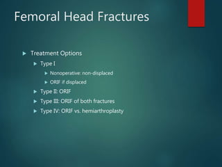
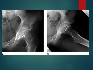
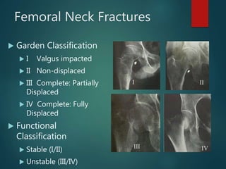
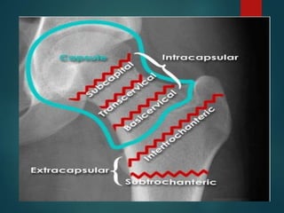
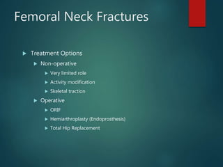
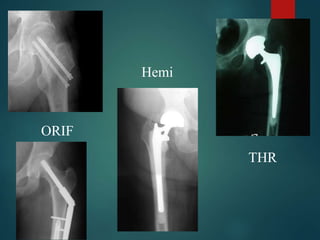
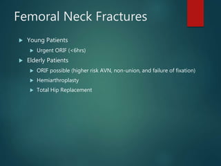
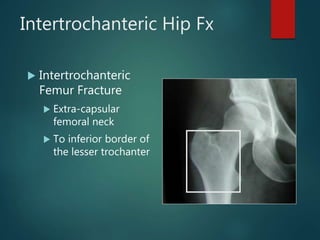
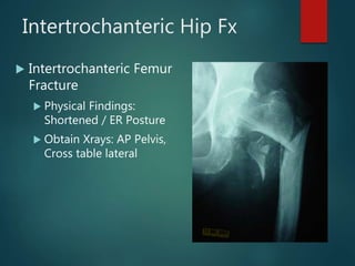
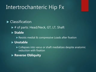
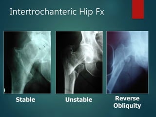
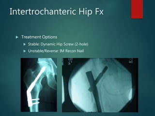
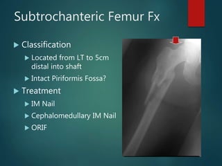
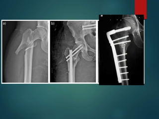
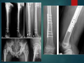
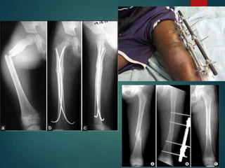
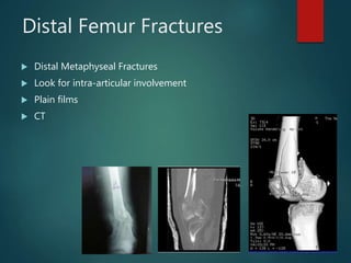
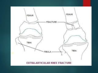
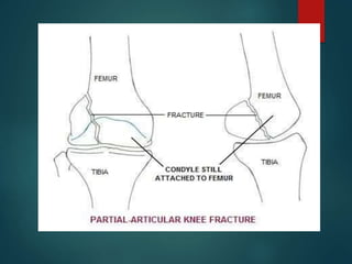
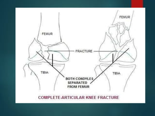
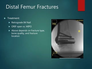
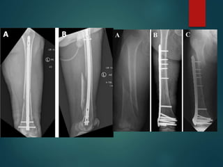
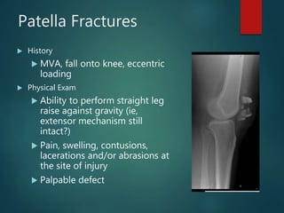
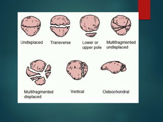
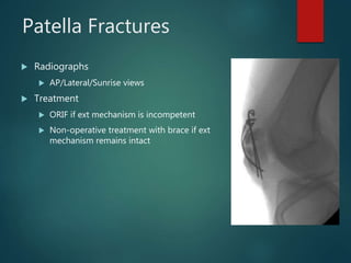
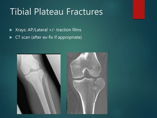
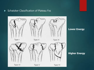
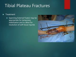
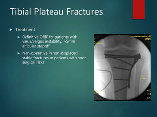
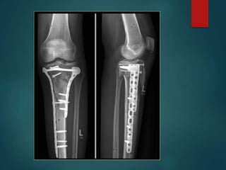
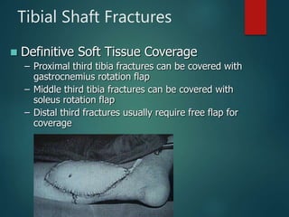
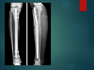
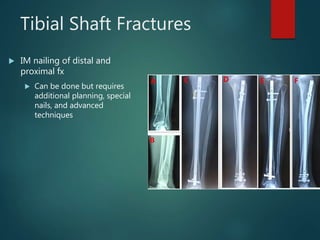
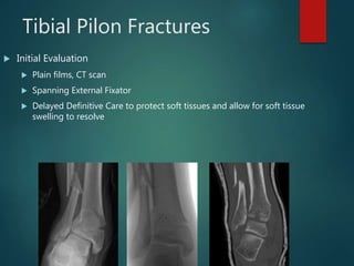
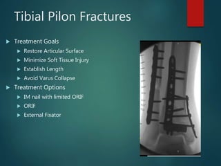
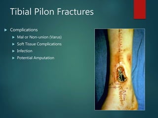
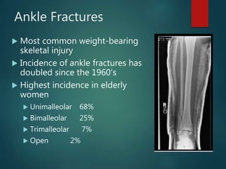
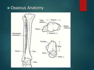
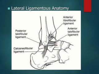
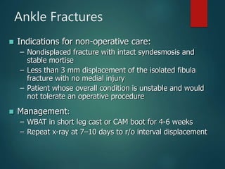
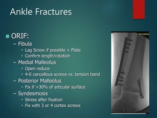
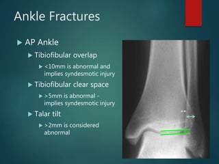
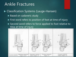
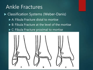
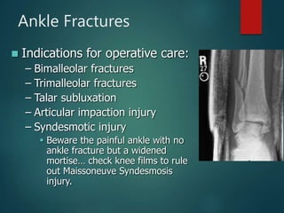
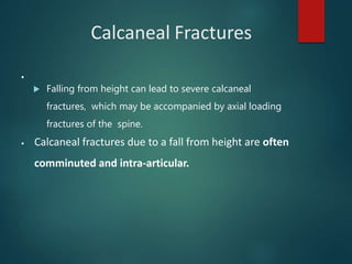
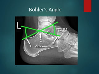
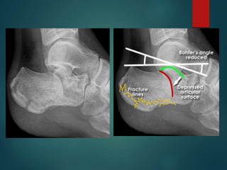
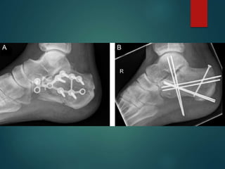
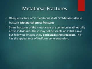
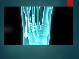
Lower Limb
Fractures
DR UDIT BISWAL
ASSISTANT PROFESSOR IN ORTHOPAEDICS
IMS & SUM
Lower Extremity Trauma
 Hip Fractures
 Femur Fractures
 Patella Fractures
 Knee Dislocations
 Tibia Fractures
 Ankle Fractures
Hip Fractures
 Femoral Head Fractures
 Femoral Neck Fractures
 Intertrochanteric Fractures
 Subtrochanteric Fractures
Epidemiology
 250,000 Hip fractures annually
 Expected to double by 2050
 At risk populations
 Elderly: poor balance & vision, osteoporosis, inactivity,
medications, malnutrition
 Young: high energy trauma
Femoral Head Fractures
 Concurrent with hip dislocation due to shear injury
Femoral Head Fractures
 Pipkin Classification
 I: Fracture inferior to fovea
 II: Fracture superior to fovea
 III: Femoral head + acetabulum fracture
 IV: Femoral head + femoral neck fracture
Femoral Head Fractures
 Treatment Options
 Type I
 Nonoperative: non-displaced
 ORIF if displaced
 Type II: ORIF
 Type III: ORIF of both fractures
 Type IV: ORIF vs. hemiarthroplasty
Femoral Neck Fractures
 Garden Classification
 I Valgus impacted
 II Non-displaced
 III Complete: Partially
Displaced
 IV Complete: Fully
Displaced
 Functional
Classification
 Stable (I/II)
 Unstable (III/IV)
I II
III IV
Intracapsular fracture – Subcapital
Intertrochanteric fracture
Subtrochanteric fracture
Grade 1 Grade 2 Grade 3
PAUWELS CLASSIFICATION
Femoral Neck Fractures
 Treatment Options
 Non-operative
 Very limited role
 Activity modification
 Skeletal traction
 Operative
 ORIF
 Hemiarthroplasty (Endoprosthesis)
 Total Hip Replacement
ORIF
Hemi
THR
Femoral Neck Fractures
 Young Patients
 Urgent ORIF (<6hrs)
 Elderly Patients
 ORIF possible (higher risk AVN, non-union, and failure of fixation)
 Hemiarthroplasty
 Total Hip Replacement
Intertrochanteric Hip Fx
 Intertrochanteric
Femur Fracture
 Extra-capsular
femoral neck
 To inferior border of
the lesser trochanter
Intertrochanteric Hip Fx
 Intertrochanteric Femur
Fracture
 Physical Findings:
Shortened / ER Posture
 Obtain Xrays: AP Pelvis,
Cross table lateral
Intertrochanteric Hip Fx
 Classification
 # of parts: Head/Neck, GT, LT, Shaft
 Stable
 Resists medial & compressive Loads after fixation
 Unstable
 Collapses into varus or shaft medializes despite anatomic
reduction with fixation
 Reverse Obliquity
Stable Reverse
Obliquity
Unstable
Intertrochanteric Hip Fx
 Treatment Options
 Stable: Dynamic Hip Screw (2-hole)
 Unstable/Reverse: IM Recon Nail
Subtrochanteric Femur Fx
 Classification
 Located from LT to 5cm
distal into shaft
 Intact Piriformis Fossa?
 Treatment
 IM Nail
 Cephalomedullary IM Nail
 ORIF
Femoral Shaft Fx
 Type 0 - No comminution
 Type 1 - Insignificant butterfly
fragment with transverse or short
oblique fracture
 Type 2 - Large butterfly of less than
50% of the bony width, > 50% of
cortex intact
 Type 3 - Larger butterfly leaving less
than 50% of the cortex in contact
 Type 4 - Segmental comminution
 Winquist and Hansen 66A,
1984
Femoral Shaft Fx
 Treatment Options
 IM Nail with locking screws
 ORIF with plate/screw construct
 External fixation
 Consider traction pin if prolonged delay to surgery
Distal Femur Fractures
 Distal Metaphyseal Fractures
 Look for intra-articular involvement
 Plain films
 CT
Distal Femur Fractures
 Treatment:
 Retrograde IM Nail
 ORIF open vs. MIPO
 Above depends on fracture type,
bone quality, and fracture
location
Patella Fractures
 History
 MVA, fall onto knee, eccentric
loading
 Physical Exam
 Ability to perform straight leg
raise against gravity (ie,
extensor mechanism still
intact?)
 Pain, swelling, contusions,
lacerations and/or abrasions at
the site of injury
 Palpable defect
Patella Fractures
 Radiographs
 AP/Lateral/Sunrise views
 Treatment
 ORIF if ext mechanism is incompetent
 Non-operative treatment with brace if ext
mechanism remains intact
Tibia Fractures
 Proximal Tibia Fractures (Tibial Plateau)
 Tibial Shaft Fractures
 Distal Tibia Fractures (Tibial Pilon/Plafond)
Tibial Plateau Fractures
 MVA, fall from height, sporting injuries
 Mechanism and energy of injury plays a major role in
determining orthopedic care
 Examine soft tissues, neurologic exam (peroneal N.),
vascular exam (esp with medial plateau injuries)
 Be aware for compartment syndrome
 Check for knee ligamentous instability
Tibial Plateau Fractures
 Xrays: AP/Lateral +/- traction films
 CT scan (after ex-fix if appropriate)
 Schatzker Classification of Plateau Fxs
Lower Energy
Higher Energy
Tibial Plateau Fractures
 Treatment
 Spanning External Fixator may be
appropriate for temporary
stabilization and to allow for
resolution of soft tissue injuries
Insert blister
Pics of ex-fix here
Tibial Plateau Fractures
 Treatment
 Definitive ORIF for patients with
varus/valgus instability, >5mm
articular stepoff
 Non-operative in non-displaced
stable fractures or patients with poor
surgical risks
Tibial Shaft Fractures
 Mechanism of Injury
 Can occur in lower energy, torsion type injury (e.g., skiing)
 More common with higher energy direct force (e.g., car bumper)
 Open fractures of the tibia are more common than in any other long
bone
Tibial Shaft Fractures
 Open Tibia Fx
 Priorities
– ABC’S
– Associated Injuries
– Tetanus
– Antibiotics
– Fixation
Tibial Shaft Fractures
 Gustilo and Anderson Classification of Open Fx
 Grade 1
 <1cm, minimal muscle contusion, usually inside out mechanism
 Grade 2
 1-10cm, extensive soft tissue damage
 Grade 3
 3a: >10cm, adequate bone coverage
 3b: >10cm, periosteal stripping requiring flap advancement or free flap
 3c: vascular injury requiring repair
Tibial Shaft Fractures
 Tscherne Classification of Soft Tissue Injury
 Grade 0- negligible soft tissue injury
 Grade 1- superficial abrasion or contusion
 Grade 2- deep contusion from direct trauma
 Grade 3- Extensive contusion and crush injury with
possible severe muscle injury
Tibial Shaft Fractures
 Management of Open Fx
Soft Tissues
 ER: initial evaluation 
wound covered with sterile
dressing and leg splinted,
tetanus prophylaxis and
appropriate antibiotics
 OR: Thorough I&D
undertaken within 6 hours
with serial debridements
as warranted followed by
definitive soft tissue cover
Tibial Shaft Fractures
 Definitive Soft Tissue Coverage
– Proximal third tibia fractures can be covered with
gastrocnemius rotation flap
– Middle third tibia fractures can be covered with
soleus rotation flap
– Distal third fractures usually require free flap for
coverage
Tibial Shaft Fractures
 Treatment Options
 IM Nail
 ORIF with Plates
 External Fixation
 Cast or Cast-Brace
Tibial Shaft Fractures
 Advantages of IM nailing
 Lower non-union rate
 Smaller incisions
 Earlier weightbearing and function
 Single surgery
Tibial Shaft Fractures
 IM nailing of distal and
proximal fx
 Can be done but requires
additional planning, special
nails, and advanced
techniques
Tibial Pilon Fractures
 Fractures involving distal tibia metaphysis and into the ankle joint
 Soft tissue management is key!
 Often occurs from fall from height or high energy injuries in MVA
 “Excellent” results are rare, “Fair to Good” is the norm outcome
 Multiple potential complications
Tibial Pilon Fractures
 Initial Evaluation
 Plain films, CT scan
 Spanning External Fixator
 Delayed Definitive Care to protect soft tissues and allow for soft tissue
swelling to resolve
Tibial Pilon Fractures
 Treatment Goals
 Restore Articular Surface
 Minimize Soft Tissue Injury
 Establish Length
 Avoid Varus Collapse
 Treatment Options
 IM nail with limited ORIF
 ORIF
 External Fixator
Tibial Pilon Fractures
 Complications
 Mal or Non-union (Varus)
 Soft Tissue Complications
 Infection
 Potential Amputation
Ankle Fractures
 Most common weight-bearing
skeletal injury
 Incidence of ankle fractures has
doubled since the 1960’s
 Highest incidence in elderly
women
 Unimalleolar 68%
 Bimalleolar 25%
 Trimalleolar 7%
 Open 2%
 Osseous Anatomy
 Lateral Ligamentous Anatomy
 Medial Ligamentous Anatomy
 Syndesmosis Anatomy
Ankle Fractures
 History
 Mechanism of injury
 Time elapsed since the injury
 Soft-tissue injury
 Has the patient ambulated on the ankle?
 Patient’s age / bone quality
 Associated injuries
 Comorbidities (DM, smoking)
Ankle Fractures
 Physical Exam
 Neurovascular exam
 Note obvious deformities
 Pain over the medial or lateral malleoli
 Palpation of ligaments about the ankle
 Palpation of proximal fibula, lateral process of talus, base
of 5th MT
 Examine the hindfoot and forefoot
Ankle Fractures
 Radiographic Studies
 AP, Lateral, Mortise of Ankle (Weight Bearing if possible)
 AP, Lateral of Knee (Maissaneve injury)
 AP, Lateral, Oblique of Foot (if painful)
Ankle Fractures
 AP Ankle
 Tibiofibular overlap
 <10mm is abnormal and
implies syndesmotic injury
 Tibiofibular clear space
 >5mm is abnormal -
implies syndesmotic injury
 Talar tilt
 >2mm is considered
abnormal
Ankle Fractures
 Ankle Mortise View
 Foot is internally rotated
and AP projection is
performed
 Abnormal findings:
 Medial joint space widening
 Talocural angle <8 or >15
degrees (compare to normal
side)
 Tibia/fibula overlap <1mm
Ankle Fractures
 Lateral View
 Posterior malleolar
fractures
 Anterior/posterior
subluxation of the talus
under the tibia
 Displacement/Shortening
of distal fibula
 Associated injuries
Ankle Fractures
 Classification Systems (Lauge-Hansen)
 Based on cadaveric study
 First word refers to position of foot at time of injury
 Second word refers to force applied to foot relative to
tibia at time of injury
Ankle Fractures
 Classification Systems (Weber-Danis)
 A: Fibula Fracture distal to mortise
 B: Fibula Fracture at the level of the mortise
 C: Fibula Fracture proximal to mortise
Ankle Fractures
 Initial Management
 Closed reduction (conscious sedation may be necessary)
 AO splint
 Delayed fixation until soft tissues stable
 Pain control
 Monitor for possible compartment syndrome in high
energy injuries
Ankle Fractures
 Indications for non-operative care:
– Nondisplaced fracture with intact syndesmosis and
stable mortise
– Less than 3 mm displacement of the isolated fibula
fracture with no medial injury
– Patient whose overall condition is unstable and would
not tolerate an operative procedure
 Management:
– WBAT in short leg cast or CAM boot for 4-6 weeks
– Repeat x-ray at 7–10 days to r/o interval displacement
Ankle Fractures
 Indications for operative care:
– Bimalleolar fractures
– Trimalleolar fractures
– Talar subluxation
– Articular impaction injury
– Syndesmotic injury
 Beware the painful ankle with no
ankle fracture but a widened
mortise… check knee films to rule
out Maissoneuve Syndesmosis
injury.
Ankle Fractures
 ORIF:
– Fibula
 Lag Screw if possible + Plate
 Confirm length/rotation
– Medial Malleolus
 Open reduce
 4-0 cancellous screws vs. tension band
– Posterior Malleolus
 Fix if >30% of articular surface
– Syndesmosis
 Stress after fixation
 Fix with 3 or 4 cortex screws
Ankle Fractures
 Most common weight-bearing
skeletal injury
 Incidence of ankle fractures has
doubled since the 1960’s
 Highest incidence in elderly
women
 Unimalleolar 68%
 Bimalleolar 25%
 Trimalleolar 7%
 Open 2%
Ankle Fractures
 History
 Mechanism of injury
 Time elapsed since the injury
 Soft-tissue injury
 Has the patient ambulated on the ankle?
 Patient’s age / bone quality
 Associated injuries
 Comorbidities (DM, smoking)
Ankle Fractures
 Physical Exam
 Neurovascular exam
 Note obvious deformities
 Pain over the medial or lateral malleoli
 Palpation of ligaments about the ankle
 Palpation of proximal fibula, lateral process of talus, base
of 5th MT
 Examine the hindfoot and forefoot
Ankle Fractures
 Radiographic Studies
 AP, Lateral, Mortise of Ankle (Weight Bearing if possible)
 AP, Lateral of Knee (Maissaneve injury)
 AP, Lateral, Oblique of Foot (if painful)
Ankle Fractures
 AP Ankle
 Tibiofibular overlap
 <10mm is abnormal and
implies syndesmotic injury
 Tibiofibular clear space
 >5mm is abnormal -
implies syndesmotic injury
 Talar tilt
 >2mm is considered
abnormal
Ankle Fractures
 Ankle Mortise View
 Foot is internally rotated
and AP projection is
performed
 Abnormal findings:
 Medial joint space widening
 Talocural angle <8 or >15
degrees (compare to normal
side)
 Tibia/fibula overlap <1mm
Ankle Fractures
 Lateral View
 Posterior malleolar
fractures
 Anterior/posterior
subluxation of the talus
under the tibia
 Displacement/Shortening
of distal fibula
 Associated injuries
Ankle Fractures
 Classification Systems (Lauge-Hansen)
 Based on cadaveric study
 First word refers to position of foot at time of injury
 Second word refers to force applied to foot relative to
tibia at time of injury
Ankle Fractures
 Classification Systems (Weber-Danis)
 A: Fibula Fracture distal to mortise
 B: Fibula Fracture at the level of the mortise
 C: Fibula Fracture proximal to mortise
Ankle Fractures
 Initial Management
 Closed reduction (conscious sedation may be necessary)
 AO splint
 Delayed fixation until soft tissues stable
 Pain control
 Monitor for possible compartment syndrome in high
energy injuries
Ankle Fractures
 Indications for non-operative care:
– Nondisplaced fracture with intact syndesmosis and
stable mortise
– Less than 3 mm displacement of the isolated fibula
fracture with no medial injury
– Patient whose overall condition is unstable and would
not tolerate an operative procedure
 Management:
– WBAT in short leg cast or CAM boot for 4-6 weeks
– Repeat x-ray at 7–10 days to r/o interval displacement
Ankle Fractures
 Indications for operative care:
– Bimalleolar fractures
– Trimalleolar fractures
– Talar subluxation
– Articular impaction injury
– Syndesmotic injury
 Beware the painful ankle with no
ankle fracture but a widened
mortise… check knee films to rule
out Maissoneuve Syndesmosis
injury.
Ankle Fractures
 ORIF:
– Fibula
 Lag Screw if possible + Plate
 Confirm length/rotation
– Medial Malleolus
 Open reduce
 4-0 cancellous screws vs. tension band
– Posterior Malleolus
 Fix if >30% of articular surface
– Syndesmosis
 Stress after fixation
 Fix with 3 or 4 cortex screws
Calcaneal Fractures
 Falling from height can lead to severe calcaneal
fractures, which may be accompanied by axial loading
fractures of the spine.
▪
▪ Calcaneal fractures due to a fall from height are often
comminuted and intra-articular.
Bohler’s Angle
▪ A line is drawn from the tuberosity to the most superior part
of the posterior facet.
▪ Another line is drawn from the most superior part of the
facet to the anterior process.
Normally the angle created is between 20 and 40 degrees.
▪
▪ If the angle is less than 20 degrees, this indicates depressed
fracture.
Bohler’s Angle
The critical angle of Gissane
 It is formed by a line along the lateral margin of the
posterior facet and another line extending anterior to
the beak of the calcaneus. The normal value is 95 to 105
degrees with an increase representing posterior facet
collapse
Types of calcaneal
fractures
 Intra and Extrarticular fractures on the basis of subtalar
joint involvement.
▪ Intrarticular fractures are more common and
involve the posterior talar articular facet of the
calcaneus.
▪ Extrarticular fractures are less common, and
located anywhere outside the subtalar joint.
The Sanders system classification
Is the most commonly used system for categorizing
intrarticular fractures.
Classifies these fractures into four types, based on the location
of the fracture at the posterior articular surface.
TYPES
▪ Type I fractures are non-displaced fractures (displacement <
2 mm).
▪ Type II fractures consist of a single intrarticular fracture that
divides the calcaneus into 2 pieces.
▪ Type III fractures consist of two intrarticular fractures that
divide the calcaneus into 3 articular pieces.
▪ Type IV fractures consist of fractures with more than three
intrarticular fractures.
Metatarsal Fractures
▪
▪
▪
Oblique fracture of 5th metatarsal shaft 5TH Metatarsal base
fracture Metatarsal stress fractures
Stress fractures of the metatarsals are common in athletically
active individuals. These may not be visible on initial X-rays
but follow up images show periosteal stress reaction. This
has the appearance of fusiform bone expansion.
THANK YOU

Lower Limb Fractures.pptx

  • 1.
    Lower Limb Fractures DR UDITBISWAL ASSISTANT PROFESSOR IN ORTHOPAEDICS IMS & SUM
  • 2.
    Lower Extremity Trauma Hip Fractures  Femur Fractures  Patella Fractures  Knee Dislocations  Tibia Fractures  Ankle Fractures
  • 3.
    Hip Fractures  FemoralHead Fractures  Femoral Neck Fractures  Intertrochanteric Fractures  Subtrochanteric Fractures
  • 4.
    Epidemiology  250,000 Hipfractures annually  Expected to double by 2050  At risk populations  Elderly: poor balance & vision, osteoporosis, inactivity, medications, malnutrition  Young: high energy trauma
  • 5.
    Femoral Head Fractures Concurrent with hip dislocation due to shear injury
  • 6.
    Femoral Head Fractures Pipkin Classification  I: Fracture inferior to fovea  II: Fracture superior to fovea  III: Femoral head + acetabulum fracture  IV: Femoral head + femoral neck fracture
  • 7.
    Femoral Head Fractures Treatment Options  Type I  Nonoperative: non-displaced  ORIF if displaced  Type II: ORIF  Type III: ORIF of both fractures  Type IV: ORIF vs. hemiarthroplasty
  • 9.
    Femoral Neck Fractures Garden Classification  I Valgus impacted  II Non-displaced  III Complete: Partially Displaced  IV Complete: Fully Displaced  Functional Classification  Stable (I/II)  Unstable (III/IV) I II III IV
  • 11.
  • 12.
  • 13.
  • 14.
    Grade 1 Grade2 Grade 3 PAUWELS CLASSIFICATION
  • 15.
    Femoral Neck Fractures Treatment Options  Non-operative  Very limited role  Activity modification  Skeletal traction  Operative  ORIF  Hemiarthroplasty (Endoprosthesis)  Total Hip Replacement
  • 16.
  • 17.
    Femoral Neck Fractures Young Patients  Urgent ORIF (<6hrs)  Elderly Patients  ORIF possible (higher risk AVN, non-union, and failure of fixation)  Hemiarthroplasty  Total Hip Replacement
  • 18.
    Intertrochanteric Hip Fx Intertrochanteric Femur Fracture  Extra-capsular femoral neck  To inferior border of the lesser trochanter
  • 19.
    Intertrochanteric Hip Fx Intertrochanteric Femur Fracture  Physical Findings: Shortened / ER Posture  Obtain Xrays: AP Pelvis, Cross table lateral
  • 20.
    Intertrochanteric Hip Fx Classification  # of parts: Head/Neck, GT, LT, Shaft  Stable  Resists medial & compressive Loads after fixation  Unstable  Collapses into varus or shaft medializes despite anatomic reduction with fixation  Reverse Obliquity
  • 21.
  • 22.
    Intertrochanteric Hip Fx Treatment Options  Stable: Dynamic Hip Screw (2-hole)  Unstable/Reverse: IM Recon Nail
  • 23.
    Subtrochanteric Femur Fx Classification  Located from LT to 5cm distal into shaft  Intact Piriformis Fossa?  Treatment  IM Nail  Cephalomedullary IM Nail  ORIF
  • 25.
    Femoral Shaft Fx Type 0 - No comminution  Type 1 - Insignificant butterfly fragment with transverse or short oblique fracture  Type 2 - Large butterfly of less than 50% of the bony width, > 50% of cortex intact  Type 3 - Larger butterfly leaving less than 50% of the cortex in contact  Type 4 - Segmental comminution  Winquist and Hansen 66A, 1984
  • 26.
    Femoral Shaft Fx Treatment Options  IM Nail with locking screws  ORIF with plate/screw construct  External fixation  Consider traction pin if prolonged delay to surgery
  • 29.
    Distal Femur Fractures Distal Metaphyseal Fractures  Look for intra-articular involvement  Plain films  CT
  • 33.
    Distal Femur Fractures Treatment:  Retrograde IM Nail  ORIF open vs. MIPO  Above depends on fracture type, bone quality, and fracture location
  • 35.
    Patella Fractures  History MVA, fall onto knee, eccentric loading  Physical Exam  Ability to perform straight leg raise against gravity (ie, extensor mechanism still intact?)  Pain, swelling, contusions, lacerations and/or abrasions at the site of injury  Palpable defect
  • 37.
    Patella Fractures  Radiographs AP/Lateral/Sunrise views  Treatment  ORIF if ext mechanism is incompetent  Non-operative treatment with brace if ext mechanism remains intact
  • 38.
    Tibia Fractures  ProximalTibia Fractures (Tibial Plateau)  Tibial Shaft Fractures  Distal Tibia Fractures (Tibial Pilon/Plafond)
  • 39.
    Tibial Plateau Fractures MVA, fall from height, sporting injuries  Mechanism and energy of injury plays a major role in determining orthopedic care  Examine soft tissues, neurologic exam (peroneal N.), vascular exam (esp with medial plateau injuries)  Be aware for compartment syndrome  Check for knee ligamentous instability
  • 40.
    Tibial Plateau Fractures Xrays: AP/Lateral +/- traction films  CT scan (after ex-fix if appropriate)
  • 41.
     Schatzker Classificationof Plateau Fxs Lower Energy Higher Energy
  • 42.
    Tibial Plateau Fractures Treatment  Spanning External Fixator may be appropriate for temporary stabilization and to allow for resolution of soft tissue injuries Insert blister Pics of ex-fix here
  • 43.
    Tibial Plateau Fractures Treatment  Definitive ORIF for patients with varus/valgus instability, >5mm articular stepoff  Non-operative in non-displaced stable fractures or patients with poor surgical risks
  • 45.
    Tibial Shaft Fractures Mechanism of Injury  Can occur in lower energy, torsion type injury (e.g., skiing)  More common with higher energy direct force (e.g., car bumper)  Open fractures of the tibia are more common than in any other long bone
  • 46.
    Tibial Shaft Fractures Open Tibia Fx  Priorities – ABC’S – Associated Injuries – Tetanus – Antibiotics – Fixation
  • 47.
    Tibial Shaft Fractures Gustilo and Anderson Classification of Open Fx  Grade 1  <1cm, minimal muscle contusion, usually inside out mechanism  Grade 2  1-10cm, extensive soft tissue damage  Grade 3  3a: >10cm, adequate bone coverage  3b: >10cm, periosteal stripping requiring flap advancement or free flap  3c: vascular injury requiring repair
  • 48.
    Tibial Shaft Fractures Tscherne Classification of Soft Tissue Injury  Grade 0- negligible soft tissue injury  Grade 1- superficial abrasion or contusion  Grade 2- deep contusion from direct trauma  Grade 3- Extensive contusion and crush injury with possible severe muscle injury
  • 49.
    Tibial Shaft Fractures Management of Open Fx Soft Tissues  ER: initial evaluation  wound covered with sterile dressing and leg splinted, tetanus prophylaxis and appropriate antibiotics  OR: Thorough I&D undertaken within 6 hours with serial debridements as warranted followed by definitive soft tissue cover
  • 50.
    Tibial Shaft Fractures Definitive Soft Tissue Coverage – Proximal third tibia fractures can be covered with gastrocnemius rotation flap – Middle third tibia fractures can be covered with soleus rotation flap – Distal third fractures usually require free flap for coverage
  • 51.
    Tibial Shaft Fractures Treatment Options  IM Nail  ORIF with Plates  External Fixation  Cast or Cast-Brace
  • 52.
    Tibial Shaft Fractures Advantages of IM nailing  Lower non-union rate  Smaller incisions  Earlier weightbearing and function  Single surgery
  • 54.
    Tibial Shaft Fractures IM nailing of distal and proximal fx  Can be done but requires additional planning, special nails, and advanced techniques
  • 55.
    Tibial Pilon Fractures Fractures involving distal tibia metaphysis and into the ankle joint  Soft tissue management is key!  Often occurs from fall from height or high energy injuries in MVA  “Excellent” results are rare, “Fair to Good” is the norm outcome  Multiple potential complications
  • 56.
    Tibial Pilon Fractures Initial Evaluation  Plain films, CT scan  Spanning External Fixator  Delayed Definitive Care to protect soft tissues and allow for soft tissue swelling to resolve
  • 57.
    Tibial Pilon Fractures Treatment Goals  Restore Articular Surface  Minimize Soft Tissue Injury  Establish Length  Avoid Varus Collapse  Treatment Options  IM nail with limited ORIF  ORIF  External Fixator
  • 58.
    Tibial Pilon Fractures Complications  Mal or Non-union (Varus)  Soft Tissue Complications  Infection  Potential Amputation
  • 59.
    Ankle Fractures  Mostcommon weight-bearing skeletal injury  Incidence of ankle fractures has doubled since the 1960’s  Highest incidence in elderly women  Unimalleolar 68%  Bimalleolar 25%  Trimalleolar 7%  Open 2%
  • 60.
  • 61.
  • 62.
  • 63.
  • 64.
    Ankle Fractures  History Mechanism of injury  Time elapsed since the injury  Soft-tissue injury  Has the patient ambulated on the ankle?  Patient’s age / bone quality  Associated injuries  Comorbidities (DM, smoking)
  • 65.
    Ankle Fractures  PhysicalExam  Neurovascular exam  Note obvious deformities  Pain over the medial or lateral malleoli  Palpation of ligaments about the ankle  Palpation of proximal fibula, lateral process of talus, base of 5th MT  Examine the hindfoot and forefoot
  • 66.
    Ankle Fractures  RadiographicStudies  AP, Lateral, Mortise of Ankle (Weight Bearing if possible)  AP, Lateral of Knee (Maissaneve injury)  AP, Lateral, Oblique of Foot (if painful)
  • 67.
    Ankle Fractures  APAnkle  Tibiofibular overlap  <10mm is abnormal and implies syndesmotic injury  Tibiofibular clear space  >5mm is abnormal - implies syndesmotic injury  Talar tilt  >2mm is considered abnormal
  • 68.
    Ankle Fractures  AnkleMortise View  Foot is internally rotated and AP projection is performed  Abnormal findings:  Medial joint space widening  Talocural angle <8 or >15 degrees (compare to normal side)  Tibia/fibula overlap <1mm
  • 69.
    Ankle Fractures  LateralView  Posterior malleolar fractures  Anterior/posterior subluxation of the talus under the tibia  Displacement/Shortening of distal fibula  Associated injuries
  • 70.
    Ankle Fractures  ClassificationSystems (Lauge-Hansen)  Based on cadaveric study  First word refers to position of foot at time of injury  Second word refers to force applied to foot relative to tibia at time of injury
  • 71.
    Ankle Fractures  ClassificationSystems (Weber-Danis)  A: Fibula Fracture distal to mortise  B: Fibula Fracture at the level of the mortise  C: Fibula Fracture proximal to mortise
  • 72.
    Ankle Fractures  InitialManagement  Closed reduction (conscious sedation may be necessary)  AO splint  Delayed fixation until soft tissues stable  Pain control  Monitor for possible compartment syndrome in high energy injuries
  • 73.
    Ankle Fractures  Indicationsfor non-operative care: – Nondisplaced fracture with intact syndesmosis and stable mortise – Less than 3 mm displacement of the isolated fibula fracture with no medial injury – Patient whose overall condition is unstable and would not tolerate an operative procedure  Management: – WBAT in short leg cast or CAM boot for 4-6 weeks – Repeat x-ray at 7–10 days to r/o interval displacement
  • 74.
    Ankle Fractures  Indicationsfor operative care: – Bimalleolar fractures – Trimalleolar fractures – Talar subluxation – Articular impaction injury – Syndesmotic injury  Beware the painful ankle with no ankle fracture but a widened mortise… check knee films to rule out Maissoneuve Syndesmosis injury.
  • 75.
    Ankle Fractures  ORIF: –Fibula  Lag Screw if possible + Plate  Confirm length/rotation – Medial Malleolus  Open reduce  4-0 cancellous screws vs. tension band – Posterior Malleolus  Fix if >30% of articular surface – Syndesmosis  Stress after fixation  Fix with 3 or 4 cortex screws
  • 76.
    Ankle Fractures  Mostcommon weight-bearing skeletal injury  Incidence of ankle fractures has doubled since the 1960’s  Highest incidence in elderly women  Unimalleolar 68%  Bimalleolar 25%  Trimalleolar 7%  Open 2%
  • 77.
    Ankle Fractures  History Mechanism of injury  Time elapsed since the injury  Soft-tissue injury  Has the patient ambulated on the ankle?  Patient’s age / bone quality  Associated injuries  Comorbidities (DM, smoking)
  • 78.
    Ankle Fractures  PhysicalExam  Neurovascular exam  Note obvious deformities  Pain over the medial or lateral malleoli  Palpation of ligaments about the ankle  Palpation of proximal fibula, lateral process of talus, base of 5th MT  Examine the hindfoot and forefoot
  • 79.
    Ankle Fractures  RadiographicStudies  AP, Lateral, Mortise of Ankle (Weight Bearing if possible)  AP, Lateral of Knee (Maissaneve injury)  AP, Lateral, Oblique of Foot (if painful)
  • 80.
    Ankle Fractures  APAnkle  Tibiofibular overlap  <10mm is abnormal and implies syndesmotic injury  Tibiofibular clear space  >5mm is abnormal - implies syndesmotic injury  Talar tilt  >2mm is considered abnormal
  • 81.
    Ankle Fractures  AnkleMortise View  Foot is internally rotated and AP projection is performed  Abnormal findings:  Medial joint space widening  Talocural angle <8 or >15 degrees (compare to normal side)  Tibia/fibula overlap <1mm
  • 82.
    Ankle Fractures  LateralView  Posterior malleolar fractures  Anterior/posterior subluxation of the talus under the tibia  Displacement/Shortening of distal fibula  Associated injuries
  • 83.
    Ankle Fractures  ClassificationSystems (Lauge-Hansen)  Based on cadaveric study  First word refers to position of foot at time of injury  Second word refers to force applied to foot relative to tibia at time of injury
  • 84.
    Ankle Fractures  ClassificationSystems (Weber-Danis)  A: Fibula Fracture distal to mortise  B: Fibula Fracture at the level of the mortise  C: Fibula Fracture proximal to mortise
  • 85.
    Ankle Fractures  InitialManagement  Closed reduction (conscious sedation may be necessary)  AO splint  Delayed fixation until soft tissues stable  Pain control  Monitor for possible compartment syndrome in high energy injuries
  • 86.
    Ankle Fractures  Indicationsfor non-operative care: – Nondisplaced fracture with intact syndesmosis and stable mortise – Less than 3 mm displacement of the isolated fibula fracture with no medial injury – Patient whose overall condition is unstable and would not tolerate an operative procedure  Management: – WBAT in short leg cast or CAM boot for 4-6 weeks – Repeat x-ray at 7–10 days to r/o interval displacement
  • 87.
    Ankle Fractures  Indicationsfor operative care: – Bimalleolar fractures – Trimalleolar fractures – Talar subluxation – Articular impaction injury – Syndesmotic injury  Beware the painful ankle with no ankle fracture but a widened mortise… check knee films to rule out Maissoneuve Syndesmosis injury.
  • 88.
    Ankle Fractures  ORIF: –Fibula  Lag Screw if possible + Plate  Confirm length/rotation – Medial Malleolus  Open reduce  4-0 cancellous screws vs. tension band – Posterior Malleolus  Fix if >30% of articular surface – Syndesmosis  Stress after fixation  Fix with 3 or 4 cortex screws
  • 89.
    Calcaneal Fractures  Fallingfrom height can lead to severe calcaneal fractures, which may be accompanied by axial loading fractures of the spine. ▪ ▪ Calcaneal fractures due to a fall from height are often comminuted and intra-articular.
  • 90.
    Bohler’s Angle ▪ Aline is drawn from the tuberosity to the most superior part of the posterior facet. ▪ Another line is drawn from the most superior part of the facet to the anterior process. Normally the angle created is between 20 and 40 degrees. ▪ ▪ If the angle is less than 20 degrees, this indicates depressed fracture.
  • 91.
  • 92.
    The critical angleof Gissane  It is formed by a line along the lateral margin of the posterior facet and another line extending anterior to the beak of the calcaneus. The normal value is 95 to 105 degrees with an increase representing posterior facet collapse
  • 94.
    Types of calcaneal fractures Intra and Extrarticular fractures on the basis of subtalar joint involvement. ▪ Intrarticular fractures are more common and involve the posterior talar articular facet of the calcaneus. ▪ Extrarticular fractures are less common, and located anywhere outside the subtalar joint.
  • 95.
    The Sanders systemclassification Is the most commonly used system for categorizing intrarticular fractures. Classifies these fractures into four types, based on the location of the fracture at the posterior articular surface.
  • 96.
    TYPES ▪ Type Ifractures are non-displaced fractures (displacement < 2 mm). ▪ Type II fractures consist of a single intrarticular fracture that divides the calcaneus into 2 pieces. ▪ Type III fractures consist of two intrarticular fractures that divide the calcaneus into 3 articular pieces. ▪ Type IV fractures consist of fractures with more than three intrarticular fractures.
  • 99.
    Metatarsal Fractures ▪ ▪ ▪ Oblique fractureof 5th metatarsal shaft 5TH Metatarsal base fracture Metatarsal stress fractures Stress fractures of the metatarsals are common in athletically active individuals. These may not be visible on initial X-rays but follow up images show periosteal stress reaction. This has the appearance of fusiform bone expansion.
  • 101.