The central nervous system (CNS) is made up of the brain and spinal cord. The brain controls most body functions, including awareness, movements, sensations, thoughts, speech and memory. The spinal cord is connected to the brain at the brain stem and is covered by the vertebrae of the spine.
The endocrine system is a messenger system comprising feedback loops of the hormones released by internal glands of an organism directly into the circulatory system, regulating distant target organs. In vertebrates, the hypothalamus is the neural control center for all endocrine systems.
There is also the quoricity about the human brain, here is the solution. This presentation give you the knowledge about the nervous system. The introduction about the neurons, neurolgia, synapse,etc.
This presentation based on a broad overview to the human central nervous system focusing over the parts of the system, different cell types present in the system, and special terminology used in the system.
The spinal cord is a long, thin, tubular structure made up of nervous tissue, which extends from the medulla oblongata in the brainstem to the lumbar region of the vertebral column. It encloses the central canal of the spinal cord, which contains cerebrospinal fluid. The brain and spinal cord together make up the central nervous system (CNS). In humans, the spinal cord begins at the occipital bone, passing through the foramen magnum and entering the spinal canal at the beginning of the cervical vertebrae.
The nervous system consists of the brain, spinal cord, sensory organs, and all of the nerves that connect these organs with the rest of the body. Together, these organs are responsible for the control of the body and communication among its parts.
An educational presentation on basics of neuroanatomy.
it define the scientific terminologies and various cells of nervous tissue. structure and function of all nervous tissue is explained. action potential generation is graphically represented.
An educational presentation on basics of neuroanatomy. It defines various cells of nervous tissue. the structure and function is well defined. It also covers various scientific terminologies and lastly their is graphical representation of action potential generation.
Nutraceutical market, scope and growth: Herbal drug technologyLokesh Patil
As consumer awareness of health and wellness rises, the nutraceutical market—which includes goods like functional meals, drinks, and dietary supplements that provide health advantages beyond basic nutrition—is growing significantly. As healthcare expenses rise, the population ages, and people want natural and preventative health solutions more and more, this industry is increasing quickly. Further driving market expansion are product formulation innovations and the use of cutting-edge technology for customized nutrition. With its worldwide reach, the nutraceutical industry is expected to keep growing and provide significant chances for research and investment in a number of categories, including vitamins, minerals, probiotics, and herbal supplements.
Comparing Evolved Extractive Text Summary Scores of Bidirectional Encoder Rep...University of Maribor
Slides from:
11th International Conference on Electrical, Electronics and Computer Engineering (IcETRAN), Niš, 3-6 June 2024
Track: Artificial Intelligence
https://www.etran.rs/2024/en/home-english/
Cancer cell metabolism: special Reference to Lactate PathwayAADYARAJPANDEY1
Normal Cell Metabolism:
Cellular respiration describes the series of steps that cells use to break down sugar and other chemicals to get the energy we need to function.
Energy is stored in the bonds of glucose and when glucose is broken down, much of that energy is released.
Cell utilize energy in the form of ATP.
The first step of respiration is called glycolysis. In a series of steps, glycolysis breaks glucose into two smaller molecules - a chemical called pyruvate. A small amount of ATP is formed during this process.
Most healthy cells continue the breakdown in a second process, called the Kreb's cycle. The Kreb's cycle allows cells to “burn” the pyruvates made in glycolysis to get more ATP.
The last step in the breakdown of glucose is called oxidative phosphorylation (Ox-Phos).
It takes place in specialized cell structures called mitochondria. This process produces a large amount of ATP. Importantly, cells need oxygen to complete oxidative phosphorylation.
If a cell completes only glycolysis, only 2 molecules of ATP are made per glucose. However, if the cell completes the entire respiration process (glycolysis - Kreb's - oxidative phosphorylation), about 36 molecules of ATP are created, giving it much more energy to use.
IN CANCER CELL:
Unlike healthy cells that "burn" the entire molecule of sugar to capture a large amount of energy as ATP, cancer cells are wasteful.
Cancer cells only partially break down sugar molecules. They overuse the first step of respiration, glycolysis. They frequently do not complete the second step, oxidative phosphorylation.
This results in only 2 molecules of ATP per each glucose molecule instead of the 36 or so ATPs healthy cells gain. As a result, cancer cells need to use a lot more sugar molecules to get enough energy to survive.
introduction to WARBERG PHENOMENA:
WARBURG EFFECT Usually, cancer cells are highly glycolytic (glucose addiction) and take up more glucose than do normal cells from outside.
Otto Heinrich Warburg (; 8 October 1883 – 1 August 1970) In 1931 was awarded the Nobel Prize in Physiology for his "discovery of the nature and mode of action of the respiratory enzyme.
WARNBURG EFFECT : cancer cells under aerobic (well-oxygenated) conditions to metabolize glucose to lactate (aerobic glycolysis) is known as the Warburg effect. Warburg made the observation that tumor slices consume glucose and secrete lactate at a higher rate than normal tissues.
Deep Behavioral Phenotyping in Systems Neuroscience for Functional Atlasing a...Ana Luísa Pinho
Functional Magnetic Resonance Imaging (fMRI) provides means to characterize brain activations in response to behavior. However, cognitive neuroscience has been limited to group-level effects referring to the performance of specific tasks. To obtain the functional profile of elementary cognitive mechanisms, the combination of brain responses to many tasks is required. Yet, to date, both structural atlases and parcellation-based activations do not fully account for cognitive function and still present several limitations. Further, they do not adapt overall to individual characteristics. In this talk, I will give an account of deep-behavioral phenotyping strategies, namely data-driven methods in large task-fMRI datasets, to optimize functional brain-data collection and improve inference of effects-of-interest related to mental processes. Key to this approach is the employment of fast multi-functional paradigms rich on features that can be well parametrized and, consequently, facilitate the creation of psycho-physiological constructs to be modelled with imaging data. Particular emphasis will be given to music stimuli when studying high-order cognitive mechanisms, due to their ecological nature and quality to enable complex behavior compounded by discrete entities. I will also discuss how deep-behavioral phenotyping and individualized models applied to neuroimaging data can better account for the subject-specific organization of domain-general cognitive systems in the human brain. Finally, the accumulation of functional brain signatures brings the possibility to clarify relationships among tasks and create a univocal link between brain systems and mental functions through: (1) the development of ontologies proposing an organization of cognitive processes; and (2) brain-network taxonomies describing functional specialization. To this end, tools to improve commensurability in cognitive science are necessary, such as public repositories, ontology-based platforms and automated meta-analysis tools. I will thus discuss some brain-atlasing resources currently under development, and their applicability in cognitive as well as clinical neuroscience.
Observation of Io’s Resurfacing via Plume Deposition Using Ground-based Adapt...Sérgio Sacani
Since volcanic activity was first discovered on Io from Voyager images in 1979, changes
on Io’s surface have been monitored from both spacecraft and ground-based telescopes.
Here, we present the highest spatial resolution images of Io ever obtained from a groundbased telescope. These images, acquired by the SHARK-VIS instrument on the Large
Binocular Telescope, show evidence of a major resurfacing event on Io’s trailing hemisphere. When compared to the most recent spacecraft images, the SHARK-VIS images
show that a plume deposit from a powerful eruption at Pillan Patera has covered part
of the long-lived Pele plume deposit. Although this type of resurfacing event may be common on Io, few have been detected due to the rarity of spacecraft visits and the previously low spatial resolution available from Earth-based telescopes. The SHARK-VIS instrument ushers in a new era of high resolution imaging of Io’s surface using adaptive
optics at visible wavelengths.
Earliest Galaxies in the JADES Origins Field: Luminosity Function and Cosmic ...Sérgio Sacani
We characterize the earliest galaxy population in the JADES Origins Field (JOF), the deepest
imaging field observed with JWST. We make use of the ancillary Hubble optical images (5 filters
spanning 0.4−0.9µm) and novel JWST images with 14 filters spanning 0.8−5µm, including 7 mediumband filters, and reaching total exposure times of up to 46 hours per filter. We combine all our data
at > 2.3µm to construct an ultradeep image, reaching as deep as ≈ 31.4 AB mag in the stack and
30.3-31.0 AB mag (5σ, r = 0.1” circular aperture) in individual filters. We measure photometric
redshifts and use robust selection criteria to identify a sample of eight galaxy candidates at redshifts
z = 11.5 − 15. These objects show compact half-light radii of R1/2 ∼ 50 − 200pc, stellar masses of
M⋆ ∼ 107−108M⊙, and star-formation rates of SFR ∼ 0.1−1 M⊙ yr−1
. Our search finds no candidates
at 15 < z < 20, placing upper limits at these redshifts. We develop a forward modeling approach to
infer the properties of the evolving luminosity function without binning in redshift or luminosity that
marginalizes over the photometric redshift uncertainty of our candidate galaxies and incorporates the
impact of non-detections. We find a z = 12 luminosity function in good agreement with prior results,
and that the luminosity function normalization and UV luminosity density decline by a factor of ∼ 2.5
from z = 12 to z = 14. We discuss the possible implications of our results in the context of theoretical
models for evolution of the dark matter halo mass function.
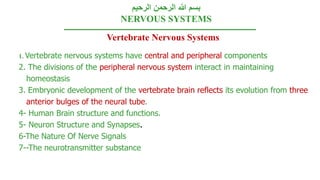
1. الرحيم الرحمن هللا بسم
NERVOUS SYSTEMS
Vertebrate Nervous Systems
1. Vertebrate nervous systems have central and peripheral components
2. The divisions of the peripheral nervous system interact in maintaining
homeostasis
3. Embryonic development of the vertebrate brain reflects its evolution from three
anterior bulges of the neural tube.
4- Human Brain structure and functions.
5- Neuron Structure and Synapses.
6-The Nature Of Nerve Signals
7--The neurotransmitter substance
2. 1. Vertebrate nervous systems compose from
central and peripheral nervous system
• A) Central nervous system (CNS):
• Form from: Brain and spinal cord.
• Both contain fluid-filled spaces which contain cerebrospinal
fluid (CSF).
• The central canal of the spinal cord is continuous with the
ventricles of the brain.
• The White matter is composed of bundles of myelinated axons
• Gray matter consists of unmyelinated axons, nuclei, and
dendrites.
• B) Peripheral nervous system. (PNS)
• All dendrites and axons fibers outside the CNS.
3. Anatomically the nervous system is divided into
Central nervous
system (CNS)
The brain
The spinal
cord
Peripheral nervous
system (PNS)
Nerve fibers
Ganglia
5. Nervous tissue
• It consists of two types of cells
1. Neurons (nerve cells).
2. Neuralgia (supporting cell).
• The neurons (receive information
and conduct it)
• The structure of neuron:
1.Cell body.
2. Processes {axon and dendrites}
6. Classification of neuron
Functionally The neurons could be :
1. Sensory neurons : where impulses are carried from receptors to the CNS
2. Motor neurons: where impulses are carried from CNS to the effector organ muscle
or gland
3. Interneurons: act as a link between sensory and motor neurons. They are present
in the CNS only.
Morphology According to the number of their processes
1. Unipolar : have only one cell process.
2. Pseudo-unipolar : have a single process that divides into two branches.
3. Bipolar :have two processes one as an axon and the other as a dendrite.
4. Multipolar :have more than two processes.
8. Inside the vertebral column form from:
1.White matter which is composed of bundles of myelinated axons, of the sensory
and motor neuron.
2. Gray matter consists of unmyelinated axons, nuclei, and dendrites. Of motor and
sensory neuron and the whole interneurons.
• A Simple Nerve Circuit – the
Reflex Arc.
• A reflex is an autonomic
response
The Spinal Cord
9. Peripheral nervous system (PNS)
• Sensory receptors a responsive to external and internal stimuli.
• Such sensory input is conveyed to integration centers at the Central nervous
system
• Where in the input is interpreted an associated with a response
10. • Motor output is the conduction of signals from integration centers to
effector cells.
• Effector cells carry out the body’s response to a stimulus.
• The central nervous system (CNS) is responsible for integration.
• The signals of the nervous system are conducted by nerves
11. • Structural composition of the PNS.
1. Paired cranial nerves that originate in the
brain and innervate the head and upper body.
2. Paired spinal nerves that originate in the spinal
cord and innervate the entire body.
3. Ganglia associated with the cranial and spinal
nerves.
2. The divisions of the peripheral nervous system (PNS) interact
in maintaining homeostasis
17. Human Brain
Conscious Brain
Cerebrum
Unconscious Brain
Corpus
Callosum
Link the two
cerebral
cortex
Cerebral Hemispheres
1-Frontal
2--Parital
3-Temporal
4- Occipital
Limbic System
Beneath cerebral
cortex
-Emotions
-Memory
-Learning
-Pain
-Pleasure
-Sorrow.
18. • The Reticular System, Arousal, and Sleep.
• The reticular activating system (RAS) of the reticular formation.
• Regulates sleep and arousal.
• Acts as a sensory filter.
19. limbic system
• In mammals, the limbic system is composed of the hippocampus,
olfactory cortex, inner portions of the cortex’s lobes, and parts of the
thalamus and hypothalamus.
• Mediates basic emotions (fear, anger), involved in emotional bonding,
establishes emotional memory
• For example, the amygdala is involved in recognizing the emotional
content of facial expression.
21. Memory and Learning
• Short-term memory stored in the frontal lobes.
• The establishment of long-term memory involves the hippocampus.
• The transfer of information from short-term to long-term memory: Is
enhanced by repetition (remember that when you are preparing for an
exam).
• The amygdala Influenced the emotional states. Influenced by association
with previously stored information.
• Different types of long-term memories are stored in different regions of the
brain.
• Memorization-type memory can be rapid. Primarily involves changes in the
strength of existing nerve connections.
• Learning of skills and procedures is slower. Appears to involves cellular
mechanisms similar to those involved in brain growth and development.
22. Conscious Brain: Cerebrum
• The cerebrum is divided into left and right cerebrum hemispheres.
• The left hemisphere is primarily responsible for the right side of the body.
• The right hemisphere is primarily responsible for the left side of the body.
• The left hemisphere.
• Specializes in language, math, logic operations, and the processing of serial
sequences of information, and visual and auditory details.
• Specializes in detailed activities required for motor control.
• The right hemisphere.
• Specializes in pattern recognition, spatial relationships, nonverbal ideation,
emotional processing, and the parallel processing of information.
The cerebrum is divided into frontal, temporal, occipital, and parietal lobes.
• Frontal lobe.: Contains the primary motor cortex.
• Parietal lobe: Contains the primary somato-sensory cortex.
23. Conscious Brain:Cerebrum
• Cerebral Hemispheres
1-Frontal
2-Parital
3-Temporal
4- Occipital
The cerebrum is derived from
the embryonic telencephalon.
24. Corpus callosum
• Link the two cerebral cortex
Cerebral cortex: outer covering of gray
matter.
Neocortex: region unique to mammals.
The more convoluted the surface of the
neocortex the more surface area the more
neurons.
Basal nuclei: internal clusters of nuclei
25.
26. • The Brainstem.
• The “lower brain.” Consists of the medulla oblongata, pons, and
midbrain.
• Derived from the embryonic hindbrain and midbrain.
• Functions in homeostasis, coordination of movement, conduction
of impulses to higher brain centers.
The Brainstem
27. The brainstem
1. Medulla oblongata:
Contains nuclei that control visceral (autonomic homeostatic) functions.
• Breathing.
• Heart and blood vessel activity.
• Swallowing.
• Vomiting.
• Digestion.
• Relays information to and from higher brain centers.
2. Pons.
Contains nuclei involved in the regulation of visceral activities such as breathing.
• Relays information to and from higher brain centers.
28. 3.The Midbrain
Contains nuclei involved in the integration of sensory information.
• Superior colliculi are involved in the regulation of visual reflexes.
• Inferior colliculi are involved in the regulation of auditory
reflexes.
• Relays information to and from higher brain centers.
29. • Sleep and wakefulness produces patterns of electrical activity in
the brain that can be recorded as an electroencephalogram
(EEG).
• Most dreaming
occurs during
REM (rapid
eye movement)
sleep.
30. The Cerebellum
• Develops from part of the metencephalon.
• Functions to error-check and coordinate motor activities, and
perceptual and cognitive factors.
• Relays sensory information about joints, muscles, sight, and
sound to the cerebrum.
• Coordinates motor commands issued by the cerebrum.
31. The thalamus and hypothalamus:
The epithalamus, thalamus, and hypothalamus are derived from the embryonic
diencephalon
• Epithalamus:
Includes a choroid plexus and the pineal gland.
• Thalamus:
• Relays all sensory information to the cerebrum.
• Contains one nucleus for each type of sensory information.
• Relays motor information from the cerebrum.
• Receives input from the cerebrum.
• Receives input from brain centers involved in the regulation of emotion and arousal.
• Hypothalamus:
• Regulates autonomic activity.
• Contains nuclei involved in thermoregulation, hunger, thirst, sexual and mating
behavior, etc.
• Regulates the pituitary gland.
32. The Hypothalamus and Circadian Rhythms.
• The biological clock is the internal timekeeper.
• The clock’s rhythm usually does not exactly match environmental events.
• Experiments in which humans have been deprived of external cues have
shown that biological clock has a period of about 25 hours.
• In mammals, the hypothalamic suprachiasmatic nuclei (SCN) function as a
biological clock.
• Produce proteins in response to light/dark cycles.
•This, and other biological clocks, may be responsive to hormonal release,
hunger, and various external stimuli.
33. 5- Neuron Structure and Synapses.
• The neuron is the structural and functional unit of the nervous system.
• Nerve impulses are conducted along a neuron.
• Dentrite cell body axon hillock axon
• Some axons are insulated by a myelin sheath.
• Axon endings are called synaptic terminals.
• They contain neurotransmitters which conduct a signal across a synapse.
• A synapse is the junction between a presynaptic and postsynaptic neuron.
• A ganglion is a cluster of nerve cell bodies within the PNS.
• A nucleus is a cluster of nerve cell bodies within the CNS.
35. Schwann cells are found within the PNS. Form a myelin sheath by insulating
axons
36. Types of Nerve Circuits.
1. Single presynaptic neuron several postsynaptic neurons.
2. Several presynaptic neurons single postsynaptic neuron.
3. Circular paths.
Supporting Cells (Glia).
There are several types of glia.
1. Astrocytes are found within the CNS.
• Structural and metabolic support.
• By inducing the formation of tight junctions between capillary cells astrocytes help form the
blood-brain barrier.
• Like neurons, astrocytes communicate with one another via chemical signals.
2. Oligodendrocytes are found within the CNS.
• Form a myelin sheath by insulating axons
37. 6- The Nature Of Nerve Signals
1.Every cell has a voltage, or membrane potential, across its plasma
membrane.
2. Changes in the membrane potential of a neuron give rise to nerve
impulses.
3. Nerve impulses propagate themselves along an axon
38. 1- Every cell has a voltage, or membrane potential, across its
plasma membrane
A membrane potential is a localized electrical gradient across membrane.
• Anions are more concentrated within a cell.
• Cations are more concentrated in the extracellular fluid.
• How a Cell Maintains a Membrane Potential?
• Anions(-) Such as: Proteins, amino acids, sulfate, and phosphate are the principal intracellular
anions.
• Cl– is principal extracellular anion.
• Cations (+) Such as: K+ the principal intracellular cation.
• Na+ is the principal extracellular cation.
40. • Ungated ion channels allow ions to diffuse across the plasma
membrane.
• These channels are always open.
• This diffusion does not achieve an equilibrium since sodium-potassium
pump transports these ions against their concentration gradients.
41. 2. Changes in the membrane potential of a neuron give rise to
nerve impulses
• Excitable cells have the ability to generate large changes in their membrane
potentials.
• Gated ion channels open or close in response to stimuli.
• The subsequent diffusion of ions leads to a change in the membrane potential.
• Types of gated ions.
1. Chemically-gated ion channels open or close in response to a chemical stimulus.
2. Voltage-gated ion channels open or close in response to a change in membrane
potential.
• Graded Potentials: Hyperpolarization and Depolarization.
• Graded potentials are changes in membrane potential.
42. • Hyperpolarization
Gated K+ channels open K+
diffuses out of the cell the
membrane potential becomes
more negative.
Depolarization.
Gated Na+ channels open
Na+ diffuses into the
cell the membrane
potential becomes less
negative.
• The Action Potential:
All or Nothing Depolarization.
• If graded potentials sum to -55mV
a threshold potential is achieved.
• This triggers an action potential.
Axons only
48. 3. Nerve impulses propagate themselves along an axon
• The action potential is repeatedly regenerated along the length of the
axon.
• An action potential achieved at one region of the membrane is
sufficient to depolarize a neighboring region above threshold.
• Thus triggering a new action potential.
• The refractory period assures that impulse conduction is unidirectional
49. Saltatory conduction.
In myelinated neurons only ,unmyelinated
regions of the axon depolarize.
Thus, the impulse moves faster than in
unmyelinated neurons
50. 4- Nerve signal across the synapse: Chemical or electrical communication
between cells occurs at synapses
• Electrical Synapses.
• Action potentials travels directly from the presynaptic to the postsynaptic
cells via gap junctions.
• Chemical Synapses.
• More common than electrical synapses.
• Postsynaptic chemically-gated channels exist for ions such as Na+, K+, and
Cl-.
• Depending on which gates open the postsynaptic neuron can depolarize or
hyperpolarize.
52. Neural integration occurs at the cellular level
• Excitatory postsynaptic potentials (EPSP) depolarize the
postsynaptic neuron.
• The binding of neurotransmitter to postsynaptic receptors open gated
channels that allow Na+ to diffuse into and K+ to diffuse out of the cell.
• Inhibitory postsynaptic potential (IPSP) hyperpolarize the
postsynaptic neuron.
• The binding of neurotransmitter to postsynaptic receptors open gated
channels that allow K+ to diffuse out of the cell and/or Cl- to diffuse into the
cell.
53. The neurotransmitter substance:
can produce different effects on different types of cells
• Acetylcholine.
• Excitatory to skeletal muscle.
• Inhibitory to cardiac muscle.
• Secreted by the CNS, PNS, and at vertebrate neuromuscular
junctions.
• Biogenic Amines.
1.Epinephrine and norepinephrine.
• Can have excitatory or inhibitory effects.
• Secreted by the CNS and PNS.
• Secreted by the adrenal glands
54. 2. Dopamine
• Generally excitatory; may be inhibitory at some sites.
• Widespread in the brain.
• Affects sleep, mood, attention, and learning.
• Secreted by the CNS and PNS.
• A lack of dopamine in the brain is associated with Parkinson’s disease.
• Excessive dopamine is linked to schizophrenia
3. Serotonin.
• Generally inhibitory.
• Widespread in the brain.
• Affects sleep, mood, attention, and learning
• Secreted by the CNS.
55. • Amino Acids
1. Gamma aminobutyric acid (GABA). : Inhibitory. Secreted by the CNS and at
invertebrate neuromuscular junctions.
2. Glycine. : Inhibitory. Secreted by the CNS.
3. Glutamate. : Excitatory. Secreted by the CNS and at
4. invertebrate neuromuscular junctions.
5. Aspartate. :Excitatory. Secreted by the CNS.
• Neuropeptides.
1. Substance P. : Excitatory. Secreted by the CNS and PNS.
2. Met-enkephalin (an endorphin) : Generally inhibitory. Secreted by the CNS.
3. Gasses that act as local regulators. Nitric oxide. Carbon monoxide.