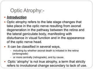
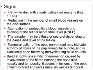
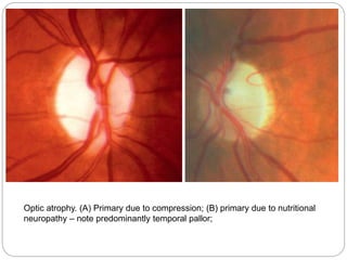
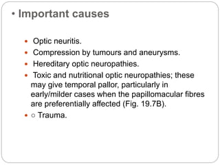
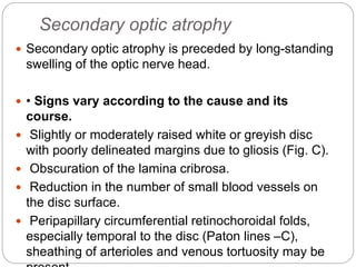
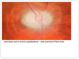
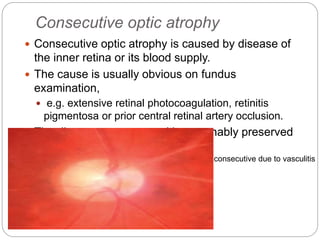
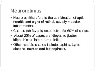
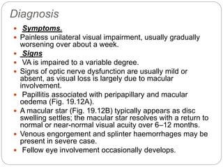
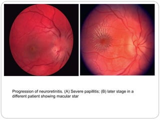
Optic atrophy refers to changes in the optic nerve resulting from axonal degeneration between the retina and lateral geniculate body, causing visual disturbance and changes in the optic nerve head appearance. It can be classified as primary, secondary, or consecutive. Primary optic atrophy occurs without prior nerve swelling and may result from lesions along the visual pathway. Secondary optic atrophy is preceded by long-term nerve swelling and includes causes like chronic papilledema. Consecutive optic atrophy is caused by diseases of the inner retina or its blood supply. Neuroretinitis refers to optic neuritis with retinal inflammation, most commonly caused by cat scratch fever, and presents with papillitis, macular edema, and sometimes a macular
A surgical procedure featuring a partial thickness scleral flap that creates a fistula between AC and subconjunctival space for filtration of aqueous and creation of conjunctival bleb in an effort to lower IOP
A systematic approach with practical tips to diagnose and manage optic disc pallor. Disc pallor is often encountered in the routine clinical practice and remains a diagnostic enigma for most ophthalmologist. I illustrate the relevant practical points to be looked out for to deal with disc pallor.
Ischemic optic neuropathy constitutes one of the major causes of blindness or seriously impaired vision among the middle-aged and elderly population.
Ischemic optic neuropathy is due to acute ischemia of the optic nerve. it can be classified into two, depending upon the part of the optic nerve involved:
1.Anterior ischemic optic neuropathy (AION)
-AION is due to acute ischemia of the front (anterior) part of the optic nerve (also called optic nerve head), which is supplied mainly by the posterior ciliary arteries.
-AION is divided into two types, depending on what causes it:
1.Arteritic AION: This is the most serious type and is due to a disease called giant cell arteritis or temporal arteritis.
2. Non-arteritic AION: This is the usual, most common type, with many different causes but not associated with giant cell arteritis.
2.Posterior ischemic optic neuropathy (PION). -
-PION is a much less common type. It is due to acute ischemia of the back (posterior) part of the optic nerve, located some distance behind the eyeball; this part of the optic nerve is NOT supplied by the posterior ciliary arteries
(Hayreh, 2009)
The presentation was made under the wise guidance of my professor DR.(prof) P. Rawat (MGMMC & M.Y. HOSPITAL, INDORE).It covers the essential aspects of optic neuritis & optic atrophy.
This ppt describe about the incidence, diagnosis and management of maculopathy in caaes of pathological myopia.
Data collected and created by Vivek Chaudhary
For queries : vivek977optom@gmail.com
Pulmonary Thromboembolism - etilogy, types, medical- Surgical and nursing man...VarunMahajani
Disruption of blood supply to lung alveoli due to blockage of one or more pulmonary blood vessels is called as Pulmonary thromboembolism. In this presentation we will discuss its causes, types and its management in depth.
Tom Selleck Health: A Comprehensive Look at the Iconic Actor’s Wellness Journeygreendigital
Tom Selleck, an enduring figure in Hollywood. has captivated audiences for decades with his rugged charm, iconic moustache. and memorable roles in television and film. From his breakout role as Thomas Magnum in Magnum P.I. to his current portrayal of Frank Reagan in Blue Bloods. Selleck's career has spanned over 50 years. But beyond his professional achievements. fans have often been curious about Tom Selleck Health. especially as he has aged in the public eye.
Follow us on: Pinterest
Introduction
Many have been interested in Tom Selleck health. not only because of his enduring presence on screen but also because of the challenges. and lifestyle choices he has faced and made over the years. This article delves into the various aspects of Tom Selleck health. exploring his fitness regimen, diet, mental health. and the challenges he has encountered as he ages. We'll look at how he maintains his well-being. the health issues he has faced, and his approach to ageing .
Early Life and Career
Childhood and Athletic Beginnings
Tom Selleck was born on January 29, 1945, in Detroit, Michigan, and grew up in Sherman Oaks, California. From an early age, he was involved in sports, particularly basketball. which played a significant role in his physical development. His athletic pursuits continued into college. where he attended the University of Southern California (USC) on a basketball scholarship. This early involvement in sports laid a strong foundation for his physical health and disciplined lifestyle.
Transition to Acting
Selleck's transition from an athlete to an actor came with its physical demands. His first significant role in "Magnum P.I." required him to perform various stunts and maintain a fit appearance. This role, which he played from 1980 to 1988. necessitated a rigorous fitness routine to meet the show's demands. setting the stage for his long-term commitment to health and wellness.
Fitness Regimen
Workout Routine
Tom Selleck health and fitness regimen has evolved. adapting to his changing roles and age. During his "Magnum, P.I." days. Selleck's workouts were intense and focused on building and maintaining muscle mass. His routine included weightlifting, cardiovascular exercises. and specific training for the stunts he performed on the show.
Selleck adjusted his fitness routine as he aged to suit his body's needs. Today, his workouts focus on maintaining flexibility, strength, and cardiovascular health. He incorporates low-impact exercises such as swimming, walking, and light weightlifting. This balanced approach helps him stay fit without putting undue strain on his joints and muscles.
Importance of Flexibility and Mobility
In recent years, Selleck has emphasized the importance of flexibility and mobility in his fitness regimen. Understanding the natural decline in muscle mass and joint flexibility with age. he includes stretching and yoga in his routine. These practices help prevent injuries, improve posture, and maintain mobilit
Flu Vaccine Alert in Bangalore Karnatakaaddon Scans
As flu season approaches, health officials in Bangalore, Karnataka, are urging residents to get their flu vaccinations. The seasonal flu, while common, can lead to severe health complications, particularly for vulnerable populations such as young children, the elderly, and those with underlying health conditions.
Dr. Vidisha Kumari, a leading epidemiologist in Bangalore, emphasizes the importance of getting vaccinated. "The flu vaccine is our best defense against the influenza virus. It not only protects individuals but also helps prevent the spread of the virus in our communities," he says.
This year, the flu season is expected to coincide with a potential increase in other respiratory illnesses. The Karnataka Health Department has launched an awareness campaign highlighting the significance of flu vaccinations. They have set up multiple vaccination centers across Bangalore, making it convenient for residents to receive their shots.
To encourage widespread vaccination, the government is also collaborating with local schools, workplaces, and community centers to facilitate vaccination drives. Special attention is being given to ensuring that the vaccine is accessible to all, including marginalized communities who may have limited access to healthcare.
Residents are reminded that the flu vaccine is safe and effective. Common side effects are mild and may include soreness at the injection site, mild fever, or muscle aches. These side effects are generally short-lived and far less severe than the flu itself.
Healthcare providers are also stressing the importance of continuing COVID-19 precautions. Wearing masks, practicing good hand hygiene, and maintaining social distancing are still crucial, especially in crowded places.
Protect yourself and your loved ones by getting vaccinated. Together, we can help keep Bangalore healthy and safe this flu season. For more information on vaccination centers and schedules, residents can visit the Karnataka Health Department’s official website or follow their social media pages.
Stay informed, stay safe, and get your flu shot today!
Prix Galien International 2024 Forum ProgramLevi Shapiro
June 20, 2024, Prix Galien International and Jerusalem Ethics Forum in ROME. Detailed agenda including panels:
- ADVANCES IN CARDIOLOGY: A NEW PARADIGM IS COMING
- WOMEN’S HEALTH: FERTILITY PRESERVATION
- WHAT’S NEW IN THE TREATMENT OF INFECTIOUS,
ONCOLOGICAL AND INFLAMMATORY SKIN DISEASES?
- ARTIFICIAL INTELLIGENCE AND ETHICS
- GENE THERAPY
- BEYOND BORDERS: GLOBAL INITIATIVES FOR DEMOCRATIZING LIFE SCIENCE TECHNOLOGIES AND PROMOTING ACCESS TO HEALTHCARE
- ETHICAL CHALLENGES IN LIFE SCIENCES
- Prix Galien International Awards Ceremony
Recomendações da OMS sobre cuidados maternos e neonatais para uma experiência pós-natal positiva.
Em consonância com os ODS – Objetivos do Desenvolvimento Sustentável e a Estratégia Global para a Saúde das Mulheres, Crianças e Adolescentes, e aplicando uma abordagem baseada nos direitos humanos, os esforços de cuidados pós-natais devem expandir-se para além da cobertura e da simples sobrevivência, de modo a incluir cuidados de qualidade.
Estas diretrizes visam melhorar a qualidade dos cuidados pós-natais essenciais e de rotina prestados às mulheres e aos recém-nascidos, com o objetivo final de melhorar a saúde e o bem-estar materno e neonatal.
Uma “experiência pós-natal positiva” é um resultado importante para todas as mulheres que dão à luz e para os seus recém-nascidos, estabelecendo as bases para a melhoria da saúde e do bem-estar a curto e longo prazo. Uma experiência pós-natal positiva é definida como aquela em que as mulheres, pessoas que gestam, os recém-nascidos, os casais, os pais, os cuidadores e as famílias recebem informação consistente, garantia e apoio de profissionais de saúde motivados; e onde um sistema de saúde flexível e com recursos reconheça as necessidades das mulheres e dos bebês e respeite o seu contexto cultural.
Estas diretrizes consolidadas apresentam algumas recomendações novas e já bem fundamentadas sobre cuidados pós-natais de rotina para mulheres e neonatos que recebem cuidados no pós-parto em unidades de saúde ou na comunidade, independentemente dos recursos disponíveis.
É fornecido um conjunto abrangente de recomendações para cuidados durante o período puerperal, com ênfase nos cuidados essenciais que todas as mulheres e recém-nascidos devem receber, e com a devida atenção à qualidade dos cuidados; isto é, a entrega e a experiência do cuidado recebido. Estas diretrizes atualizam e ampliam as recomendações da OMS de 2014 sobre cuidados pós-natais da mãe e do recém-nascido e complementam as atuais diretrizes da OMS sobre a gestão de complicações pós-natais.
O estabelecimento da amamentação e o manejo das principais intercorrências é contemplada.
Recomendamos muito.
Vamos discutir essas recomendações no nosso curso de pós-graduação em Aleitamento no Instituto Ciclos.
Esta publicação só está disponível em inglês até o momento.
Prof. Marcus Renato de Carvalho
www.agostodourado.com
HOT NEW PRODUCT! BIG SALES FAST SHIPPING NOW FROM CHINA!! EU KU DB BK substit...GL Anaacs
Contact us if you are interested:
Email / Skype : kefaya1771@gmail.com
Threema: PXHY5PDH
New BATCH Ku !!! MUCH IN DEMAND FAST SALE EVERY BATCH HAPPY GOOD EFFECT BIG BATCH !
Contact me on Threema or skype to start big business!!
Hot-sale products:
NEW HOT EUTYLONE WHITE CRYSTAL!!
5cl-adba precursor (semi finished )
5cl-adba raw materials
ADBB precursor (semi finished )
ADBB raw materials
APVP powder
5fadb/4f-adb
Jwh018 / Jwh210
Eutylone crystal
Protonitazene (hydrochloride) CAS: 119276-01-6
Flubrotizolam CAS: 57801-95-3
Metonitazene CAS: 14680-51-4
Payment terms: Western Union,MoneyGram,Bitcoin or USDT.
Deliver Time: Usually 7-15days
Shipping method: FedEx, TNT, DHL,UPS etc.Our deliveries are 100% safe, fast, reliable and discreet.
Samples will be sent for your evaluation!If you are interested in, please contact me, let's talk details.
We specializes in exporting high quality Research chemical, medical intermediate, Pharmaceutical chemicals and so on. Products are exported to USA, Canada, France, Korea, Japan,Russia, Southeast Asia and other countries.
Lung Cancer: Artificial Intelligence, Synergetics, Complex System Analysis, S...Oleg Kshivets
RESULTS: Overall life span (LS) was 2252.1±1742.5 days and cumulative 5-year survival (5YS) reached 73.2%, 10 years – 64.8%, 20 years – 42.5%. 513 LCP lived more than 5 years (LS=3124.6±1525.6 days), 148 LCP – more than 10 years (LS=5054.4±1504.1 days).199 LCP died because of LC (LS=562.7±374.5 days). 5YS of LCP after bi/lobectomies was significantly superior in comparison with LCP after pneumonectomies (78.1% vs.63.7%, P=0.00001 by log-rank test). AT significantly improved 5YS (66.3% vs. 34.8%) (P=0.00000 by log-rank test) only for LCP with N1-2. Cox modeling displayed that 5YS of LCP significantly depended on: phase transition (PT) early-invasive LC in terms of synergetics, PT N0—N12, cell ratio factors (ratio between cancer cells- CC and blood cells subpopulations), G1-3, histology, glucose, AT, blood cell circuit, prothrombin index, heparin tolerance, recalcification time (P=0.000-0.038). Neural networks, genetic algorithm selection and bootstrap simulation revealed relationships between 5YS and PT early-invasive LC (rank=1), PT N0—N12 (rank=2), thrombocytes/CC (3), erythrocytes/CC (4), eosinophils/CC (5), healthy cells/CC (6), lymphocytes/CC (7), segmented neutrophils/CC (8), stick neutrophils/CC (9), monocytes/CC (10); leucocytes/CC (11). Correct prediction of 5YS was 100% by neural networks computing (area under ROC curve=1.0; error=0.0).
CONCLUSIONS: 5YS of LCP after radical procedures significantly depended on: 1) PT early-invasive cancer; 2) PT N0--N12; 3) cell ratio factors; 4) blood cell circuit; 5) biochemical factors; 6) hemostasis system; 7) AT; 8) LC characteristics; 9) LC cell dynamics; 10) surgery type: lobectomy/pneumonectomy; 11) anthropometric data. Optimal diagnosis and treatment strategies for LC are: 1) screening and early detection of LC; 2) availability of experienced thoracic surgeons because of complexity of radical procedures; 3) aggressive en block surgery and adequate lymph node dissection for completeness; 4) precise prediction; 5) adjuvant chemoimmunoradiotherapy for LCP with unfavorable prognosis.
NVBDCP.pptx Nation vector borne disease control programSapna Thakur
NVBDCP was launched in 2003-2004 . Vector-Borne Disease: Disease that results from an infection transmitted to humans and other animals by blood-feeding arthropods, such as mosquitoes, ticks, and fleas. Examples of vector-borne diseases include Dengue fever, West Nile Virus, Lyme disease, and malaria.
Couples presenting to the infertility clinic- Do they really have infertility...Sujoy Dasgupta
Dr Sujoy Dasgupta presented the study on "Couples presenting to the infertility clinic- Do they really have infertility? – The unexplored stories of non-consummation" in the 13th Congress of the Asia Pacific Initiative on Reproduction (ASPIRE 2024) at Manila on 24 May, 2024.
Report Back from SGO 2024: What’s the Latest in Cervical Cancer?bkling
Are you curious about what’s new in cervical cancer research or unsure what the findings mean? Join Dr. Emily Ko, a gynecologic oncologist at Penn Medicine, to learn about the latest updates from the Society of Gynecologic Oncology (SGO) 2024 Annual Meeting on Women’s Cancer. Dr. Ko will discuss what the research presented at the conference means for you and answer your questions about the new developments.
1. By
Mutahir Shah
Resident M Phil VS
Pakistan Institute of Community
Ophthalmology
Optic Atrophy and Neuroretinitis
2. Optic Atrophy:-
Introduction
Optic atrophy refers to the late stage changes that
take place in the optic nerve resulting from axonal
degeneration in the pathway between the retina and
the lateral geniculate body, manifesting with
disturbance in visual function and in the appearance
of the optic nerve head.
It can be classified in several ways,
including by whether axonal death is initiated in the retina
(anterograde)
or more centrally (retrograde), and by cause.
Optic ‘atrophy’ is not true atrophy, a term that strictly
refers to involutional change secondary to lack of use.
3. Primary Optic Atrophy
Primary optic atrophy
Primary optic atrophy occurs without antecedent
swelling of the optic nerve head.
It may be caused by lesions affecting the visual
pathways at any point from the retrolaminar portion of
the optic nerve to the lateral geniculate body.
Lesions anterior to the optic chiasm result in
unilateral optic atrophy, whereas those involving the
chiasm and optic tract will cause bilateral changes.
4. • Signs
Flat white disc with clearly delineated margins (Fig.
19.7A).
Reduction in the number of small blood vessels on
the disc surface.
Attenuation of peripapillary blood vessels and
thinning of the retinal nerve fibre layer (RNFL).
The atrophy may be diffuse or sectoral depending on
the cause and level of the lesion.
Temporal pallor of the optic nerve head may indicate
atrophy of fibres of the papillomacular bundle, and is
classically seen following demyelinating optic neuritis.
Band atrophy is a similar phenomenon caused by
involvement of the fibres entering the optic disc
nasally and temporally; it occurs in lesions of the optic
chiasm or tract and gives nasal as well as temporal
5. Optic atrophy. (A) Primary due to compression; (B) primary due to nutritional
neuropathy – note predominantly temporal pallor;
6. • Important causes
Optic neuritis.
Compression by tumours and aneurysms.
Hereditary optic neuropathies.
Toxic and nutritional optic neuropathies; these
may give temporal pallor, particularly in
early/milder cases when the papillomacular fibres
are preferentially affected (Fig. 19.7B).
○ Trauma.
7. Secondary optic atrophy
Secondary optic atrophy is preceded by long-standing
swelling of the optic nerve head.
• Signs vary according to the cause and its
course.
Slightly or moderately raised white or greyish disc
with poorly delineated margins due to gliosis (Fig. C).
Obscuration of the lamina cribrosa.
Reduction in the number of small blood vessels on
the disc surface.
Peripapillary circumferential retinochoroidal folds,
especially temporal to the disc (Paton lines –C),
sheathing of arterioles and venous tortuosity may be
8. secondary due to chronic papilloedema – note prominent Paton lines
9. Causes
Include
chronic papilloedema,
anterior ischaemic optic neuropathy
papillitis.
Intraocular inflammatory causes of marked disc
swelling are sometimes considered to cause
secondary rather than consecutive atrophy
10. Consecutive optic atrophy
Consecutive optic atrophy is caused by disease of
the inner retina or its blood supply.
The cause is usually obvious on fundus
examination,
e.g. extensive retinal photocoagulation, retinitis
pigmentosa or prior central retinal artery occlusion.
The disc appears waxy, with reasonably preserved
architecture
consecutive due to vasculitis
11. Neuroretinitis
Neuroretinitis refers to the combination of optic
neuritis and signs of retinal, usually macular,
inflammation.
Cat-scratch fever is responsible for 60% of cases.
About 25% of cases are idiopathic (Leber
idiopathic stellate neuroretinitis).
Other notable causes include syphilis, Lyme
disease, mumps and leptospirosis.
12. Diagnosis
Symptoms.
Painless unilateral visual impairment, usually gradually
worsening over about a week.
Signs
VA is impaired to a variable degree.
Signs of optic nerve dysfunction are usually mild or
absent, as visual loss is largely due to macular
involvement.
Papillitis associated with peripapillary and macular
oedema (Fig. 19.12A).
A macular star (Fig. 19.12B) typically appears as disc
swelling settles; the macular star resolves with a return to
normal or near-normal visual acuity over 6–12 months.
Venous engorgement and splinter haemorrhages may be
present in severe case.
Fellow eye involvement occasionally develops.
14. Investigation and Treatment
Optical coherence tomography (OCT)
demonstrates sub- and intraretinal fluid to a
variable extent.
Fluorescein angiography (FA) shows diffuse
leakage from superficial disc vessels.
Blood tests may include serology for Bartonella
and other causes according to clinical suspicion
Treatment
This is specific to the cause, and often consists of
antibiotics. Recurrent idiopathic cases may
require treatment with steroids and/or other
immunosuppressants.