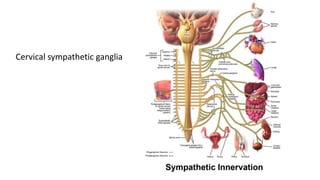
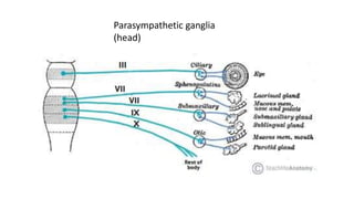
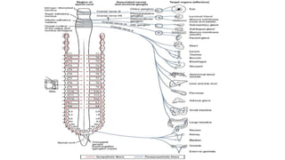
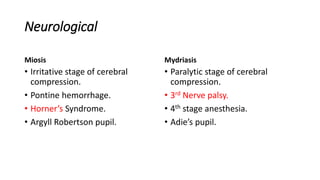
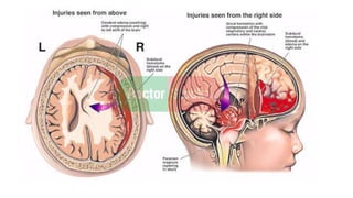
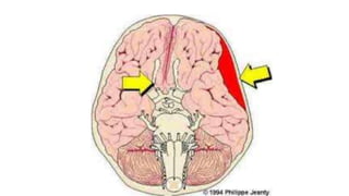
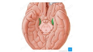
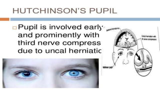
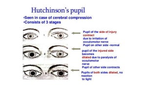
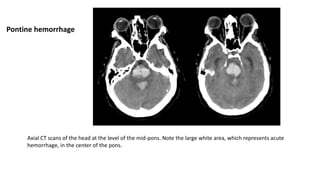
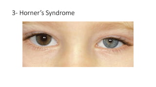
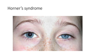
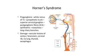
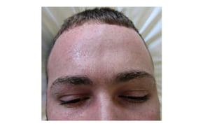
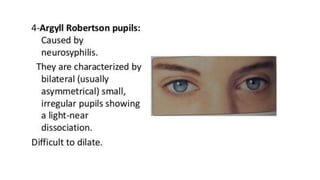
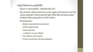
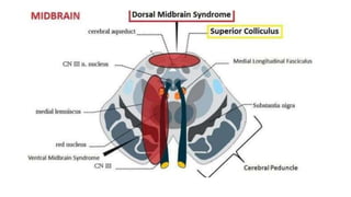
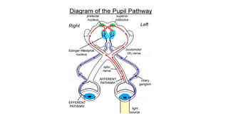
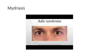
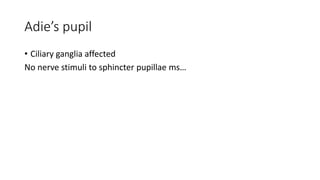
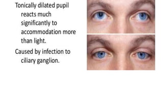
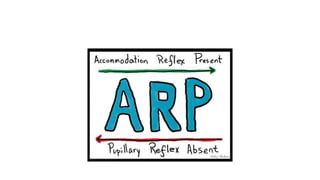
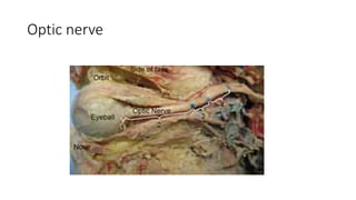
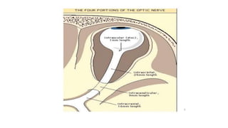
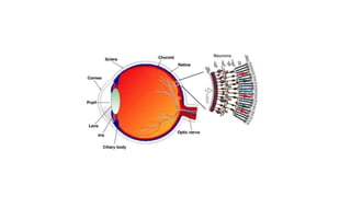
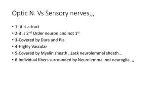
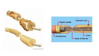
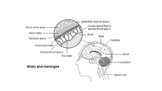
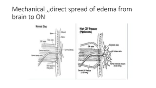
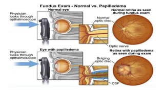
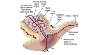
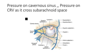
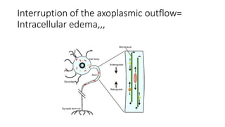
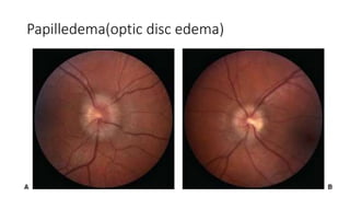
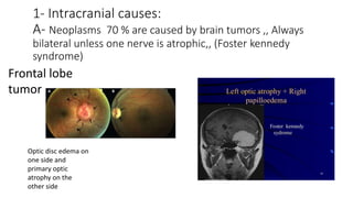
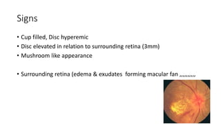
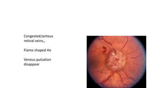
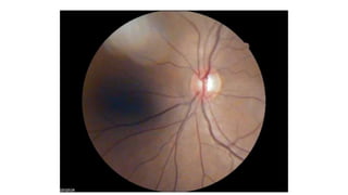
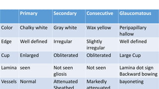
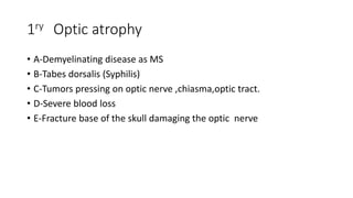
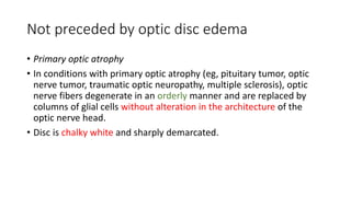
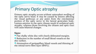
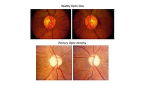
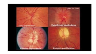
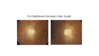
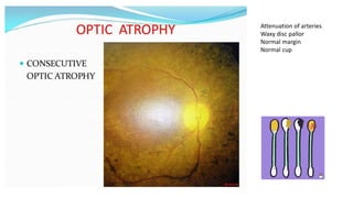
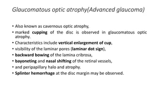
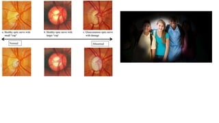
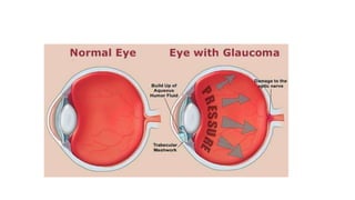
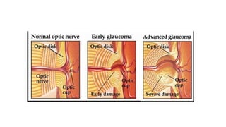
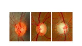
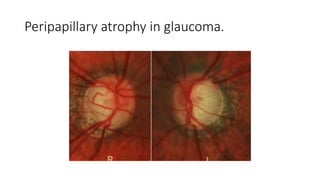
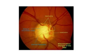
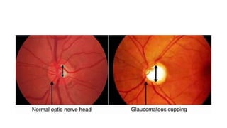
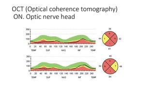
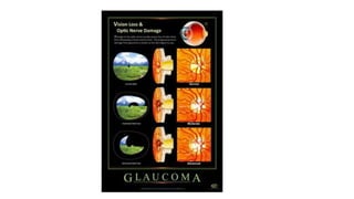
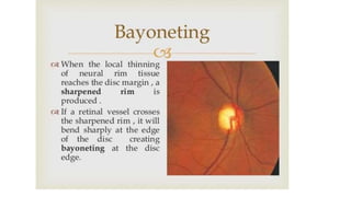
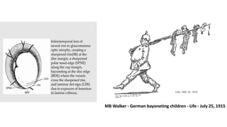
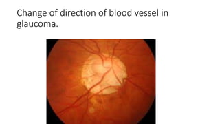
Neuro Ophthalmology
Ahmed Osama Hashem
FRCS ophth
PhD,MD Ain Shams university
Lecturer of ophthalmology
Mydriasis
• Dark
• Excitement
• Light colored iris, Myopic
• Anesthesia 2nd stage
• Ciliospinal reflex (pain applied to head ,neck and upper trunk)
(pupillary-skin reflex) consists of dilation of the ipsilateral pupil in response to pain applied to the
neck, face, and upper trunk. If the right side of the neck is subjected to a painful stimulus, the right
pupil dilates (increases in size 1-2mm from baseline).
Ocular causes
-Miosis
• Acute Iritis
• Trauma
• Paracentesis and hypotony
-Mydriasis
. Acute congestive glaucoma
. Trauma
. Blind eye
Cervical sympathetic ganglia
Parasympathetic ganglia
(head)
Medications
-Miosis
Local miotics
Opium (Heroin)
-Mydriasis
Local mydriatics
Atropine & Datura
Cocaine (Atypical nor epinephrine)
Neurological
Miosis
• Irritative stage of cerebral
compression.
• Pontine hemorrhage.
• Horner’s Syndrome.
• Argyll Robertson pupil.
Mydriasis
• Paralytic stage of cerebral
compression.
• 3rd Nerve palsy.
• 4th stage anesthesia.
• Adie’s pupil.
Axial CT scans of the head at the level of the mid-pons. Note the large white area, which represents acute
hemorrhage, in the center of the pons.
Pontine hemorrhage
3- Horner’s Syndrome
Horner’s syndrome
Mydriasis
Adie’s pupil
• Ciliary ganglia affected
No nerve stimuli to sphincter pupillae ms…
Optic nerve
• Structure. The term pyramidal tracts refers to upper motor neurons
that originate in the cerebral cortex and terminate in the spinal cord
(corticospinal) or brainstem (corticobulbar)…
Optic N. Vs Sensory nerves,,,
• 1- it is a tract
• 2-it is 2nd Order neuron and not 1st
• 3-Covered by Dura and Pia
• 4-Highly Vascular
• 5-Covered by Myelin sheath ,,Lack neurolemmal sheath…
• 6-individual fibers surrounded by Neurolemmal not neuroglia ,,,
• Optic disc edema
• Optic Neuritis
• Optic atrophy
Papilledema: Bilateral
• Swelling of the optic nerve head
(Non inflammatory) dt increase ICT
Localized to ON head or diffuse edema…
Mechanical ,,direct spread of edema from
brain to ON
Pressure on cavernous sinus ,, Pressure on
CRV as it cross subarachnoid space
10-12 mm behind the globe,,
Interruption of the axoplasmic outflow=
Intracellular edema,,,
Papilledema(optic disc edema)
Etiology
1- Intracranial causes:
A- Neoplasms 70 % are caused by brain tumors ,, Always bilateral
unless one nerve is atrophic,, (Foster kennedy syndrome)
1- Intracranial causes:
A- Neoplasms 70 % are caused by brain tumors ,, Always
bilateral unless one nerve is atrophic,, (Foster kennedy
syndrome)
Frontal lobe
tumor
Optic disc edema on
one side and
primary optic
atrophy on the
other side
1-Intracranial causes:
A-Neoplasms(70%)
B- Inflammatory:Meningitis,tuberculoma,brain abscess
C- Vascular: Subarachnoid Hge and cavernous sinus thrombosis
D-Pseudotumour cerebri:Benign increase in intracranial tension
Young Female (Hypervitaminosis A & D ! … Oral contraceptive
pills,,Tetracycline, Addison’s disease ,, cushing’s syndrome)
2-Systemic causes
• Malignant hypertension,eclampsia(pregnancy) and renal retinopathy
• Polycythemia
• Anemia:iron-deficiency contributes to thrombosis by inducing
reactive thrombocytosis, possibly via erythropoietin production,,
Optic disc edema (unilateral)
• 3-orbital causes:
-retrobulbar mass (tumour or cyst)
-inflammation (orbital cellulitis)
10-12 mm behind the globe,,
4-Ocular causes
• Sudden lowering to IOP (cornea perforatioin,after glaucoma surgery)
• Central retinal vein occlusion
Clinical picture
• Symptoms
-Asymptomatic
-Amarousis
-Associated symptoms (Projectile vomiting(without nausea)
,headache,diplopia (dt 6th N palsy)
Diminution of vision is late
Signs
• Pupil: Early ,,Normal in size reactivity (MCQ-Case)
• But late with occurrence of optic neuropathy : loss of direct light
reflex
• If edema is asymmetrical there will be RAPD
Signs
• VF (confrontation) enlargement of the blind spot ?
• Late there is contraction of the field
Signs
• Cup filled, Disc hyperemic
• Disc elevated in relation to surrounding retina (3mm)
• Mushroom like appearance
• Surrounding retina (edema & exudates forming macular fan ,,,,,,,,,,
Congested,tortous
retinal veins,,
Flame shaped He
Venous pulsation
disappear
Investigation
• Perimetry (field of vision) ,,, central scotoma for blue
• Radiology : X ray – CT – MRI
• VEP
Complication
• Post papilledemic optic atrophy
Optic Atrophy: Optic Neuropathy
• Definition: Degeneration of optic nerve fibers
• Interruption of nerve fibers at any point between
A- retinal ganglion cell
B-lateral geniculate body
According to presence of edema in ON Head
• primary if it occurs without any preceding optic nerve head edema
• secondary if it is preceded by edema
Primary Secondary Consecutive Glaucomatous
Color Chalky white Gray white Wax yellow Peripapillary
hallow
Edge Well defined Irregular Slightly
irregular
Well defined
Cup Enlarged Obliterated Obliterated Large Cup
Lamina seen Not seen
gliosis
Not seen Lamina dot sign
Backward bowing
Vessels Normal Attenuated Markedly bayoneting
1ry Optic atrophy
• A-Demyelinating disease as MS
• B-Tabes dorsalis (Syphilis)
• C-Tumors pressing on optic nerve ,chiasma,optic tract.
• D-Severe blood loss
• E-Fracture base of the skull damaging the optic nerve
Not preceded by optic disc edema
• Primary optic atrophy
• In conditions with primary optic atrophy (eg, pituitary tumor, optic
nerve tumor, traumatic optic neuropathy, multiple sclerosis), optic
nerve fibers degenerate in an orderly manner and are replaced by
columns of glial cells without alteration in the architecture of the
optic nerve head.
• Disc is chalky white and sharply demarcated.
2ry optic atrophy
• Post papilledemic optic atrophy
Preceded by optic disc edema
• In conditions with secondary optic atrophy (eg, papilledema,
papillitis), the atrophy is secondary to disc edema.
• Optic nerve fibers exhibit marked degeneration, with excessive
proliferation of glial tissue.
• Architecture is lost, ill defined margins.
• Disc is grey or dirty grey.
• Lamina cribrosa is obscured due to proliferating fibroglial tissue.
Consecutive optic atrophy
• In consecutive optic atrophy (eg, retinitis pigmentosa, myopia,
central retinal artery occlusion), the disc is waxy pale with a
normal disc margin, marked attenuation of arteries, and a
normal physiologic cup.
Attenuation of arteries
Waxy disc pallor
Normal margin
Normal cup
Glaucomatous optic atrophy(Advanced glaucoma)
• Also known as cavernous optic atrophy,
• marked cupping of the disc is observed in glaucomatous optic
atrophy.
• Characteristics include vertical enlargement of cup,
• visibility of the laminar pores (laminar dot sign),
• backward bowing of the lamina cribrosa,
• bayoneting and nasal shifting of the retinal vessels,
• and peripapillary halo and atrophy.
• Splinter hemorrhage at the disc margin may be observed.
Peripapillary atrophy in glaucoma.
OCT (Optical coherence tomography)
ON. Optic nerve head
MB Walker - German bayoneting children - Life - July 25, 1915
Change of direction of blood vessel in
glaucoma.
Thank You

Optic Nerve and pupil tutorial

  • 1.
    Neuro Ophthalmology Ahmed OsamaHashem FRCS ophth PhD,MD Ain Shams university Lecturer of ophthalmology
  • 2.
    Mydriasis • Dark • Excitement •Light colored iris, Myopic • Anesthesia 2nd stage • Ciliospinal reflex (pain applied to head ,neck and upper trunk) (pupillary-skin reflex) consists of dilation of the ipsilateral pupil in response to pain applied to the neck, face, and upper trunk. If the right side of the neck is subjected to a painful stimulus, the right pupil dilates (increases in size 1-2mm from baseline).
  • 3.
    Ocular causes -Miosis • AcuteIritis • Trauma • Paracentesis and hypotony -Mydriasis . Acute congestive glaucoma . Trauma . Blind eye
  • 5.
  • 6.
  • 8.
    Medications -Miosis Local miotics Opium (Heroin) -Mydriasis Localmydriatics Atropine & Datura Cocaine (Atypical nor epinephrine)
  • 9.
    Neurological Miosis • Irritative stageof cerebral compression. • Pontine hemorrhage. • Horner’s Syndrome. • Argyll Robertson pupil. Mydriasis • Paralytic stage of cerebral compression. • 3rd Nerve palsy. • 4th stage anesthesia. • Adie’s pupil.
  • 15.
    Axial CT scansof the head at the level of the mid-pons. Note the large white area, which represents acute hemorrhage, in the center of the pons. Pontine hemorrhage
  • 16.
  • 17.
  • 24.
  • 25.
    Adie’s pupil • Ciliaryganglia affected No nerve stimuli to sphincter pupillae ms…
  • 28.
  • 30.
    • Structure. Theterm pyramidal tracts refers to upper motor neurons that originate in the cerebral cortex and terminate in the spinal cord (corticospinal) or brainstem (corticobulbar)…
  • 34.
    Optic N. VsSensory nerves,,, • 1- it is a tract • 2-it is 2nd Order neuron and not 1st • 3-Covered by Dura and Pia • 4-Highly Vascular • 5-Covered by Myelin sheath ,,Lack neurolemmal sheath… • 6-individual fibers surrounded by Neurolemmal not neuroglia ,,,
  • 37.
    • Optic discedema • Optic Neuritis • Optic atrophy
  • 38.
    Papilledema: Bilateral • Swellingof the optic nerve head (Non inflammatory) dt increase ICT Localized to ON head or diffuse edema…
  • 40.
    Mechanical ,,direct spreadof edema from brain to ON
  • 43.
    Pressure on cavernoussinus ,, Pressure on CRV as it cross subarachnoid space
  • 44.
    10-12 mm behindthe globe,,
  • 45.
    Interruption of theaxoplasmic outflow= Intracellular edema,,,
  • 46.
  • 47.
    Etiology 1- Intracranial causes: A-Neoplasms 70 % are caused by brain tumors ,, Always bilateral unless one nerve is atrophic,, (Foster kennedy syndrome)
  • 48.
    1- Intracranial causes: A-Neoplasms 70 % are caused by brain tumors ,, Always bilateral unless one nerve is atrophic,, (Foster kennedy syndrome) Frontal lobe tumor Optic disc edema on one side and primary optic atrophy on the other side
  • 49.
    1-Intracranial causes: A-Neoplasms(70%) B- Inflammatory:Meningitis,tuberculoma,brainabscess C- Vascular: Subarachnoid Hge and cavernous sinus thrombosis D-Pseudotumour cerebri:Benign increase in intracranial tension Young Female (Hypervitaminosis A & D ! … Oral contraceptive pills,,Tetracycline, Addison’s disease ,, cushing’s syndrome)
  • 50.
    2-Systemic causes • Malignanthypertension,eclampsia(pregnancy) and renal retinopathy • Polycythemia • Anemia:iron-deficiency contributes to thrombosis by inducing reactive thrombocytosis, possibly via erythropoietin production,,
  • 51.
    Optic disc edema(unilateral) • 3-orbital causes: -retrobulbar mass (tumour or cyst) -inflammation (orbital cellulitis)
  • 52.
    10-12 mm behindthe globe,,
  • 53.
    4-Ocular causes • Suddenlowering to IOP (cornea perforatioin,after glaucoma surgery) • Central retinal vein occlusion
  • 54.
    Clinical picture • Symptoms -Asymptomatic -Amarousis -Associatedsymptoms (Projectile vomiting(without nausea) ,headache,diplopia (dt 6th N palsy) Diminution of vision is late
  • 55.
    Signs • Pupil: Early,,Normal in size reactivity (MCQ-Case) • But late with occurrence of optic neuropathy : loss of direct light reflex • If edema is asymmetrical there will be RAPD
  • 56.
    Signs • VF (confrontation)enlargement of the blind spot ? • Late there is contraction of the field
  • 57.
    Signs • Cup filled,Disc hyperemic • Disc elevated in relation to surrounding retina (3mm) • Mushroom like appearance • Surrounding retina (edema & exudates forming macular fan ,,,,,,,,,,
  • 58.
  • 60.
    Investigation • Perimetry (fieldof vision) ,,, central scotoma for blue • Radiology : X ray – CT – MRI • VEP
  • 61.
  • 62.
    Optic Atrophy: OpticNeuropathy • Definition: Degeneration of optic nerve fibers • Interruption of nerve fibers at any point between A- retinal ganglion cell B-lateral geniculate body
  • 63.
    According to presenceof edema in ON Head • primary if it occurs without any preceding optic nerve head edema • secondary if it is preceded by edema
  • 64.
    Primary Secondary ConsecutiveGlaucomatous Color Chalky white Gray white Wax yellow Peripapillary hallow Edge Well defined Irregular Slightly irregular Well defined Cup Enlarged Obliterated Obliterated Large Cup Lamina seen Not seen gliosis Not seen Lamina dot sign Backward bowing Vessels Normal Attenuated Markedly bayoneting
  • 65.
    1ry Optic atrophy •A-Demyelinating disease as MS • B-Tabes dorsalis (Syphilis) • C-Tumors pressing on optic nerve ,chiasma,optic tract. • D-Severe blood loss • E-Fracture base of the skull damaging the optic nerve
  • 66.
    Not preceded byoptic disc edema • Primary optic atrophy • In conditions with primary optic atrophy (eg, pituitary tumor, optic nerve tumor, traumatic optic neuropathy, multiple sclerosis), optic nerve fibers degenerate in an orderly manner and are replaced by columns of glial cells without alteration in the architecture of the optic nerve head. • Disc is chalky white and sharply demarcated.
  • 70.
    2ry optic atrophy •Post papilledemic optic atrophy
  • 72.
    Preceded by opticdisc edema • In conditions with secondary optic atrophy (eg, papilledema, papillitis), the atrophy is secondary to disc edema. • Optic nerve fibers exhibit marked degeneration, with excessive proliferation of glial tissue. • Architecture is lost, ill defined margins. • Disc is grey or dirty grey. • Lamina cribrosa is obscured due to proliferating fibroglial tissue.
  • 74.
    Consecutive optic atrophy •In consecutive optic atrophy (eg, retinitis pigmentosa, myopia, central retinal artery occlusion), the disc is waxy pale with a normal disc margin, marked attenuation of arteries, and a normal physiologic cup.
  • 75.
    Attenuation of arteries Waxydisc pallor Normal margin Normal cup
  • 76.
    Glaucomatous optic atrophy(Advancedglaucoma) • Also known as cavernous optic atrophy, • marked cupping of the disc is observed in glaucomatous optic atrophy. • Characteristics include vertical enlargement of cup, • visibility of the laminar pores (laminar dot sign), • backward bowing of the lamina cribrosa, • bayoneting and nasal shifting of the retinal vessels, • and peripapillary halo and atrophy. • Splinter hemorrhage at the disc margin may be observed.
  • 82.
  • 85.
    OCT (Optical coherencetomography) ON. Optic nerve head
  • 88.
    MB Walker -German bayoneting children - Life - July 25, 1915
  • 89.
    Change of directionof blood vessel in glaucoma.
  • 90.