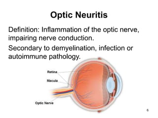
This document discusses optic neuritis, papilledema, and optic atrophy. It defines each condition, describes their signs and symptoms, classification, etiology, investigations, treatment and prognosis. Optic neuritis is inflammation of the optic nerve that can be caused by demyelination, infection or autoimmunity. Papilledema is bilateral swelling of the optic disc due to increased intracranial pressure. Optic atrophy is degeneration of the optic nerve fibers leading to pallor of the optic disc. The document provides detailed information and images to help understand and differentiate between these three important optic nerve conditions.

![Acknowledgement
• Yanoff and Duker. Papilledema. 2016 In: Ophthalmology. Mosby Inc.
• Khurana AK. Optic atrophy 2014 In: Comprehensive Ophthalmology. CBS Publishers
• Weerasinghe, D., & Lueck, C.J. (2016). Mimics and chameleons of optic neuritis. Practical
neurology;16(2):96-110.
• CME: Optic Neuritis: Diagnosis, Treatment, and Prognosis.
https://www.medscape.org/viewarticle/571660_2
• Kahloun R et al. Infectious optic neuropathies: a clinical update. Eye and Brain 2015;7:59-81
• Creel D. Visually Evoked Potentials. 2012 Mar 1. In: Kolb H, Fernandez E, Nelson R, editors. Webvision:
The Organization of the Retina and Visual System [Internet]. Salt Lake City (UT): University of Utah
Health Sciences Center; 1995-. Available from: https://www.ncbi.nlm.nih.gov/books/NBK107218/
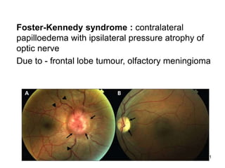
• Pastora-Salvador N et al. Foster Kennedy syndrome: papilledema in one eye with optic atrophy in the
other eye. CMAJ 2011;183(18):2135
• Reynolds SA. Pinpointing Papilledema. Optometric Management 2015:82-4
https://www.optometricmanagement.com/issues/2015/september-2015/clinical-posterior
• Schiffmann J et al. Evaluation and treatment of papilledema in pregnancy. Comprehensive ophthalmology
update 2006;7(4):187-202
• Toosy AT. Optic Neuritis. Lancet Neurol. 2014 Jan;13(1):83-99
2](https://image.slidesharecdn.com/830opticnervefinal-230524062307-9532ad31/85/830_Optic_nerve_final-ppt-2-320.jpg)