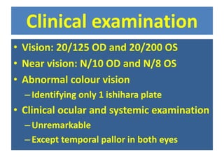
2. Presenting complaint
⢠A 30 year old male
⢠Complaint of loss of central vision
âslow
âpainless
âprogressive
⢠Since the age of 10 years
⢠Currently in both eyes.
3. Family history
⢠Two year younger sister
having similar loss of vision
since her age 10 years
4. Clinical examination
⢠Vision: 20/125 OD and 20/200 OS
⢠Near vision: N/10 OD and N/8 OS
⢠Abnormal colour vision
âIdentifying only 1 ishihara plate
⢠Clinical ocular and systemic examination
âUnremarkable
âExcept temporal pallor in both eyes
7. Referral letter: First Diagnosis
ghjgggggggggggggggggggggggggg
ggggggggggggggggggggggggggggg
gggggg
8. Differential Diagnosis
⢠Of the complaint of progressive
symmetric, bilateral, central
visual loss
â Comprise a group of disorders in
which the cause of optic nerve
dysfunction appears to be
hereditable.
1. Leberâs hereditary optic
neuropathy-LHON
2. Dominant optic atrophy-DOA
3. Hereditary optic atrophy with
other consistent neurologic or
systemic findings
4. Optic neuropathy as a
manifestation of hereditary
degenerative or developmental
diseases
9. Whatâs thought by first consultant
⢠Neuromyelitis optica (NMO), Devic's
disease or Devic's syndrome
â Is a heterogeneous condition consisting of the
simultaneous inflammation and demyelination of
the optic nerve (optic neuritis) and the spinal
cord (myelitis
â It can be monophasic or recurrent
10. Why it can not be Devicâs
â Is not a condition to be considered in
differential diagnosis
⢠As Spinal cord lesions lead to varying degrees
âOf weakness or paralysis in the legs or arms,
loss of sensation (including blindness),
and/or bladder and bowel dysfunction
19. LHON
⢠Men are affected with
visual loss more
frequently than
women, with a male
predominance of about
80 to 90% in most
pedigrees
⢠20 to 60% of men at
risk for LHON
experience visual loss.
⢠onset of visual loss
typically occurs
between the ages of 15
and 35 years, but
otherwise classic LHON
has been reported in
many individuals both
younger and older
20. Visual loss
⢠typically begins
painlessly and centrally
in one eye. The second
eye is usually affected
weeks to months later
⢠symptoms at the time of
visual loss are usually
minor and nonspecific,
such as headache; eye
discomfort; flashes of
light, colour, or both;
limb paresthesias; and
dizziness
⢠Colour vision is affected
severely, often early in
the course, but rarely
before significant visual
loss
⢠Pupillary light responses
may be relatively
preserved when
compared with the
responses in patients
with optic neuropathies
from other causes
21. Visual field defects
⢠are typically central or
cecocentral
⢠the earliest descriptions of
this disease noted
funduscopic abnormalities
other than optic atrophy
⢠Especially during the acute
phase of visual loss,
hyperaemia of the optic nerve
head, dilation and tortuosity
of vessels, retinal and disc
haemorrhages, macular
oedema, exudates, retinal
striations, and obscuration of
the disc margin were
recognized
⢠Triad of signs
â Believed pathognomonic for
LHON:
â 1. circumpapillary
telangiectatic microangiopathy
â 2. Swelling of the nerve fibre
layer around the disc
(pseudoedema)
â 3. Absence of leakage from
the disc or papillary region on
fluorescein angiography
(distinguishing the LHON
nerve head from truly
oedematous discs
22. LHON
classicâ LHON
⢠âophthalmoscopic
appearance may be helpful
in suggesting the diagnosis
if recognized in patients or
their maternal relatives
⢠Its absenceFeven during
the period of acute visual
lossFdoes not exclude the
diagnosis of LHON
Lack of pain, has led to the misdiagnosis of
nonorganic visual loss in some LHON patients
⢠Course
â Telangiectatic vessels
disappear and the pseudo-
oedema of the disc resolves
â Because of the initial
hyperaemia, the optic discs
of patients with LHON may
not appear pale for some
time
â Relatively preserved
pupillary responses
23. âLeberâs Plusâ pedigrees
⢠Visual dysfunction is the
only significant
manifestation of the
disease
⢠Minor neurologic
abnormalities, such as
exaggerated or pathologic
reflexes, mild cerebellar
ataxia, tremor, movement
disorders, myoclonus,
seizures, muscle wasting,
distal sensory neuropathy,
motor neuropathy, and
migraine
⢠Less commonly, pedigrees
have been described in
which multiple maternal
members demonstrate the
clinical features of LHON in
addition to more severe
neurologic abnormalities.
⢠Some pedigrees have
members with associated
cardiac conduction
abnormalities, especially
the pre-excitation
syndrome
24. LHON & MS
⢠Disease clinically
indistinguishable from
multiple sclerosis may occur
in families with LHON
⢠Pedigrees of LHON with
individuals, especially
females, exhibiting symptoms
and signs of demyelinating
disease combined with
â Nonremitting visual loss
typical of LHON
â Cerebrospinal fluid and MR
imaging findings were
characteristic of multiple
sclerosis.
⢠In multiple sclerosis patients
prominent early optic
neuropathy, the primary
LHON mutations may be
found more frequently
⢠It is possible that this
association between LHON
and multiple sclerosis is no
greater than the prevalence
of the two diseases.
⢠An underlying LHON
mutation, however, may
worsen the prognosis of optic
neuritis in patients with
multiple sclerosis
25. Dominant optic atrophy:
⢠Autosomal dominant optic
atrophy
⢠Most common of the
hereditary optic
⢠Abiotrophy with usual onset
in the 1st decade of life
⢠Pts usually unaware of visual
difficulties
⢠with imperceptible onset in
childhood, often mild degree
of visual dysfunction, absence
of night blindness, and
absence of acute or sub-acute
progression.
⢠Inability to perceive blue
colours
â Tritanopia is the characteristic
colour vision defect in patients
with dominant optic atrophy
â Generalized dyschromatopsia,
with both blueâyellow and
redâgreen defects, is even
more common than an
isolated tritanopia
â Visual fields show central,
paracentral, or cecocentral
scotomas.
26. DOA
Ophthalmoscopic findings
⢠The optic atrophy may be
subtle temporal only Or
⢠Involving the entire optic
disc
⢠Sometimes changes like
translucent pallor
⢠Absence of fine superficial
capillaries of the temporal
aspect of the disc
(Kirshenbaum)
⢠Triangular excavation of
the temporal portion of the
disc are found.
â Peripapillary atrophy
â Absent foveal reflex
â Mild macular
pigmentary changes
â Arterial attenuation
â Nonglaucomatous
cupping
â With absence of a
healthy neuroretinal rim
â Shallow saucerization of
the cup.
27. DOA from those with normal tension
glaucoma
⢠Several clinical features
â Early age of onse
â Preferential loss of central vision
â Sparing of the peripheral fields
â Pallor of the remaining neuroretinal rim
â Family history of unexplained visual loss
â Or
â Optic atrophy
29. III. Hereditary optic atrophy with other
consistent neurologic or systemic findings:
⢠Autosomal dominant optic
atrophy and deafness
⢠Autosomal dominant optic
atrophy with hearing loss
and ataxia
⢠Hereditary optic atrophy
with hearing loss and
polyneuropathy
⢠Spastic quadriplegia, mental
deterioration and death
(opticocochleodentate
degeneration)
⢠Opticoacoustic nerve
atrophy with dementia
⢠DIDMOAD (Wolframâs syndrome)
â Diabetes insipidus, diabetes
mellitus, optic atrophy and
deafness
⢠Diabetes mellitus precede
the development of optic
atrophy.
⢠Optic atrophy is uniformly
severe
⢠May be mild to moderate
cupping of the disc
30. Behrâs syndrome:
⢠Heterogeneous, progressive encephalopathy with
oedema
⢠Hypsarrhythmia and optic atrophy
⢠Autosomal recessive complicated hereditary infantile
optic atrophy
⢠Ataxia, mental retardation, urinary incontinence, and
pes cavus, Visual loss usually manifests before age 10
years
⢠Moderate to severe, and is frequently accompanied
by nystagmus
⢠Neuroimaging may demonstrate diffuse symmetric
white matter abnormalities
31. IV. Optic neuropathy as a manifestation of hereditary
degenerative or developmental diseases:
⢠Inherited neurologic or systemic diseases
with multisystem degeneration
manifestations, optic atrophy as a secondary
and inconsistent finding include
32. Hereditary ataxias:
⢠Friedrichâs ataxia
â starts in second decade of
life progressive, and includes
progressive ataxia,
dysarthria, loss of joint
position and vibratory
sensation, absence of lower
extremity tendon reflexes,
and extensor plantar
responses. Scoliosis, foot
deformity, diabetes mellitus,
and cardiac disease,
deafness, eye movement
abnormalities consistent
with abnormal cerebellar
function, and optic atrophy
⢠spinocerebellar ataxias
(SCA
â Group of hereditary ataxic
disorders in which the ataxia
is due to degeneration of the
cerebellum than the spinal
cord. Loss of vision is usually
mild but may be a prominent
symptom, occurring in
association with constricted
visual fields and diffuse optic
atrophy
33. Hereditary polyneuropathies:
⢠CharcotMarie-Tooth
disease (CMT)
â Encompasses a group of
heredofamilial disorders,
with progressive muscular
weakness and atrophy
that begins during the first
two decades of life,
afferent visual pathway
dysfunction, subclinical
optic neuropathy or optic
atrophy, mild associated
visual loss.
⢠Familial dysautonomia
(Riley-Day syndrome)
â Autosomal recessive
disease affects Ashkenazi
Jews, abnormalities of the
peripheral nervous system
cause the clinical
manifestations of sensory
and autonomic
dysfunction
⢠Optic atrophy is very
common
34. Hereditary spastic paraplegias:
⢠The Strumpell-Lorrain disease, autosomal
dominant progressive spasticity of the lower
limbs and pathological reflexes with
degeneration or demyelination of the cortico-
spinal system and of the spinocerebellar system.
Optic neuropathies.
⢠Hereditary muscular dystrophies: myotonic
dystrophy autosomal dominant progressive
myopathy, ptosis, cataracts, cardiomyopathy
with conduction defects, frontal balding, bifacial
weakness, and diabetes mellitus.
35. Storage diseases:
⢠Inherited metabolic diseases with ocular
manifestations, including optic atrophy.
selective vulnerability of the ganglion cell or
its axon to both hereditary and acquired
mitochondrial abnormalities
36. Mucopolysaccharidoses and the
lipidoses,
⢠Hereditary leukodystrophie
â Krabbeâs disease, mucosulphatidosis,
metachromatic leukodystrophy,
adrenoleukodystrophy and
adrenomyeloneuropathy
â Zellweger syndrome
â PelizeusâMerzbacher disease, infantile
neuroaxonal dystrophy
â HallervordenâSpatz diseas
â Menkes syndrome
â Canavanâs disease
â Cockayne syndrome
â Cerebro-oculo-facio-skeletal syndrome
â SmithâLemliâ Opitz syndrome
â Allgrove syndrome
â GAPO (growth retardation, alopecia,
pseudoanodontia, and optic atrophy)
â Syndrome. y,
â Kjerâs dominant optic atrophy likely
results from perturbations of
mitochondrial function secondary to
abnormalities in a nuclear gene whose
product is destined for the
mitochondria.
â Wolframâs syndrome, multisystem
hereditary disorders with prominent
optic nerve involvement, such have a
final common pathway in
mitochondrial dysfunction.