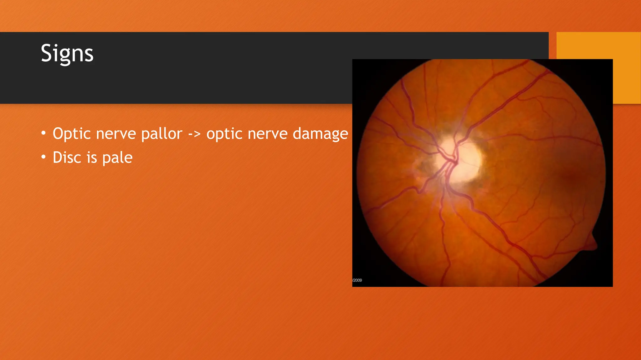
Optic Atrophy
• The death of the retinal ganglion cell axons that comprise the
optic nerve with the resulting picture of a pale optic nerve on
fundoscopy.
• Optic atrophy is an end stage that arises from myriad causes of
optic nerve damage anywhere along the path from the retina to
the lateral geniculate
Etiology
• Damage within the eye
• Optic nerve to the brain
• Congenital
Risk Factors
• Increased intraocular pressure
• Ischemia
• Compression
• Inflammation
• Infection
Pathology
• The optic nerve is a bundle of 1.2 million axons of retinal ganglion
cells that carries visual information from the retina to the brain.
• The optic nerve is myelinated by oligodendrocytes that do not
regenerate after damage.
Diagnosis
• Vision Loss
• Loss of contrast or color vision.
• Brightness test loss
• Color test loss
• Physical examination
Signs
• Optic nerve pallor -> optic nerve damage
• Disc is pale
The following work up should be considered for patients presenting
with unexplained optic atrophy:
• Check for afferent pupil
• Visual fields; Color vision
• MRI of brain and orbit with contrast
• CT with contrast (check bony disease, sinuses)
• Blood pressure and check of cardiovascular health
Tatalaksana
• The management goal is to intervene before optic atrophy is noted
or to save remaining function
• This will depend on the underlying cause for the optic nerve
damage
THANKYOU

Optic Atrophy ajajajajajajns aajaja.pptx

  • 1.
    Optic Atrophy • Thedeath of the retinal ganglion cell axons that comprise the optic nerve with the resulting picture of a pale optic nerve on fundoscopy. • Optic atrophy is an end stage that arises from myriad causes of optic nerve damage anywhere along the path from the retina to the lateral geniculate
  • 2.
    Etiology • Damage withinthe eye • Optic nerve to the brain • Congenital
  • 3.
    Risk Factors • Increasedintraocular pressure • Ischemia • Compression • Inflammation • Infection
  • 4.
    Pathology • The opticnerve is a bundle of 1.2 million axons of retinal ganglion cells that carries visual information from the retina to the brain. • The optic nerve is myelinated by oligodendrocytes that do not regenerate after damage.
  • 5.
    Diagnosis • Vision Loss •Loss of contrast or color vision. • Brightness test loss • Color test loss • Physical examination
  • 6.
    Signs • Optic nervepallor -> optic nerve damage • Disc is pale
  • 7.
    The following workup should be considered for patients presenting with unexplained optic atrophy: • Check for afferent pupil • Visual fields; Color vision • MRI of brain and orbit with contrast • CT with contrast (check bony disease, sinuses) • Blood pressure and check of cardiovascular health
  • 8.
    Tatalaksana • The managementgoal is to intervene before optic atrophy is noted or to save remaining function • This will depend on the underlying cause for the optic nerve damage
  • 9.

Editor's Notes

  • #1 Kematian akson sel ganglion retina yang terdiri dari saraf optik dengan gambaran yang dihasilkan dari saraf optik pucat pada funduskopi. Atrofi optik adalah tahap akhir yang muncul dari berbagai penyebab kerusakan saraf optik di mana saja di sepanjang jalan dari retina ke genikulatum lateral.
  • #2 glaucoma, optic neuritis, papilledema, etc.) tumor, neurodegenerative disorder, trauma, etc.)
  • #4  Saraf optik adalah kumpulan 1,2 juta akson sel ganglion retina yang membawa informasi visual dari retina ke otak. Saraf optik bermielin oleh oligodendrosit yang tidak beregenerasi setelah kerusakan.
  • #6 Lapisan serat saraf optik menipis atau tidak ada tepi diskus yang muncul Mungkin mencerminkan tidak adanya pembuluh darah kecil di kepala cakram.: Optic nerve fiber layer is thinned or absent the disc margins appear Probably reflecting absence of small vessels in the disc head.
  • #7 Pemeriksaan berikut harus dipertimbangkan untuk pasien dengan atrofi optik yang tidak dapat dijelaskan penyebabnya: Periksa pupil aferen bidang visual; Penglihatan warna MRI otak dan orbit dengan kontras CT dengan kontras (periksa penyakit tulang, sinus) Tekanan darah dan pemeriksaan kesehatan jantung
  • #8 Tujuan penatalaksanaan adalah untuk mengintervensi sebelum terjadi atrofi optik atau untuk menyelamatkan fungsi yang tersisa Ini akan tergantung pada penyebab yang mendasari kerusakan saraf optik