Papillitis isdisk swelling caused by inflammation at the nerve
head (intraocular optic nerve).
Cause:
Opticneuritis
multiplesclerosis
Loss of vision is the cardinal symptom of optic neuritis and is
particularly useful in differentiating papillitis from papilledema
Papillitis
6.
Optic Atrophy
• Introduction
Opticatrophy refers to the late stage changes that take place in the
optic nerve resulting from axonal degeneration in the pathway between
the retina and the lateral geniculate body, manifesting with disturbance
in visual function and in the appearance of the optic nerve head.
It can be classified in several ways,
. including by whether axonal death is initiated in the retina
(anterograde)
or more centrally (retrograde), and by cause.
Optic ‘atrophy’ is not true atrophy, a term that strictly refers to
involutional change secondary to lack of use.
7.
Primary Optic Atrophy
Primary optic atrophy
Primary optic atrophy occurs without antecedent
swelling of the optic nerve head.
It may be caused by lesions affecting the visual pathways at any point
from the retrolaminar portion of the optic nerve to the lateral geniculate
body.
Lesions anterior to the optic chiasm result in unilateral optic atrophy,
whereas those involving the chiasm and optic tract will cause bilateral
changes.
8.
• Signs:
• Flatwhite disc with clearly delineated margins.
• Reduction in the number of small blood vessels on the disc surface.
• Attenuationofperipapillarybloodvesselsand thinning of the retinal
nerve fibre layer (RNFL).
• The atrophy may be diffuse or sectoral depending on the cause and
level of the lesion.
• Temporal pallor of the optic nerve head may indicate atrophy of
fibres of the papillomacular bundle, and is classically seen following
demyelinating optic neuritis.
• Band atrophy is a similar phenomenon caused by involvement of
the fibres entering the optic disc nasally and temporally; it occurs in
lesions of the optic chiasm or tract and gives nasal as well as
temporal.
10.
• Important causes
Opticneuritis.
Compression by tumours and aneurysms. Hereditary optic neuropathies.
Toxic and nutritional optic neuropathies; these may give temporal pallor, pa
Trauma.
11.
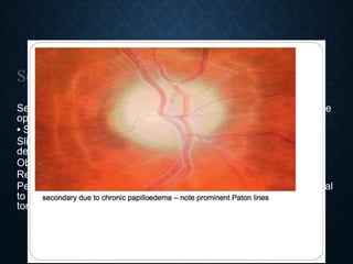
Secondary optic atrophy
Secondaryoptic atrophy is preceded by long-standing swelling of the
optic nerve head.
• Signs vary according to the cause and its course.
Slightly or moderately raised white or greyish disc with poorly
delineated margins due to gliosis.
Obscuration of the lamina cribrosa.
Reduction in the number of small blood vessels on the disc surface.
Peripapillary circumferential retinochoroidal folds, especially temporal
to the disc (Paton lines –C), sheathing of arterioles and venous
tortuosity may be
12.
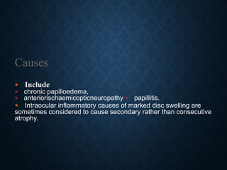
Causes
Include
chronicpapilloedema,
anteriorischaemicopticneuropathy papillitis.
Intraocular inflammatory causes of marked disc swelling are
sometimes considered to cause secondary rather than consecutive
atrophy.
13.
Consecutive optic atrophy
Consecutive optic atrophy is caused by disease of the inner retina or
its blood supply.
The cause is usually obvious on fundus examination,
e.g. extensive retinal photocoagulation, retinitis pigmentosa or prior
central retinal artery occlusion.
The disc appears waxy, with reasonably preserved architecture
consecutive due to vasculitis
14.
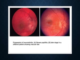
Neuroretinitis refersto the combination of optic neuritis and signs of
retinal, usually macular, inflammation.
Cat-scratch fever is responsible for 60% of cases.
About 25% of cases are idiopathic (Leber
idiopathic stellate neuroretinitis).
Other notable causes include syphilis, Lyme disease, mumps and
leptospirosis.
Neuroretinitis
15.
Diagnosis:
Symptoms:
Painless unilateral visualimpairment, usually gradually worsening over
about a week.
Signs:
• VA is impaired to a variable degree.
• Signs of optic nerve dysfunction are usually mild or absent, as visual
loss is largely due to macular involvement.
• Papillitis associated with peripapillary and macular oedema
• A macular star typically appears as disc swelling settles; the macular
star resolves with a return to normal or near-normal visual acuity over
6–12 months.
• Venous engorgement and splinter haemorrhages may be present in
severe case.
• Fellow eye involvement occasionally develops.
17.
Optical coherence tomography(OCT) demonstrates sub- and
intraretinal fluid to a variable extent.
Fluorescein angiography (FA) shows diffuse leakage from superficial
disc vessels.
Blood tests may include serology for Bartonella and other causes
according to clinical suspicion
Treatment
This is specific to the cause, and often consists of antibiotics.
Recurrent idiopathic cases may require treatment with steroids and/or
other immunosuppressants.
Investigation and Treatment