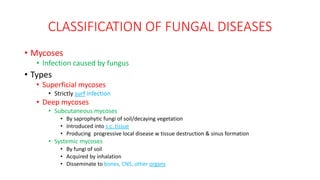
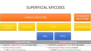
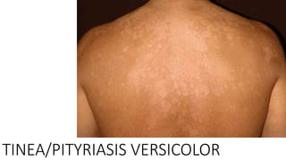
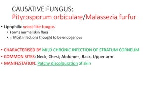
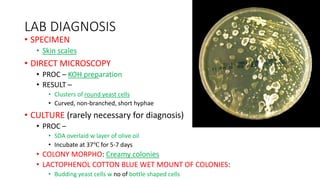
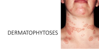
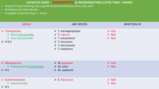
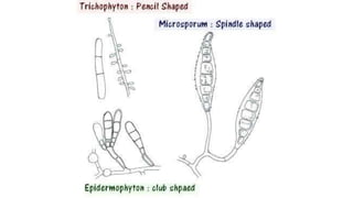
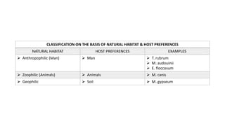
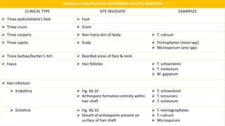
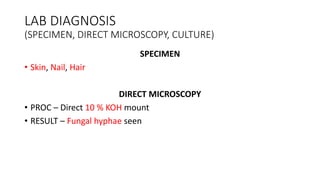
CHAP 66
MEDICAL MYCOLOGY
PART 2.1
CLASSIFICATION OF FUNGAL DISEASES
• Mycoses
• Infection caused by fungus
• Types
• Superficial mycoses
• Strictly surf infection
• Deep mycoses
• Subcutaneous mycoses
• By saprophytic fungi of soil/decaying vegetation
• Introduced into s.c. tissue
• Producing progressive local disease w tissue destruction & sinus formation
• Systemic mycoses
• By fungi of soil
• Acquired by inhalation
• Disseminate to bones, CNS, other organs
SUPERFICIAL MYCOSES
SURFACE INFECTIONS
Tinea/Pityriasis
versicolor
Tinea nigra Piedra
Black White
CUTANEOUS
INFECTIONS
Dermatophytoses
SURFACE INFECTIONS CUTANEOUS INFECTIONS
 Fungi live on dead layers of skin & its appendages  Confined to cornified layer of skin & its appendages
 No inflammatory response
 As no contact w living tissue
 Inflammatory & allergic responses induced
 Due to fungi and their metabolic products
TINEA/PITYRIASIS VERSICOLOR
CAUSATIVE FUNGUS:
Pityrosporum orbiculare/Malassezia furfur
• Lipophilic yeast-like fungus
• Forms normal skin flora
• ∴ Most infections thought to be endogenous
• CHARACTERISED BY MILD CHRONIC INFECTION OF STRATUM CORNEUM
• COMMON SITES: Neck, Chest, Abdomen, Back, Upper arm
• MANIFESTATION: Patchy discolouration of skin
LAB DIAGNOSIS
• SPECIMEN
• Skin scales
• DIRECT MICROSCOPY
• PROC – KOH preparation
• RESULT –
• Clusters of round yeast cells
• Curved, non-branched, short hyphae
• CULTURE (rarely necessary for diagnosis)
• PROC –
• SDA overlaid w layer of olive oil
• Incubate at 37℃ for 5-7 days
• COLONY MORPHO: Creamy colonies
• LACTOPHENOL COTTON BLUE WET MOUNT OF COLONIES:
• Budding yeast cells w no of bottle shaped cells
DERMATOPHYTOSES
CAUSATIVE AGENT – DERMATOPHYTES @ RINGWORM/TINEA (LATIN; TINEA = WORM)
• Group of fungi infecting only superficial keratinized tissue (hair, nail, skin)
• Breakdown & utilize keratin
• Incapable of penetrating s.c. tissue
GENUS IMP. SPECIES INFECTION OF
 Trichophyton
 More microconidia
 Few macroconidia
 H N S
 T. mentagrophytes
 T. rubrum
 T. schoenleinii
 T. tonsurans
 T. verrucosum
 T. violaceum
 Hair
 Nail
 Skin
 Microsporum
 Predominant macroconidia
 H S
 M. gypseum
 M. canis
 M. audouinii
 Hair
 Skin
 Epidermophyton
 Macroconidia
 N S
 E. floccosum  Nail
 Skin
CLASSIFICATION ON THE BASIS OF NATURAL HABITAT & HOST PREFERENCES
NATURAL HABITAT HOST PREFERENCES EXAMPLES
 Anthropophilic (Man)  Man  T. rubrum
 M. audouinii
 E. floccosum
 Zoophilic (Animals)  Animals  M. canis
 Geophilic  Soil  M. gypseum
CLINICAL CLASSIFICATION DEPENDING ON SITE INVOLVED
CLINICAL TYPE SITE INVOLVED EXAMPLES
 Tinea pedis/athlete’s foot  Foot
 Tinea cruris  Groin
 Tinea corporis  Non-hairy skin of body  T. rubrum
 Tinea capitis  Scalp  Trichophyton (most spp)
 Microsporum (any spp)
 Tinea barbae/barber’s itch  Bearded areas of face & neck
 Favus  Hair follicles  T. schoenleinii
 T. violaceum
 M. gypseum
 Hair infection
 Endothrix  Fig. 66.10
 Arthospore formation entirely within
hair shaft
 T. schoenleinii
 T. tonsurans
 T. violaceum
 Ectothrix  Fig. 66.10
 Sheath of arthospores present on
surface of hair shaft
 T. mentagrophytes
 T. rubrum
 Microsporum
LAB DIAGNOSIS
(SPECIMEN, DIRECT MICROSCOPY, CULTURE)
SPECIMEN
• Skin, Nail, Hair
DIRECT MICROSCOPY
• PROC – Direct 10 % KOH mount
• RESULT – Fungal hyphae seen
CULTURE
• SDA, SDA w antibiotics
• PROC –
• Culture media incubated at 25-30℃ for 3 weeks
• Identify dermatophytes based on
• Colony morphology
• Pigment production
• Microconidia & Macroconidia
• Lactophenol cotton blue prep from colony reveals microconidia & macroconidia
• RESULT –
• Colony morphology
• White to creamy cottony growth
• Pigment production
• Microconidia & Macroconidia
• Genus Trichophyton
• More microconidia
• Few macroconidia
• Genus Microsporum
• Predominant macroconidia
• Genus Epidermophyton
• Macroconidia
TREATMENT
• DOC
• Oral griseofulvin
• GENERALLY USED
• Topical antifungal agents

SUPERFICIAL MYCOSES

  • 1.
  • 2.
    CLASSIFICATION OF FUNGALDISEASES • Mycoses • Infection caused by fungus • Types • Superficial mycoses • Strictly surf infection • Deep mycoses • Subcutaneous mycoses • By saprophytic fungi of soil/decaying vegetation • Introduced into s.c. tissue • Producing progressive local disease w tissue destruction & sinus formation • Systemic mycoses • By fungi of soil • Acquired by inhalation • Disseminate to bones, CNS, other organs
  • 3.
    SUPERFICIAL MYCOSES SURFACE INFECTIONS Tinea/Pityriasis versicolor Tineanigra Piedra Black White CUTANEOUS INFECTIONS Dermatophytoses SURFACE INFECTIONS CUTANEOUS INFECTIONS  Fungi live on dead layers of skin & its appendages  Confined to cornified layer of skin & its appendages  No inflammatory response  As no contact w living tissue  Inflammatory & allergic responses induced  Due to fungi and their metabolic products
  • 4.
  • 5.
    CAUSATIVE FUNGUS: Pityrosporum orbiculare/Malasseziafurfur • Lipophilic yeast-like fungus • Forms normal skin flora • ∴ Most infections thought to be endogenous • CHARACTERISED BY MILD CHRONIC INFECTION OF STRATUM CORNEUM • COMMON SITES: Neck, Chest, Abdomen, Back, Upper arm • MANIFESTATION: Patchy discolouration of skin
  • 6.
    LAB DIAGNOSIS • SPECIMEN •Skin scales • DIRECT MICROSCOPY • PROC – KOH preparation • RESULT – • Clusters of round yeast cells • Curved, non-branched, short hyphae • CULTURE (rarely necessary for diagnosis) • PROC – • SDA overlaid w layer of olive oil • Incubate at 37℃ for 5-7 days • COLONY MORPHO: Creamy colonies • LACTOPHENOL COTTON BLUE WET MOUNT OF COLONIES: • Budding yeast cells w no of bottle shaped cells
  • 7.
  • 8.
    CAUSATIVE AGENT –DERMATOPHYTES @ RINGWORM/TINEA (LATIN; TINEA = WORM) • Group of fungi infecting only superficial keratinized tissue (hair, nail, skin) • Breakdown & utilize keratin • Incapable of penetrating s.c. tissue GENUS IMP. SPECIES INFECTION OF  Trichophyton  More microconidia  Few macroconidia  H N S  T. mentagrophytes  T. rubrum  T. schoenleinii  T. tonsurans  T. verrucosum  T. violaceum  Hair  Nail  Skin  Microsporum  Predominant macroconidia  H S  M. gypseum  M. canis  M. audouinii  Hair  Skin  Epidermophyton  Macroconidia  N S  E. floccosum  Nail  Skin
  • 10.
    CLASSIFICATION ON THEBASIS OF NATURAL HABITAT & HOST PREFERENCES NATURAL HABITAT HOST PREFERENCES EXAMPLES  Anthropophilic (Man)  Man  T. rubrum  M. audouinii  E. floccosum  Zoophilic (Animals)  Animals  M. canis  Geophilic  Soil  M. gypseum
  • 11.
    CLINICAL CLASSIFICATION DEPENDINGON SITE INVOLVED CLINICAL TYPE SITE INVOLVED EXAMPLES  Tinea pedis/athlete’s foot  Foot  Tinea cruris  Groin  Tinea corporis  Non-hairy skin of body  T. rubrum  Tinea capitis  Scalp  Trichophyton (most spp)  Microsporum (any spp)  Tinea barbae/barber’s itch  Bearded areas of face & neck  Favus  Hair follicles  T. schoenleinii  T. violaceum  M. gypseum  Hair infection  Endothrix  Fig. 66.10  Arthospore formation entirely within hair shaft  T. schoenleinii  T. tonsurans  T. violaceum  Ectothrix  Fig. 66.10  Sheath of arthospores present on surface of hair shaft  T. mentagrophytes  T. rubrum  Microsporum
  • 12.
    LAB DIAGNOSIS (SPECIMEN, DIRECTMICROSCOPY, CULTURE) SPECIMEN • Skin, Nail, Hair DIRECT MICROSCOPY • PROC – Direct 10 % KOH mount • RESULT – Fungal hyphae seen
  • 13.
    CULTURE • SDA, SDAw antibiotics • PROC – • Culture media incubated at 25-30℃ for 3 weeks • Identify dermatophytes based on • Colony morphology • Pigment production • Microconidia & Macroconidia • Lactophenol cotton blue prep from colony reveals microconidia & macroconidia • RESULT – • Colony morphology • White to creamy cottony growth • Pigment production • Microconidia & Macroconidia • Genus Trichophyton • More microconidia • Few macroconidia • Genus Microsporum • Predominant macroconidia • Genus Epidermophyton • Macroconidia
  • 14.
    TREATMENT • DOC • Oralgriseofulvin • GENERALLY USED • Topical antifungal agents