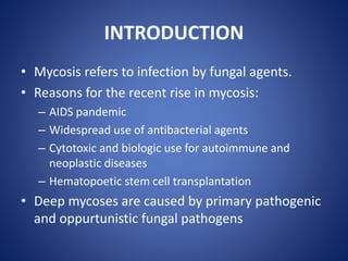
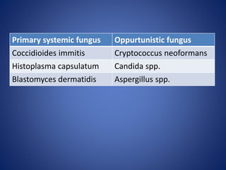
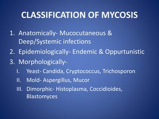
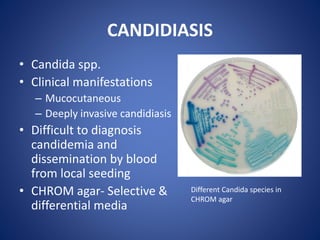
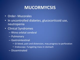
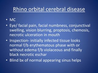
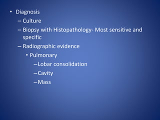
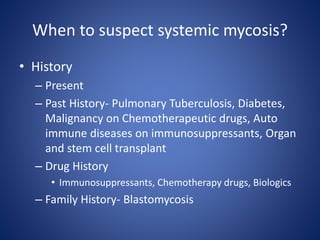
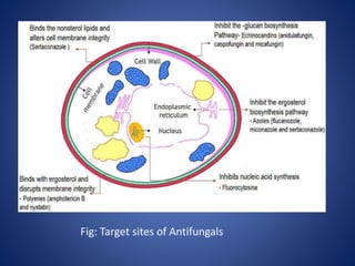
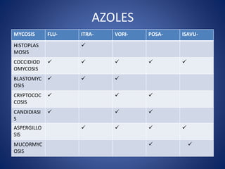
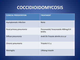
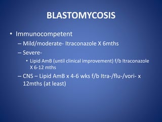
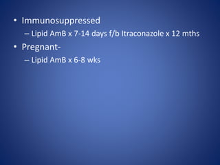
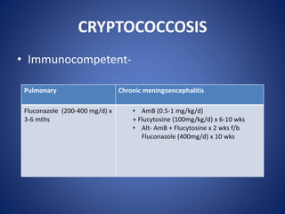
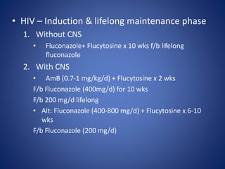
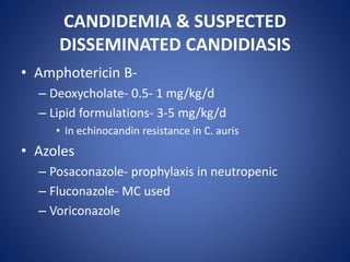
APPROACH TO SYSTEMIC
MYCOSIS
MODERATOR
Dr. Rima Moni Doley,
Associate Professor,
Department of Medicine,
AMCH
PRESENTER
Dr. Swdwamshree Boro,
PGT 3rd Year,
Department of
Medicine,
AMCH
INTRODUCTION
• Mycosis refers to infection by fungal agents.
• Reasons for the recent rise in mycosis:
– AIDS pandemic
– Widespread use of antibacterial agents
– Cytotoxic and biologic use for autoimmune and
neoplastic diseases
– Hematopoetic stem cell transplantation
• Deep mycoses are caused by primary pathogenic
and oppurtunistic fungal pathogens
Primary systemic fungus Oppurtunistic fungus
Coccidioides immitis Cryptococcus neoformans
Histoplasma capsulatum Candida spp.
Blastomyces dermatidis Aspergillus spp.
CLASSIFICATION OF MYCOSIS
1. Anatomically- Mucocutaneous &
Deep/Systemic infections
2. Epidemiologically- Endemic & Oppurtunistic
3. Morphologically-
I. Yeast- Candida, Cryptococcus, Trichosporon
II. Mold- Aspergillus, Mucor
III. Dimorphic- Histoplasma, Coccidioides,
Blastomyces
HISTOPLASMOSIS
• Etiologic agent- Histoplasma
• Clinical Features
– Asymptomatic
– Flu like symptoms- fever, chills, headache, myalgia,
anorexia, dry cough, dyspnea, chest pain
• a/w arthritis, arthralgia, erythema nodosum
• Clinical Manifestations:
1. Acute pulmonary
2. Chronic/ cavitary pulmonary
• Smokers with structural lung disease
• CF- Productive cough, dyspnea, low grade fever, night
sweats, weight loss
3. Progressive disseminated
• RF- Immunocompromised
• CF- RF, Shock, Coagulopathy
4. Fibrosing Mediastinitis
• Uncommon
COCCIDIODOMYCOSIS
• Genus- Coccidioides; Species- C. immitis, C.
posadasii
• Transmission- Direct exposure to soil
– Inhalation of spores
• Clinical manifestations-
– Primary pulmonary coccidioidomycosis
– Disseminated disease- skin, bones, joints, soft
tissue, meninges( Coccidioidal meningitis)
Primary Pulmonary Coccidioidomycosis
– Fever, cough, pleuritic chest pain, night sweats,
fatigue, failure to improve by antibiotics
– Immunologic response manifestations
• Toxic erythema- diffuse, maculopapular, erythematous
• Symmetrical arthralgia esp lower limbs
• Erythema nodosum
• Erythema multiforme
– Eosinophilia
– Hilar, mediastinal lymphadenopathy, pleural
nodules radiologically
• D/d- CAP
Coccidioidal meningitis
– RF:
• Immunosuppressed
• Second or third trimester pregnancy
– Persistent headache, lethargy, confusion
– CSF
• Lymphocytic pleocytosis
– Increased protein
– Hypoglycorrhachia
BLASTOMYCOSIS
• Transmission- Inhalation of spores
• Clinical Manifestations
1. Pulmonary Blastomycosis
• Asymptomatic
• Flu like presentation
• Acute pneumonia
• Chronic pneumonia
• ARDS
– Severe in diabetics, immunosuppressed and
multilobular pneumonia
– Radiology-
• Lobar consolidation
• Mass lesion
• Interstitial infiltrates
• Milliary pattern
• Nodules
• Cavitation
• Multiple lobe involvement
*Hilar adenopathy, pleural effusion, empyema are
uncommon
2. Disseminated Blastomycosis
• Mainly skin
• Rarely- CNS, GU tract
– D/D- Pulmonary TB, CAP, Malignancy
CRYPTOCOCCOSIS
• Genus- Cryptococcus; Species- C. neoformans,
C. gattii
• RF-
– C. gattii in immunocompetent
– C. neoformans-
• Immunocompromised
• hematologic malignancies
• HIV (CD4+ <200/µl)
• Clinical manifestation:
1. Chronic meningoencephalitis-
immunosuppressed
• Headache
• Fever
• neurologic symptoms
• Vision loss
• Subacute dementia (indolent)
2. Pulmonary Cryptococcosis
• Cough, increased sputum, chest pain
• a/w diabetes, malignancy, TB
3. Cutaneous (in disseminated)- variable lesions
• CSF Analysis
– Lymphocytic pleocytosis
– Increased protein
• Cryptococcal antigen + in CSF/ blood
• Culture- Cryptococcal cells
– Blood, CSF
*IRIS
CANDIDIASIS
• Candida spp.
• Clinical manifestations
– Mucocutaneous
– Deeply invasive candidiasis
• Difficult to diagnosis
candidemia and
dissemination by blood
from local seeding
• CHROM agar- Selective &
differential media
Different Candida species in
CHROM agar
ASPERGILLOSIS
• RF-
– Neutropenia
– ICU
– Severe COVID 19
– Underlying respiratory disease- Pulmonary TB,
COPD, Asthma
– ECMO
– Immunomodulatory agents
• Clinical manifestations:
– Lung, sinus, heart, eye, brain & skin are involved
1. Invasive aspergillosis
2. Chronic
1. Chronic pulmonry aspergillosis
2. Chronic Aspergillus sinusitis
3. Allergic
1. Allergic Bronchopulmonary Aspergillosis (MC)
2. Severe asthma with fungal sensitization (SAFS)
4. Saprophytic
1. Aspergilloma
2. Maxillary fungal ball
ALLERGIC BRONCOPULMONARY ASPERGILLOSIS
• CF:
– Recurrent exacerbations of asthma
– Uncontrolled with usual anti asthmatic therapy
– Sputum- brownish mucus plugs, haemoptysis
• Diagnosis:
– Asthmatics
– Peripheral blood eosinophilia >500/microL
– Pulmonary infiltrates in chest radiograph
– HRCT thorax- string of pearl, signet ring
– Sputum culture +
– Total IgE against A. fumigatus >1000 IU/ml
• D/D- Pulmonary TB, Bronchiectasis, CAP
MUCORMYCSIS
• Order- Mucorales
• In uncontrolled diabetes, glucocorticoid use,
neutropenia
• Clinical Syndromes
– Rhino orbital cerebral
– Pulmonary
– Gastrointestinal
• GI bleed, pain and distension, may progress to perforation
• Endoscopy- Fungating mass in stomach
– Disseminated
Rhino orbital cerebral disease
• MC
• Eye/ facial pain, facial numbness, conjunctival
swelling, vision blurring, proptosis, chemosis,
necrotic ulceration in mouth
• Inspection- initially infected tissue looks
normal f/b erythematous phase with or
without edema f/b violaceous and finally
black necrotic eschar
• Blind bx of normal appearing sinus helps
• Diagnosis
– Culture
– Biopsy with Histopathology- Most sensitive and
specific
– Radiographic evidence
• Pulmonary
–Lobar consolidation
–Cavity
–Mass
When to suspect systemic mycosis?
• History
– Present
– Past History- Pulmonary Tuberculosis, Diabetes,
Malignancy on Chemotherapeutic drugs, Auto
immune diseases on immunosuppressants, Organ
and stem cell transplant
– Drug History
• Immunosuppressants, Chemotherapy drugs, Biologics
– Family History- Blastomycosis
• Exposure History
– Soil- Coccidiodomycosis, Aspergillosis,
Blastomycosis
• Travel History
• Occupational History
INVESTIGATIONS
1. Specimen collection- depends on the site
involved
2. Direct Microscopy:
a) Wet mounts:
• KOH- Yeast cells, hyphae, pseudohyphae
• Gram stain- yeast & yeast like fungus
• India ink- negative stain
–Cryptococcal capsule- CSF
• Calcoflour white stain- Blastomyces & mixed
infection
C. neoformans capsule in
India Ink
Hyphae seen in KOH stain
b) Histopathological examination:
• PAS, Gommori Methenamine silver, H&E stain
• Fungal elements from tissue biopsy
c) Frozen section Biopsy:
• Intra operative diagnosis of suspected
malignancy
d) Lactophenol cotton blue:
• microscopic appearance of fungi from isolates
grown in culture
3. Fungal Culture- Blood, CSF, Sputum, BAL fluid
– Isolation and identification
– SDA (mc)
– niger seed agar- selective media for cryptococcus
– CHROM agar- selective & differential media for
Candida species
4. Immunological methods
– Antibody detection- ELISA, Agglutination test, CFT
• Prognosis & response to anti fungals
– Antigen detection- Latex agglutination
* In early stages & impaired immunity or immunity not
sufficient to raise significant antibody levels
• Cryptoccus- Serum, CSF
• Blastomyces- Urine, blood, CSF, BAL fluid
• Aspergillus- BAL
• Histoplasma
– Immunohistochemistry-
• Ag detection on the cells of tissue sections
4. Antifungal Susceptibility Testing
5. New Techniques
– AccuProbe
– PNA Fish
– MALDI TOF mass spectrometry
TREATMENT
• Antifungals
– Antifungal antibiotics
• Polyene- Amphotericin B
– Azoles
– Echinocandins- Caspofungin, Anidulafungin,
Micafungin
– Flucytosine
Fig: Target sites of Antifungals
AZOLES
MYCOSIS FLU- ITRA- VORI- POSA- ISAVU-
HISTOPLAS
MOSIS

COCCIDIOD
OMYCOSIS
    
BLASTOMYC
OSIS
  
CRYPTOCOC
COSIS
  
CANDIDIASI
S
  
ASPERGILLO
SIS
   
MUCORMYC
OSIS
 
COCCIDIOIDOMYCOSIS
CLINICAL PRESENTATION TREATMENT
Asymptomatic infection None
Focal primary pneumonia Fluconazole/ Itraconazole 400mg/d X
6mths
Diffuse pneumonia AmB f/b Triazole x6mths to a yr
Chronic pneumonia Triazole X 1 y
Meningitis Lifelong triazole
BLASTOMYCOSIS
• Immunocompetent
– Mild/moderate- Itraconazole X 6mths
– Severe-
• Lipid AmB (until clinical improvement) f/b Itraconazole
X 6-12 mths
– CNS – Lipid AmB x 4-6 wks f/b Itra-/flu-/vori- x
12mths (at least)
• Immunosuppressed
– Lipid AmB x 7-14 days f/b Itraconazole x 12 mths
• Pregnant-
– Lipid AmB x 6-8 wks
CRYPTOCOCCOSIS
• Immunocompetent-
Pulmonary Chronic meningoencephalitis
Fluconazole (200-400 mg/d) x
3-6 mths
• AmB (0.5-1 mg/kg/d)
+ Flucytosine (100mg/kg/d) x 6-10 wks
• Alt- AmB + Flucytosine x 2 wks f/b
Fluconazole (400mg/d) x 10 wks
• HIV – Induction & lifelong maintenance phase
1. Without CNS
• Fluconazole+ Flucytosine x 10 wks f/b lifelong
fluconazole
2. With CNS
• AmB (0.7-1 mg/kg/d) + Flucytosine x 2 wks
F/b Fluconazole (400mg/d) for 10 wks
F/b 200 mg/d lifelong
• Alt: Fluconazole (400-800 mg/d) + Flucytosine x 6-10
wks
F/b Fluconazole (200 mg/d)
CANDIDEMIA & SUSPECTED
DISSEMINATED CANDIDIASIS
• Amphotericin B-
– Deoxycholate- 0.5- 1 mg/kg/d
– Lipid formulations- 3-5 mg/kg/d
• In echinocandin resistance in C. auris
• Azoles
– Posaconazole- prophylaxis in neutropenic
– Fluconazole- MC used
– Voriconazole
• Echinocandins
– First choice if concern for resistance
– First line for multi drug resistant C. auris
– Caspofungin, Anidulafungin, Micafungin
• Candida meningoencephalitis
– Polyene+ flucytosine
ASPERGILLOSIS
Primary treatment Secondary treatment
Invasive Vori-, isa-, posa- Lipid AmB, Caspofungin,
micafungin
Prophylaxis Posa-, itra- Mica-, aerosolised AmB
Chronic
pulmonary
Vori-,itra- Posa-, IV Mica-, IV AmB
Fungal asthma Itra- Vori-, Posa-
Single
aspergilloma
Surgical resection Itra-, Vori-, AmB
intracavitary
*Oral corticosteroid is the cornerstone of ABPA
MUCORMYCOSIS
Drugs Dose
Amphotericin B deoxycholate 1-1.5 mg/kg/d
Liposomal AmB 5-10 mg/kg/d
AmB lipid complex 5 mg/kg /d
First line anti fungal
Second line anti fungal
1. Isavuconazole
2. Posaconazole
• Combination therapy
– Echinocandin + lipid polyene
– Lipid polyene + azole
– Triple therapy
• 38 YO male, CRPF by occupation came with
– Weakness of all the limbs for 1 month which was gradually
progressive
– Urinary and stool incontinence for 15 days
– Fever and headache for 15 days
• ICTC positive
• CSF – raised protein, lymphocyte pleocytosis
• Serum Cryptoccal antigen- positive
• MRI Brain with screening of whole spine- Toxoplasmosis and
Syrinx formation
• Diagnosis- CRYPTOCOCCAL MENINGITIS
CASE DISCUSSION
• He was started on Amphotericin B1 mg/kg/day
IV for 2 weeks and Tab Trimethoprim+
Sulphamethoxazole followed by Tab
Fluconazole 400 mg/ day for 8 weeks.
• ART was started after 1mth
• He could slowly walk with residual weakness
post therapy after about 3 weeks with a
walking stick.
REFERENCES
• Harrison’s Principles of Internal Medicine 21st
Edition
• Textbook of Medical Mycology 4th edition
APPROACH TO SYSTEMIC MYCOSIS.pptx

APPROACH TO SYSTEMIC MYCOSIS.pptx

  • 1.
    APPROACH TO SYSTEMIC MYCOSIS MODERATOR Dr.Rima Moni Doley, Associate Professor, Department of Medicine, AMCH PRESENTER Dr. Swdwamshree Boro, PGT 3rd Year, Department of Medicine, AMCH
  • 2.
    INTRODUCTION • Mycosis refersto infection by fungal agents. • Reasons for the recent rise in mycosis: – AIDS pandemic – Widespread use of antibacterial agents – Cytotoxic and biologic use for autoimmune and neoplastic diseases – Hematopoetic stem cell transplantation • Deep mycoses are caused by primary pathogenic and oppurtunistic fungal pathogens
  • 3.
    Primary systemic fungusOppurtunistic fungus Coccidioides immitis Cryptococcus neoformans Histoplasma capsulatum Candida spp. Blastomyces dermatidis Aspergillus spp.
  • 4.
    CLASSIFICATION OF MYCOSIS 1.Anatomically- Mucocutaneous & Deep/Systemic infections 2. Epidemiologically- Endemic & Oppurtunistic 3. Morphologically- I. Yeast- Candida, Cryptococcus, Trichosporon II. Mold- Aspergillus, Mucor III. Dimorphic- Histoplasma, Coccidioides, Blastomyces
  • 5.
    HISTOPLASMOSIS • Etiologic agent-Histoplasma • Clinical Features – Asymptomatic – Flu like symptoms- fever, chills, headache, myalgia, anorexia, dry cough, dyspnea, chest pain • a/w arthritis, arthralgia, erythema nodosum
  • 6.
    • Clinical Manifestations: 1.Acute pulmonary 2. Chronic/ cavitary pulmonary • Smokers with structural lung disease • CF- Productive cough, dyspnea, low grade fever, night sweats, weight loss 3. Progressive disseminated • RF- Immunocompromised • CF- RF, Shock, Coagulopathy 4. Fibrosing Mediastinitis • Uncommon
  • 7.
    COCCIDIODOMYCOSIS • Genus- Coccidioides;Species- C. immitis, C. posadasii • Transmission- Direct exposure to soil – Inhalation of spores • Clinical manifestations- – Primary pulmonary coccidioidomycosis – Disseminated disease- skin, bones, joints, soft tissue, meninges( Coccidioidal meningitis)
  • 8.
    Primary Pulmonary Coccidioidomycosis –Fever, cough, pleuritic chest pain, night sweats, fatigue, failure to improve by antibiotics – Immunologic response manifestations • Toxic erythema- diffuse, maculopapular, erythematous • Symmetrical arthralgia esp lower limbs • Erythema nodosum • Erythema multiforme – Eosinophilia – Hilar, mediastinal lymphadenopathy, pleural nodules radiologically • D/d- CAP
  • 9.
    Coccidioidal meningitis – RF: •Immunosuppressed • Second or third trimester pregnancy – Persistent headache, lethargy, confusion – CSF • Lymphocytic pleocytosis – Increased protein – Hypoglycorrhachia
  • 10.
    BLASTOMYCOSIS • Transmission- Inhalationof spores • Clinical Manifestations 1. Pulmonary Blastomycosis • Asymptomatic • Flu like presentation • Acute pneumonia • Chronic pneumonia • ARDS – Severe in diabetics, immunosuppressed and multilobular pneumonia
  • 11.
    – Radiology- • Lobarconsolidation • Mass lesion • Interstitial infiltrates • Milliary pattern • Nodules • Cavitation • Multiple lobe involvement *Hilar adenopathy, pleural effusion, empyema are uncommon
  • 12.
    2. Disseminated Blastomycosis •Mainly skin • Rarely- CNS, GU tract – D/D- Pulmonary TB, CAP, Malignancy
  • 13.
    CRYPTOCOCCOSIS • Genus- Cryptococcus;Species- C. neoformans, C. gattii • RF- – C. gattii in immunocompetent – C. neoformans- • Immunocompromised • hematologic malignancies • HIV (CD4+ <200/µl)
  • 14.
    • Clinical manifestation: 1.Chronic meningoencephalitis- immunosuppressed • Headache • Fever • neurologic symptoms • Vision loss • Subacute dementia (indolent) 2. Pulmonary Cryptococcosis • Cough, increased sputum, chest pain • a/w diabetes, malignancy, TB 3. Cutaneous (in disseminated)- variable lesions
  • 15.
    • CSF Analysis –Lymphocytic pleocytosis – Increased protein • Cryptococcal antigen + in CSF/ blood • Culture- Cryptococcal cells – Blood, CSF *IRIS
  • 16.
    CANDIDIASIS • Candida spp. •Clinical manifestations – Mucocutaneous – Deeply invasive candidiasis • Difficult to diagnosis candidemia and dissemination by blood from local seeding • CHROM agar- Selective & differential media Different Candida species in CHROM agar
  • 17.
    ASPERGILLOSIS • RF- – Neutropenia –ICU – Severe COVID 19 – Underlying respiratory disease- Pulmonary TB, COPD, Asthma – ECMO – Immunomodulatory agents
  • 18.
    • Clinical manifestations: –Lung, sinus, heart, eye, brain & skin are involved 1. Invasive aspergillosis 2. Chronic 1. Chronic pulmonry aspergillosis 2. Chronic Aspergillus sinusitis 3. Allergic 1. Allergic Bronchopulmonary Aspergillosis (MC) 2. Severe asthma with fungal sensitization (SAFS) 4. Saprophytic 1. Aspergilloma 2. Maxillary fungal ball
  • 19.
    ALLERGIC BRONCOPULMONARY ASPERGILLOSIS •CF: – Recurrent exacerbations of asthma – Uncontrolled with usual anti asthmatic therapy – Sputum- brownish mucus plugs, haemoptysis • Diagnosis: – Asthmatics – Peripheral blood eosinophilia >500/microL – Pulmonary infiltrates in chest radiograph – HRCT thorax- string of pearl, signet ring – Sputum culture + – Total IgE against A. fumigatus >1000 IU/ml • D/D- Pulmonary TB, Bronchiectasis, CAP
  • 20.
    MUCORMYCSIS • Order- Mucorales •In uncontrolled diabetes, glucocorticoid use, neutropenia • Clinical Syndromes – Rhino orbital cerebral – Pulmonary – Gastrointestinal • GI bleed, pain and distension, may progress to perforation • Endoscopy- Fungating mass in stomach – Disseminated
  • 21.
    Rhino orbital cerebraldisease • MC • Eye/ facial pain, facial numbness, conjunctival swelling, vision blurring, proptosis, chemosis, necrotic ulceration in mouth • Inspection- initially infected tissue looks normal f/b erythematous phase with or without edema f/b violaceous and finally black necrotic eschar • Blind bx of normal appearing sinus helps
  • 22.
    • Diagnosis – Culture –Biopsy with Histopathology- Most sensitive and specific – Radiographic evidence • Pulmonary –Lobar consolidation –Cavity –Mass
  • 23.
    When to suspectsystemic mycosis? • History – Present – Past History- Pulmonary Tuberculosis, Diabetes, Malignancy on Chemotherapeutic drugs, Auto immune diseases on immunosuppressants, Organ and stem cell transplant – Drug History • Immunosuppressants, Chemotherapy drugs, Biologics – Family History- Blastomycosis
  • 24.
    • Exposure History –Soil- Coccidiodomycosis, Aspergillosis, Blastomycosis • Travel History • Occupational History
  • 25.
    INVESTIGATIONS 1. Specimen collection-depends on the site involved 2. Direct Microscopy: a) Wet mounts: • KOH- Yeast cells, hyphae, pseudohyphae • Gram stain- yeast & yeast like fungus • India ink- negative stain –Cryptococcal capsule- CSF • Calcoflour white stain- Blastomyces & mixed infection
  • 26.
    C. neoformans capsulein India Ink Hyphae seen in KOH stain
  • 27.
    b) Histopathological examination: •PAS, Gommori Methenamine silver, H&E stain • Fungal elements from tissue biopsy c) Frozen section Biopsy: • Intra operative diagnosis of suspected malignancy d) Lactophenol cotton blue: • microscopic appearance of fungi from isolates grown in culture
  • 28.
    3. Fungal Culture-Blood, CSF, Sputum, BAL fluid – Isolation and identification – SDA (mc) – niger seed agar- selective media for cryptococcus – CHROM agar- selective & differential media for Candida species
  • 29.
    4. Immunological methods –Antibody detection- ELISA, Agglutination test, CFT • Prognosis & response to anti fungals – Antigen detection- Latex agglutination * In early stages & impaired immunity or immunity not sufficient to raise significant antibody levels • Cryptoccus- Serum, CSF • Blastomyces- Urine, blood, CSF, BAL fluid • Aspergillus- BAL • Histoplasma
  • 30.
    – Immunohistochemistry- • Agdetection on the cells of tissue sections 4. Antifungal Susceptibility Testing 5. New Techniques – AccuProbe – PNA Fish – MALDI TOF mass spectrometry
  • 31.
    TREATMENT • Antifungals – Antifungalantibiotics • Polyene- Amphotericin B – Azoles – Echinocandins- Caspofungin, Anidulafungin, Micafungin – Flucytosine
  • 32.
    Fig: Target sitesof Antifungals
  • 33.
    AZOLES MYCOSIS FLU- ITRA-VORI- POSA- ISAVU- HISTOPLAS MOSIS  COCCIDIOD OMYCOSIS      BLASTOMYC OSIS    CRYPTOCOC COSIS    CANDIDIASI S    ASPERGILLO SIS     MUCORMYC OSIS  
  • 34.
    COCCIDIOIDOMYCOSIS CLINICAL PRESENTATION TREATMENT Asymptomaticinfection None Focal primary pneumonia Fluconazole/ Itraconazole 400mg/d X 6mths Diffuse pneumonia AmB f/b Triazole x6mths to a yr Chronic pneumonia Triazole X 1 y Meningitis Lifelong triazole
  • 35.
    BLASTOMYCOSIS • Immunocompetent – Mild/moderate-Itraconazole X 6mths – Severe- • Lipid AmB (until clinical improvement) f/b Itraconazole X 6-12 mths – CNS – Lipid AmB x 4-6 wks f/b Itra-/flu-/vori- x 12mths (at least)
  • 36.
    • Immunosuppressed – LipidAmB x 7-14 days f/b Itraconazole x 12 mths • Pregnant- – Lipid AmB x 6-8 wks
  • 37.
    CRYPTOCOCCOSIS • Immunocompetent- Pulmonary Chronicmeningoencephalitis Fluconazole (200-400 mg/d) x 3-6 mths • AmB (0.5-1 mg/kg/d) + Flucytosine (100mg/kg/d) x 6-10 wks • Alt- AmB + Flucytosine x 2 wks f/b Fluconazole (400mg/d) x 10 wks
  • 38.
    • HIV –Induction & lifelong maintenance phase 1. Without CNS • Fluconazole+ Flucytosine x 10 wks f/b lifelong fluconazole 2. With CNS • AmB (0.7-1 mg/kg/d) + Flucytosine x 2 wks F/b Fluconazole (400mg/d) for 10 wks F/b 200 mg/d lifelong • Alt: Fluconazole (400-800 mg/d) + Flucytosine x 6-10 wks F/b Fluconazole (200 mg/d)
  • 39.
    CANDIDEMIA & SUSPECTED DISSEMINATEDCANDIDIASIS • Amphotericin B- – Deoxycholate- 0.5- 1 mg/kg/d – Lipid formulations- 3-5 mg/kg/d • In echinocandin resistance in C. auris • Azoles – Posaconazole- prophylaxis in neutropenic – Fluconazole- MC used – Voriconazole
  • 40.
    • Echinocandins – Firstchoice if concern for resistance – First line for multi drug resistant C. auris – Caspofungin, Anidulafungin, Micafungin • Candida meningoencephalitis – Polyene+ flucytosine
  • 41.
    ASPERGILLOSIS Primary treatment Secondarytreatment Invasive Vori-, isa-, posa- Lipid AmB, Caspofungin, micafungin Prophylaxis Posa-, itra- Mica-, aerosolised AmB Chronic pulmonary Vori-,itra- Posa-, IV Mica-, IV AmB Fungal asthma Itra- Vori-, Posa- Single aspergilloma Surgical resection Itra-, Vori-, AmB intracavitary *Oral corticosteroid is the cornerstone of ABPA
  • 42.
    MUCORMYCOSIS Drugs Dose Amphotericin Bdeoxycholate 1-1.5 mg/kg/d Liposomal AmB 5-10 mg/kg/d AmB lipid complex 5 mg/kg /d First line anti fungal Second line anti fungal 1. Isavuconazole 2. Posaconazole
  • 43.
    • Combination therapy –Echinocandin + lipid polyene – Lipid polyene + azole – Triple therapy
  • 44.
    • 38 YOmale, CRPF by occupation came with – Weakness of all the limbs for 1 month which was gradually progressive – Urinary and stool incontinence for 15 days – Fever and headache for 15 days • ICTC positive • CSF – raised protein, lymphocyte pleocytosis • Serum Cryptoccal antigen- positive • MRI Brain with screening of whole spine- Toxoplasmosis and Syrinx formation • Diagnosis- CRYPTOCOCCAL MENINGITIS CASE DISCUSSION
  • 45.
    • He wasstarted on Amphotericin B1 mg/kg/day IV for 2 weeks and Tab Trimethoprim+ Sulphamethoxazole followed by Tab Fluconazole 400 mg/ day for 8 weeks. • ART was started after 1mth • He could slowly walk with residual weakness post therapy after about 3 weeks with a walking stick.
  • 46.
    REFERENCES • Harrison’s Principlesof Internal Medicine 21st Edition • Textbook of Medical Mycology 4th edition