SlideShare a Scribd company logo
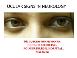
OCULAR SIGNS IN NEUROLOGY
DR. SUBODH KUMAR MAHTO,
DEPT. OF MEDICINE,
PGIMER,DR.RML HOSPITAL.
NEW Delhi
Ocular examination: objectives
– Recognize and interpret the common signs and symptoms
of neuro-ophthalmic disorders
– Obtain appropriate history
– Measure visual acuity
– Evaluate the visual fields
– Examine pupillary reaction
– Test the function of the extraocular muscles
– Inspect the optic nerve head ( fundoscopy)
Systematic examination
• Eye opening
• Pupil
• Ocular motor system
• Visual system
• Visual fields
• Common neuro-ophthalmic disorders
Glasgow coma scale
• Eye response:
• Four grades:
1. No eye opening
2. Eye opening in response to pain stimulus
3. Eye opening to speech
4. Eyes opening spontaneously
Pupil
• Size,
• Shape,
• Position and
• Equality.
• Light reflex
• Accommodation reflex
Pupil
• Pupillary size is determined by number of
factors including
– Age
– Level of retinal illumination
– Accommodative effort
Pupil
Pinpoint pupils
• Old age
• Alcohol abuse
• Neurosyphilis
• Drug effects
• Diabetes
• Horner syndrome and
• Pontine hematoma.
• Opthalmologic causes
include iridocyclitis,
miotic drops chronic
anterior segment
ischemia and an old
adies pupil.
Dilated pupils
• Anxiety
• Fear
• Pain
• Anticholinergics
• Bilateral lesions of
retina
• Post cardiac arrest
• Cerebral anoxia
Anisocoria
Anisocoria
Pupillary inequality greatest
In bright light
(large pupil)
In dim light
(small pupil)
3rd nerve palsy
Trauma
Tumor
Temporal lobe herniation
Aneurysm
No 3rd nerve palsy
Drug induced
Adie’s pupil
Iris damage (trauma/surgery/laser)
Ptosis
Horner syndrome
• An elderly male presented with fever, dry
cough and significant wt loss since 3 months.
Patient also complains of hoarseness of voice
and pain in the neck and rt. shoulder since 1
month. He used to smoke around 30
cigarettes since last 30 years.
Horner syndrome
Components
• Ptosis
• Anhydrosis
• Miosis
• Enophthalmos
• Loss of ciliospinal reflex
Causes
• Brainstem lesions eg. Wallenberg syndrome
• Cluster headache
• Internal carotid artery thrombosis/ dissection
• Cavernous sinus disease
• Apical lung tumors
• Neck trauma
• Syringomelia
Pathway of pupillary reaction to light
Pupillary reflex
• Direct
• Consensual
• Swinging flash light test
• Accommodation reflex
Afferent pupillary defect (APD)
Causes of APD
• Optic nerve disease
• Significant retinal disease
• Amblyopia
Grading Scale: APD
Grade 1+: A weak initial pupillary constriction
followed by greater redilation
Grade 2+: An initial pupillary stall followed by
greater redilation
Grade 3+: An immediate pupillary dilation
Grade 4+: No reaction to light – Amaurotic
pupil
Accommodation reflex
• Miosis
• Increased convexity of lens
• Convergence of eyes
Argyll robertson pupil
Holmes-Adie pupil
Pupil that is larger than normal and
constricts slowly in bright light (tonic
pupil),
Along with the absence of deep
tendon reflexes, usually in the
Achilles tendon
Causes for light near dissociation
• Neurosyphilis
• Diabetic autonomic neuropathy
• Dorsal rostral mid brain syndrome
• Lymes disease
• Chronic alcoholism
• Chiasmal lesions
• Myotonic muscular dystrophy
• Amyloidosis, adies pupil, ms and sarcoidosis
Efferent pupillary defect
3rd Nerve palsy pupil
Pupil was non-reactive
Associated findings in the right
eye included complete ptosis,
exotropia, hypotropia, and
impaired adduction, elevation
and depression
Mid brain pupils
Moderately dilated
Neither responded to direct light
stimulation
During fixation on a near target,
both pupils briskly constrict to 2.5
mm
Light-near dissociation of the pupils
is a feature of the dorsal midbrain
syndrome
OCULAR MOVEMENTS
Ocular motor system
Eye Movements
• Saccades—rapid shift in gaze
• Pursuit—stabilize image of moving object
• Fixation—stabilize image of still object
• VOR—stabilize image during head motion
• OKN—backup for when VOR decays to
cont’d head rotation
• Vergent movements—change depth of
focus
OCULOMOTOR NERVE
Ocular motor system
Third nerve palsy
• Internal ophthalmoplegia
• external ophthalmoplegia
• Complete/ Incomplete
• Common causes
 Ischemia
 Aneurysm
 Tumor
 Trauma
• Pupillary sparing
• Oculomotor synkinesis
Localization of oculomotor nerve
lesion
• Brainstem- 13%
• Subarachanoid space- 32%
• Cavernous signs-23%
• Peripheral component-18%
• Uncertain location-13%
TROCHLEAR NERVE
• 74 Year old male presented with a three day
history of sudden onset vertical diplopia. This
was worse when reading. He had had a mild
left cerebral vascular accident (CVA) one week
previously resulting in hand weakness which
later resolved.
Parks 3 step test
• Which eye is hyper deviated in primary gaze?
• Is the vertical deviation greater in right gaze or
left gaze?
• Is the vertical deviation greater with right
head tilt or left head tilt?
Signs of right fourth nerve palsy
• Right overaction on left gaze
• Right underaction on depression
in adduction
• Vertical diplopia
• Right hyperdeviation in primary
position when left eye fixating
• Excyclotorsion
Positive Bielschowsky test in right fourth nerve pals
Absence of right
hyperdeviation on
contralateral head tilt
Increase in right
hyperdeviation on ipsilateral
head tilt
Abducens nerve
SUPRANUCLEAR CONTROL OF
GAZE
Internuclear Ophthalmoplegia (INO)
SEVENTH NERVE
Facial nerve innervates the
orbicularis
Lid closure is impossible and the
eye turns upward (bell's
phenomenon)
Gaze palsies and gaze deviations
Horizontal gaze abnormalities
• Frontal lobe seizure activity
• Frontal lobe destructive lesion
• Pontine destructive lesion
Vertical gaze abnormalities
• Perinaud’s syndrome
– Upgaze palsy
– Convergence retraction nystagmus
– Tectal pupils
– Collier’s sign
– Mass lesion invloving posterior third ventricle and
upper dorsal mid brain such as pinealoma
• Progressive supranuclear palsy
Eyelids
NYSTAGMUS
Nystagmus
• a rythmic oscillation of the eyes. It has many
different patterns, and may arise in 3 situations
– Physiologically
– Sensory Deprivation
– Motor Imbalance
Nystagmus - selected types
• May be benign or indicate ocular and/or central nervous system
disease
• Definition according to fast phase
• End-point Nystagmus
– seen only in extreme positions of eye movement
• Drug-induced Nystagmus
– Anticonvulsants, Barbiturates/Other sedatives
• Searching/Pendular Nystagmus
– common with congenital severe visual impairment
• Nystagmus associated with INO
Classification
Nystagmus can be:
– Jerk - fast one direction, slow the other
– Pendular - equal velocity in both directions
– Mixed - of above
And can be
• Horizontal/Vertical/Oblique or Rotary
• In overwhelming majority of cases both eyes move in a co-
ordinated manner
Classification
Pendular
jerk
Physiological Nystagmus
• Not due to a disease process
• Has no benefit, except as a diagnostic tool
• Not associated with reduced VA
• Examples include
– End point nystagmus
– Postrotational nystagmus
– Induced caloric testing
– Optokinetic nystagmus
– Voluntary nystagmus
Sensory deprivation
• Due to a defect in the neural control of fixation
• Poor macular function that cannot be restored and
therefore of little clinical significance
• Typically pendular and horizontal
• Reduced by convergence and head posture
• If a child loses vision before 2 yrs they will invariably
develop nystagmus
• After 6 yrs they do not
• In between ??? Less predictable
Motor Imbalance
• Congential
• Spasmus Nutans
• Latent nystagmus
• Ataxic nystagmus
• Downbeat nystagmus
• Upbeat nystagmus
• Convergence retraction nystagmus
• See-Saw nystagmus
• Periodic alternating nystagmus
CONGENITAL NYSTAGMUS
• Due to a congenital anomaly of the motor system or to a
congenital disorder of vision
• Inherited as x-link recessive or autosomal dominant trait
• May appear during early childhood but is rarely present
at birth.
• Generally horizontal jerk type
• Absent in sleep
• Visual impairment is variable
Latent Nystagmus
• Horizontal jerk nystagmus presents when the light
stimulus is reduced to either eye (e.g. by occluding).
• In latent, no observable movement is present on
uncovering and full BSV is restored.
• Jerk nystagmus with fast phase towards the uncovered
eye
• Often noted in early childhood but can be observed in
adults (especially if they have had strabismus surgery
or in DVD)
Ataxic Nystagmus
• Occurs in abducting eye in internuclear ophthalmoplegia
Downbeat Nystagmus
• Has a fast downward beat
• Pathognomic of a brain lesion at the cervicomedullary junction
at the foramen magnum
Upbeat Nystagmus
• Commonly caused by drug intoxication (e.g. phenytoin - used
a san anticonvulsant)
• May be associated with a brain lesion at the posterior fossa
Convergence Retraction Nystagmus
• Jerk nystagmus
• Fast phase generating convergence and retraction of globe into
orbit
• Usually associated with brain lesion in the pretectal area
See-Saw nystagmus
• Usually an acquired motility disorder
associated with chiasmal lesions
• Where one eye elevates and intorts followed
by depression and extorsion of the other eye
• May be associated with a chiasmal lesion
(bitemporal hemianopia could be present)
• Rare
Periodic Alternating nystagmus
• Very rare jerk nystagmus
• Nystagmus changes amplitude and direction
• Associated with vascular or demylinating
brainstem disease
Fundamental questions when you see a
Patient with nystagmus
• What type
– May help provisional diagnosis
• How long has it been present
– Recent = refer
• The cause
• The activity of the lesion
– Some may produce deficient inhibitory neural activity
leading to neurologic hypofunction other excessive
excitory neural activity - hyperfunction
• Any form of nystagmus which is of recent
onset requires fairly urgent referral for an
ophthalmic opinion and if necessary further
neurological investigation
Clinical procedure for nystagmus
cases
• Close questioning as to the onset of the nystagmus, family
history, general health, medication, history of CNS disorders,
associated symptoms (oscillopsia, vertigo, unsteadiness and
loss of vision all imply acquired forms)
• Carefully note the type of nystagmus, distance/near, latency,
AHP etc
• VA recorded uni- and binocularly, with and without AHP, dist
and near and compared
• Full ophthalmoscopic, slit-lamp (transillumination), and
binocular vision assessment
Visual system
Visual system
Visual system
Visual fields defect
The swollen optic disc
•Papilloedema
•Papillitis
•Malignant hypertension
•Ischaemic optic neuropathy
•Diabetic optic neuropathy
•CRVO
•Intraocular inflammation
The pale optic disc
•Congenital
•Secondary to
•raised ICP
•vascular
retinal disease
•optic neuritis
•optic nerve
compression
•trauma
•Glaucoma
Papilloedema
• Disc swelling secondary to raised ICP
• Headache
– Worse in the morning
– Valsalva manouver
• Nausea and projectile vomiting
• Horizontal diplopia (VI palsy)
• Causes
– Space occupying lesion
– Intracranial hypertension
• Idiopathic
• Drugs
• Endocrine
– Severe hypertension
Haemorrhages
CWS
Blurred optic
disc margin
Small optic
cup
Disc pallor
Vessel attenuation
Differences between papilledema and
pseudopapilledema
Amaurosis Fugax
– Transient monocular visual loss or dimming
– May last from 2-3 minutes to 30 minutes or more
– Due to decrease blood flow to the eye
– Causes:
• Carotid atheroma
• Cardiac valvular disease
• Atrial myxoma
• Retinal migraine
• Giant cell arteritis
• Hyperviscosity syndromes
Myasthenia Gravis (MG)
– Chronic auto-immune disorder characterized by presence
of antibodies which block the ACH receptor sites
– It can affect any muscle
– Eye signs are the presenting signs in 50% of the patients
• Ptosis
• Any ocular motility disturbances
• INO
• Variability is the hallmark
Myasthenia Gravis (MG)
– Diagnosis
• Clinically
• Pharmacologically (Tensilon test)
• Serologically
• Sleep test
• Ice-pack test
• CT chest
• Thyroid function test
• ANA
– Treatment
• Acetylcholinesterase inhibitors
• Steroid
• Immunosuppressant
• Plasmapheresis
• Thymectomy
Myasthenia Gravis (MG)
PRE & POST TENSILON TEST
Ocular signs in I.C. Bleed
TYPE OF
INTRACEREBRAL
HEMORRHAGE
HOMONYMO
US VISUAL
DEFECTS
HORIZONTAL
GAZE PALSY
VERTICAL
GAZE PALSY
Putamen In large
hematoma
Contralateral NO
CAUDATE NO Absent No
THALAMIC In large
hematoma
Contralateral,
occasionally
Ipsilateral
Upward
LOBAR In occipital
hematoma
Contralateral in
frontal
hematoma
No
CEREBELLAR No Ipsilateral No
PONTINE No Bilateral No
MESENCEPHALIC No No Occasionally
upward
MEDULLARY No No no
Comatose patients - Eye examination
• Observation of the cornea, conjunctiva, sclera, iris,
lens, and eyelids.
• Edema of the conjunctiva and eyelids may occur in
congestive heart failure and nephrotic syndrome.
• Congestion and inflammation of the conjunctiva may
occur in the comatose patient from exposure.
• Enophthalmos indicates dehydration.
• Scleral icterus is seen with liver disease, and yellowish
discoloration of the skin without scleral involvement
may be due to drugs such as rifampin.
Comatose patients - Eye examination
• Band keratopathy is caused by hypercalcemia,
whereas hypocalcemia is associated with
cataracts.
• Kayser-fleischer rings are seen in progressive
lenticular degeneration (wilson's disease).
• Arcus senilis is seen in normal aging but also in
hyperlipidemia.
• Fat embolism may cause petechiae in
conjunctiva and eye grounds.
Comatose patient - Fundus Examination
• Evidence of hypertension or diabetes.
• Grayish deposits surrounding the optic disc have been
reported in lead poisoning.
• Retina congested and edematous in methyl alcohol
poisoning, and the disc margin may be blurred.
• Subhyaloid hemorrhage appears occasionally as a
consequence of a rapid increase in icp due to
subarachnoid hemorrhage (terson's syndrome).
• Papilledema results from increased icp and may be
indicative of an intracranial mass lesion or hypertensive
Multiple sclerosis
• Patients with multiple sclerosis (MS)
frequently have visual complaints
– Cerebellar dysfunction
– Motor symptoms
– Sensory symptoms
– Mental changes
– Sphincter disturbances
Multiple sclerosis
• Ocular complications:
– Optic neuritis
– Chiasmal and retro chiasmal abnormalities
– Ocular motility disturbances
• Treatment
– Steroid
– Interferon
Optic Neuritis
Common Symptoms
•Monocular
•Central Vision loss
•Pain (eye movement)
•Altered colour vision
•Recovery common
•Uhthoff’s symptom
•Flashes
•Pulfrich
phenomenon
Optic Neuritis: Physical
Findings
•Decreased visual acuity
•VF defect
•(Central/Altitudinal 29% )
•Dyschromatopsia
•Afferent Pupil Defect
(RAPD)
•Optic disc swelling 35%
•Abnormal Contrast
Sensitivity
•Abnormal VEP
•Altered Flicker
Perception
•Altered depth
perception
•Optic disc pallor
Optic Neuritis: Optic Disc
Herpes zoster ophthalmicus
Cysticercosis
Cavernous sinus thrombosis
Approach to examination
1. External examination
2. Visual acuity
3. Pupil function
4. Eye movements
5. Visual field testing
6. Eye pressure
7. Fundus examination
8. Slit lamp examination
THANKYOU

More Related Content

What's hot

Saccadic Eye Movement
Saccadic Eye Movement Saccadic Eye Movement
Saccadic Eye Movement
Ade Wijaya
 
Supranuclear pathways and lesions
Supranuclear pathways and lesionsSupranuclear pathways and lesions
Supranuclear pathways and lesions
Laxmi Eye Institute
 
Anterior ischemic optic neuropathy
Anterior ischemic optic neuropathyAnterior ischemic optic neuropathy
Anterior ischemic optic neuropathyJagdish Dukre
 
The pupillary pathway and its clinical aspects
The pupillary pathway and its clinical aspectsThe pupillary pathway and its clinical aspects
The pupillary pathway and its clinical aspects
Laxmi Eye Institute
 
Defects of visual pathway
Defects of visual pathwayDefects of visual pathway
Defects of visual pathway
Rohit Rao
 
Nystagmus
NystagmusNystagmus
Abnormalities of pupil
Abnormalities of pupilAbnormalities of pupil
Abnormalities of pupil
Imrul Morshed
 
Pupil anomalies and disorders shiva ppt
Pupil anomalies and disorders shiva pptPupil anomalies and disorders shiva ppt
Pupil anomalies and disorders shiva ppt
Shiva Ram
 
Supranuclear disorders of ocular motility
Supranuclear disorders of ocular motilitySupranuclear disorders of ocular motility
Supranuclear disorders of ocular motility
SSSIHMS-PG
 
Papilledema - Dr Shylesh Dabke
Papilledema - Dr Shylesh DabkePapilledema - Dr Shylesh Dabke
Papilledema - Dr Shylesh Dabke
Shylesh Dabke
 
6th Nerve Palsy
6th Nerve Palsy6th Nerve Palsy
6th Nerve Palsy
Hasnain Pasha
 
Gaze palcy
Gaze palcy Gaze palcy
Gaze palcy
shovon2026
 
Ptosis
PtosisPtosis
Optic atrophy (b)
Optic atrophy (b)Optic atrophy (b)
Optic atrophy (b)
Muhammad AbdulWahidKarim
 
Case presentation: Third nerve palsy
Case presentation: Third nerve palsyCase presentation: Third nerve palsy
Case presentation: Third nerve palsy
Anis Suzanna Mohamad
 
Oculomotor nerve palsy
Oculomotor nerve palsyOculomotor nerve palsy
Oculomotor nerve palsy
Dr. Mahziba Rahman
 
Abnormal Pupil Reactions
Abnormal Pupil Reactions Abnormal Pupil Reactions
Abnormal Pupil Reactions
Hatlan Al Hatlan
 
Approach to vision loss
Approach to vision lossApproach to vision loss
Approach to vision loss
Kirat Singh Grewal
 
Nystagmus
NystagmusNystagmus
Nystagmus
Sheim Elteb
 

What's hot (20)

Saccadic Eye Movement
Saccadic Eye Movement Saccadic Eye Movement
Saccadic Eye Movement
 
Supranuclear pathways and lesions
Supranuclear pathways and lesionsSupranuclear pathways and lesions
Supranuclear pathways and lesions
 
Oculomotor palsy
Oculomotor palsyOculomotor palsy
Oculomotor palsy
 
Anterior ischemic optic neuropathy
Anterior ischemic optic neuropathyAnterior ischemic optic neuropathy
Anterior ischemic optic neuropathy
 
The pupillary pathway and its clinical aspects
The pupillary pathway and its clinical aspectsThe pupillary pathway and its clinical aspects
The pupillary pathway and its clinical aspects
 
Defects of visual pathway
Defects of visual pathwayDefects of visual pathway
Defects of visual pathway
 
Nystagmus
NystagmusNystagmus
Nystagmus
 
Abnormalities of pupil
Abnormalities of pupilAbnormalities of pupil
Abnormalities of pupil
 
Pupil anomalies and disorders shiva ppt
Pupil anomalies and disorders shiva pptPupil anomalies and disorders shiva ppt
Pupil anomalies and disorders shiva ppt
 
Supranuclear disorders of ocular motility
Supranuclear disorders of ocular motilitySupranuclear disorders of ocular motility
Supranuclear disorders of ocular motility
 
Papilledema - Dr Shylesh Dabke
Papilledema - Dr Shylesh DabkePapilledema - Dr Shylesh Dabke
Papilledema - Dr Shylesh Dabke
 
6th Nerve Palsy
6th Nerve Palsy6th Nerve Palsy
6th Nerve Palsy
 
Gaze palcy
Gaze palcy Gaze palcy
Gaze palcy
 
Ptosis
PtosisPtosis
Ptosis
 
Optic atrophy (b)
Optic atrophy (b)Optic atrophy (b)
Optic atrophy (b)
 
Case presentation: Third nerve palsy
Case presentation: Third nerve palsyCase presentation: Third nerve palsy
Case presentation: Third nerve palsy
 
Oculomotor nerve palsy
Oculomotor nerve palsyOculomotor nerve palsy
Oculomotor nerve palsy
 
Abnormal Pupil Reactions
Abnormal Pupil Reactions Abnormal Pupil Reactions
Abnormal Pupil Reactions
 
Approach to vision loss
Approach to vision lossApproach to vision loss
Approach to vision loss
 
Nystagmus
NystagmusNystagmus
Nystagmus
 

Similar to Ocular signs in medicine/ neurology

ocular.pptx
ocular.pptxocular.pptx
ocular.pptx
devashish416090
 
Strabismus
StrabismusStrabismus
Strabismus
gladismathew
 
strabismus-190706104054.pptx
strabismus-190706104054.pptxstrabismus-190706104054.pptx
strabismus-190706104054.pptx
ugonnanwoke
 
Diplopia
Diplopia Diplopia
Diplopia
Anisha Rathod
 
Pupil ppt
Pupil  pptPupil  ppt
HISTORY TAKING SKILLS ---1.pptx
HISTORY TAKING SKILLS ---1.pptxHISTORY TAKING SKILLS ---1.pptx
HISTORY TAKING SKILLS ---1.pptx
LavanyaMadabushi
 
Nystagmus assessments and management mehedi
Nystagmus assessments and management  mehediNystagmus assessments and management  mehedi
Nystagmus assessments and management mehedi
Mehedi Hasan
 
Supranuclear disorders of ocular motility
Supranuclear disorders of ocular motilitySupranuclear disorders of ocular motility
Supranuclear disorders of ocular motility
erameshita
 
Anomalies of accommodation, convergence & its management
Anomalies of accommodation, convergence & its managementAnomalies of accommodation, convergence & its management
Anomalies of accommodation, convergence & its management
Mohammad Arman Bin Aziz
 
PUPILARY REFLEX PATHWAYS.pptx
PUPILARY REFLEX PATHWAYS.pptxPUPILARY REFLEX PATHWAYS.pptx
PUPILARY REFLEX PATHWAYS.pptx
Naveen Challa
 
Approach to vision loss
Approach to vision lossApproach to vision loss
Approach to vision loss
NeurologyKota
 
Approach to Neurological causes of Vision loss.pptx
Approach to Neurological causes of Vision loss.pptxApproach to Neurological causes of Vision loss.pptx
Approach to Neurological causes of Vision loss.pptx
NeurologyKota
 
Nystagmus and Nystagmoid Movements
Nystagmus and Nystagmoid MovementsNystagmus and Nystagmoid Movements
Nystagmus and Nystagmoid Movements
neurophq8
 
NYSTAGMUS.pptx
NYSTAGMUS.pptxNYSTAGMUS.pptx
NYSTAGMUS.pptx
LavanyaMadabushi
 
pupil ppt dr kavitha.pptx
pupil ppt dr kavitha.pptxpupil ppt dr kavitha.pptx
pupil ppt dr kavitha.pptx
SreedharNaik6
 
paediatric ophthalmology and strabismus
paediatric ophthalmology and strabismuspaediatric ophthalmology and strabismus
paediatric ophthalmology and strabismus
OphthalmicDocs Chiong
 
non paralytic and paralytic strabismus- 20.07.16
 non paralytic and paralytic strabismus- 20.07.16 non paralytic and paralytic strabismus- 20.07.16
non paralytic and paralytic strabismus- 20.07.16
ophthalmgmcri
 
Nystagmus By Praneet Mishra .pptx
Nystagmus By Praneet Mishra .pptxNystagmus By Praneet Mishra .pptx
Nystagmus By Praneet Mishra .pptx
Praneet MISHRA
 

Similar to Ocular signs in medicine/ neurology (20)

ocular.pptx
ocular.pptxocular.pptx
ocular.pptx
 
Strabismus
StrabismusStrabismus
Strabismus
 
strabismus-190706104054.pptx
strabismus-190706104054.pptxstrabismus-190706104054.pptx
strabismus-190706104054.pptx
 
Accommodation and its anomalies
Accommodation and its anomaliesAccommodation and its anomalies
Accommodation and its anomalies
 
Diplopia
Diplopia Diplopia
Diplopia
 
Pupil ppt
Pupil  pptPupil  ppt
Pupil ppt
 
HISTORY TAKING SKILLS ---1.pptx
HISTORY TAKING SKILLS ---1.pptxHISTORY TAKING SKILLS ---1.pptx
HISTORY TAKING SKILLS ---1.pptx
 
Nystagmus assessments and management mehedi
Nystagmus assessments and management  mehediNystagmus assessments and management  mehedi
Nystagmus assessments and management mehedi
 
Supranuclear disorders of ocular motility
Supranuclear disorders of ocular motilitySupranuclear disorders of ocular motility
Supranuclear disorders of ocular motility
 
Pupil
PupilPupil
Pupil
 
Anomalies of accommodation, convergence & its management
Anomalies of accommodation, convergence & its managementAnomalies of accommodation, convergence & its management
Anomalies of accommodation, convergence & its management
 
PUPILARY REFLEX PATHWAYS.pptx
PUPILARY REFLEX PATHWAYS.pptxPUPILARY REFLEX PATHWAYS.pptx
PUPILARY REFLEX PATHWAYS.pptx
 
Approach to vision loss
Approach to vision lossApproach to vision loss
Approach to vision loss
 
Approach to Neurological causes of Vision loss.pptx
Approach to Neurological causes of Vision loss.pptxApproach to Neurological causes of Vision loss.pptx
Approach to Neurological causes of Vision loss.pptx
 
Nystagmus and Nystagmoid Movements
Nystagmus and Nystagmoid MovementsNystagmus and Nystagmoid Movements
Nystagmus and Nystagmoid Movements
 
NYSTAGMUS.pptx
NYSTAGMUS.pptxNYSTAGMUS.pptx
NYSTAGMUS.pptx
 
pupil ppt dr kavitha.pptx
pupil ppt dr kavitha.pptxpupil ppt dr kavitha.pptx
pupil ppt dr kavitha.pptx
 
paediatric ophthalmology and strabismus
paediatric ophthalmology and strabismuspaediatric ophthalmology and strabismus
paediatric ophthalmology and strabismus
 
non paralytic and paralytic strabismus- 20.07.16
 non paralytic and paralytic strabismus- 20.07.16 non paralytic and paralytic strabismus- 20.07.16
non paralytic and paralytic strabismus- 20.07.16
 
Nystagmus By Praneet Mishra .pptx
Nystagmus By Praneet Mishra .pptxNystagmus By Praneet Mishra .pptx
Nystagmus By Praneet Mishra .pptx
 

More from PGIMER,DR.RML HOSPITAL

Disorders of the Neuromuscular junction
Disorders of the Neuromuscular junction Disorders of the Neuromuscular junction
Disorders of the Neuromuscular junction
PGIMER,DR.RML HOSPITAL
 
PATHOPHYSIOLOGY AND MANAGEMEMENT OF PAIN
PATHOPHYSIOLOGY AND   MANAGEMEMENT	OF PAINPATHOPHYSIOLOGY AND   MANAGEMEMENT	OF PAIN
PATHOPHYSIOLOGY AND MANAGEMEMENT OF PAIN
PGIMER,DR.RML HOSPITAL
 
Speech disorder .
Speech disorder .Speech disorder .
Speech disorder .
PGIMER,DR.RML HOSPITAL
 
NERVE CONDUCTION STUDIES, ELECTROMYOGRAPHY
NERVE CONDUCTION STUDIES, ELECTROMYOGRAPHYNERVE CONDUCTION STUDIES, ELECTROMYOGRAPHY
NERVE CONDUCTION STUDIES, ELECTROMYOGRAPHY
PGIMER,DR.RML HOSPITAL
 
Influenza h1 n1 pdf
Influenza h1 n1 pdfInfluenza h1 n1 pdf
Influenza h1 n1 pdf
PGIMER,DR.RML HOSPITAL
 
Infective endocarditis pdf
Infective endocarditis pdfInfective endocarditis pdf
Infective endocarditis pdf
PGIMER,DR.RML HOSPITAL
 
Rickettsial ds
Rickettsial dsRickettsial ds
Rickettsial ds
PGIMER,DR.RML HOSPITAL
 
Organophosphorus poisoning final
Organophosphorus poisoning finalOrganophosphorus poisoning final
Organophosphorus poisoning final
PGIMER,DR.RML HOSPITAL
 
Neurocysticercosis
NeurocysticercosisNeurocysticercosis
Neurocysticercosis
PGIMER,DR.RML HOSPITAL
 
Acute kidney injury defnition, causes,
Acute kidney injury   defnition, causes,Acute kidney injury   defnition, causes,
Acute kidney injury defnition, causes,
PGIMER,DR.RML HOSPITAL
 
General physical examination of CVS
General physical examination of CVSGeneral physical examination of CVS
General physical examination of CVS
PGIMER,DR.RML HOSPITAL
 

More from PGIMER,DR.RML HOSPITAL (17)

Disorders of the Neuromuscular junction
Disorders of the Neuromuscular junction Disorders of the Neuromuscular junction
Disorders of the Neuromuscular junction
 
PATHOPHYSIOLOGY AND MANAGEMEMENT OF PAIN
PATHOPHYSIOLOGY AND   MANAGEMEMENT	OF PAINPATHOPHYSIOLOGY AND   MANAGEMEMENT	OF PAIN
PATHOPHYSIOLOGY AND MANAGEMEMENT OF PAIN
 
Speech disorder .
Speech disorder .Speech disorder .
Speech disorder .
 
NCV AND EMG
NCV AND EMGNCV AND EMG
NCV AND EMG
 
NERVE CONDUCTION STUDIES, ELECTROMYOGRAPHY
NERVE CONDUCTION STUDIES, ELECTROMYOGRAPHYNERVE CONDUCTION STUDIES, ELECTROMYOGRAPHY
NERVE CONDUCTION STUDIES, ELECTROMYOGRAPHY
 
Influenza h1 n1 pdf
Influenza h1 n1 pdfInfluenza h1 n1 pdf
Influenza h1 n1 pdf
 
Infective endocarditis pdf
Infective endocarditis pdfInfective endocarditis pdf
Infective endocarditis pdf
 
Dengue final pdf
Dengue final pdfDengue final pdf
Dengue final pdf
 
Rickettsial ds
Rickettsial dsRickettsial ds
Rickettsial ds
 
Organophosphorus poisoning final
Organophosphorus poisoning finalOrganophosphorus poisoning final
Organophosphorus poisoning final
 
Neurocysticercosis
NeurocysticercosisNeurocysticercosis
Neurocysticercosis
 
Malabsorption syndrome
Malabsorption syndromeMalabsorption syndrome
Malabsorption syndrome
 
Cogenital heart ds.
Cogenital heart ds.Cogenital heart ds.
Cogenital heart ds.
 
Acute kidney injury defnition, causes,
Acute kidney injury   defnition, causes,Acute kidney injury   defnition, causes,
Acute kidney injury defnition, causes,
 
ABG and spirometry
ABG and spirometryABG and spirometry
ABG and spirometry
 
ABG
ABGABG
ABG
 
General physical examination of CVS
General physical examination of CVSGeneral physical examination of CVS
General physical examination of CVS
 

Recently uploaded

Supporting (UKRI) OA monographs at Salford.pptx
Supporting (UKRI) OA monographs at Salford.pptxSupporting (UKRI) OA monographs at Salford.pptx
Supporting (UKRI) OA monographs at Salford.pptx
Jisc
 
How to Break the cycle of negative Thoughts
How to Break the cycle of negative ThoughtsHow to Break the cycle of negative Thoughts
How to Break the cycle of negative Thoughts
Col Mukteshwar Prasad
 
CLASS 11 CBSE B.St Project AIDS TO TRADE - INSURANCE
CLASS 11 CBSE B.St Project AIDS TO TRADE - INSURANCECLASS 11 CBSE B.St Project AIDS TO TRADE - INSURANCE
CLASS 11 CBSE B.St Project AIDS TO TRADE - INSURANCE
BhavyaRajput3
 
How to Split Bills in the Odoo 17 POS Module
How to Split Bills in the Odoo 17 POS ModuleHow to Split Bills in the Odoo 17 POS Module
How to Split Bills in the Odoo 17 POS Module
Celine George
 
special B.ed 2nd year old paper_20240531.pdf
special B.ed 2nd year old paper_20240531.pdfspecial B.ed 2nd year old paper_20240531.pdf
special B.ed 2nd year old paper_20240531.pdf
Special education needs
 
Additional Benefits for Employee Website.pdf
Additional Benefits for Employee Website.pdfAdditional Benefits for Employee Website.pdf
Additional Benefits for Employee Website.pdf
joachimlavalley1
 
MARUTI SUZUKI- A Successful Joint Venture in India.pptx
MARUTI SUZUKI- A Successful Joint Venture in India.pptxMARUTI SUZUKI- A Successful Joint Venture in India.pptx
MARUTI SUZUKI- A Successful Joint Venture in India.pptx
bennyroshan06
 
Unit 8 - Information and Communication Technology (Paper I).pdf
Unit 8 - Information and Communication Technology (Paper I).pdfUnit 8 - Information and Communication Technology (Paper I).pdf
Unit 8 - Information and Communication Technology (Paper I).pdf
Thiyagu K
 
Cambridge International AS A Level Biology Coursebook - EBook (MaryFosbery J...
Cambridge International AS  A Level Biology Coursebook - EBook (MaryFosbery J...Cambridge International AS  A Level Biology Coursebook - EBook (MaryFosbery J...
Cambridge International AS A Level Biology Coursebook - EBook (MaryFosbery J...
AzmatAli747758
 
Sha'Carri Richardson Presentation 202345
Sha'Carri Richardson Presentation 202345Sha'Carri Richardson Presentation 202345
Sha'Carri Richardson Presentation 202345
beazzy04
 
Thesis Statement for students diagnonsed withADHD.ppt
Thesis Statement for students diagnonsed withADHD.pptThesis Statement for students diagnonsed withADHD.ppt
Thesis Statement for students diagnonsed withADHD.ppt
EverAndrsGuerraGuerr
 
TESDA TM1 REVIEWER FOR NATIONAL ASSESSMENT WRITTEN AND ORAL QUESTIONS WITH A...
TESDA TM1 REVIEWER  FOR NATIONAL ASSESSMENT WRITTEN AND ORAL QUESTIONS WITH A...TESDA TM1 REVIEWER  FOR NATIONAL ASSESSMENT WRITTEN AND ORAL QUESTIONS WITH A...
TESDA TM1 REVIEWER FOR NATIONAL ASSESSMENT WRITTEN AND ORAL QUESTIONS WITH A...
EugeneSaldivar
 
Palestine last event orientationfvgnh .pptx
Palestine last event orientationfvgnh .pptxPalestine last event orientationfvgnh .pptx
Palestine last event orientationfvgnh .pptx
RaedMohamed3
 
Unit 2- Research Aptitude (UGC NET Paper I).pdf
Unit 2- Research Aptitude (UGC NET Paper I).pdfUnit 2- Research Aptitude (UGC NET Paper I).pdf
Unit 2- Research Aptitude (UGC NET Paper I).pdf
Thiyagu K
 
Mule 4.6 & Java 17 Upgrade | MuleSoft Mysore Meetup #46
Mule 4.6 & Java 17 Upgrade | MuleSoft Mysore Meetup #46Mule 4.6 & Java 17 Upgrade | MuleSoft Mysore Meetup #46
Mule 4.6 & Java 17 Upgrade | MuleSoft Mysore Meetup #46
MysoreMuleSoftMeetup
 
The Roman Empire A Historical Colossus.pdf
The Roman Empire A Historical Colossus.pdfThe Roman Empire A Historical Colossus.pdf
The Roman Empire A Historical Colossus.pdf
kaushalkr1407
 
2024.06.01 Introducing a competency framework for languag learning materials ...
2024.06.01 Introducing a competency framework for languag learning materials ...2024.06.01 Introducing a competency framework for languag learning materials ...
2024.06.01 Introducing a competency framework for languag learning materials ...
Sandy Millin
 
Phrasal Verbs.XXXXXXXXXXXXXXXXXXXXXXXXXX
Phrasal Verbs.XXXXXXXXXXXXXXXXXXXXXXXXXXPhrasal Verbs.XXXXXXXXXXXXXXXXXXXXXXXXXX
Phrasal Verbs.XXXXXXXXXXXXXXXXXXXXXXXXXX
MIRIAMSALINAS13
 
Fish and Chips - have they had their chips
Fish and Chips - have they had their chipsFish and Chips - have they had their chips
Fish and Chips - have they had their chips
GeoBlogs
 
1.4 modern child centered education - mahatma gandhi-2.pptx
1.4 modern child centered education - mahatma gandhi-2.pptx1.4 modern child centered education - mahatma gandhi-2.pptx
1.4 modern child centered education - mahatma gandhi-2.pptx
JosvitaDsouza2
 

Recently uploaded (20)

Supporting (UKRI) OA monographs at Salford.pptx
Supporting (UKRI) OA monographs at Salford.pptxSupporting (UKRI) OA monographs at Salford.pptx
Supporting (UKRI) OA monographs at Salford.pptx
 
How to Break the cycle of negative Thoughts
How to Break the cycle of negative ThoughtsHow to Break the cycle of negative Thoughts
How to Break the cycle of negative Thoughts
 
CLASS 11 CBSE B.St Project AIDS TO TRADE - INSURANCE
CLASS 11 CBSE B.St Project AIDS TO TRADE - INSURANCECLASS 11 CBSE B.St Project AIDS TO TRADE - INSURANCE
CLASS 11 CBSE B.St Project AIDS TO TRADE - INSURANCE
 
How to Split Bills in the Odoo 17 POS Module
How to Split Bills in the Odoo 17 POS ModuleHow to Split Bills in the Odoo 17 POS Module
How to Split Bills in the Odoo 17 POS Module
 
special B.ed 2nd year old paper_20240531.pdf
special B.ed 2nd year old paper_20240531.pdfspecial B.ed 2nd year old paper_20240531.pdf
special B.ed 2nd year old paper_20240531.pdf
 
Additional Benefits for Employee Website.pdf
Additional Benefits for Employee Website.pdfAdditional Benefits for Employee Website.pdf
Additional Benefits for Employee Website.pdf
 
MARUTI SUZUKI- A Successful Joint Venture in India.pptx
MARUTI SUZUKI- A Successful Joint Venture in India.pptxMARUTI SUZUKI- A Successful Joint Venture in India.pptx
MARUTI SUZUKI- A Successful Joint Venture in India.pptx
 
Unit 8 - Information and Communication Technology (Paper I).pdf
Unit 8 - Information and Communication Technology (Paper I).pdfUnit 8 - Information and Communication Technology (Paper I).pdf
Unit 8 - Information and Communication Technology (Paper I).pdf
 
Cambridge International AS A Level Biology Coursebook - EBook (MaryFosbery J...
Cambridge International AS  A Level Biology Coursebook - EBook (MaryFosbery J...Cambridge International AS  A Level Biology Coursebook - EBook (MaryFosbery J...
Cambridge International AS A Level Biology Coursebook - EBook (MaryFosbery J...
 
Sha'Carri Richardson Presentation 202345
Sha'Carri Richardson Presentation 202345Sha'Carri Richardson Presentation 202345
Sha'Carri Richardson Presentation 202345
 
Thesis Statement for students diagnonsed withADHD.ppt
Thesis Statement for students diagnonsed withADHD.pptThesis Statement for students diagnonsed withADHD.ppt
Thesis Statement for students diagnonsed withADHD.ppt
 
TESDA TM1 REVIEWER FOR NATIONAL ASSESSMENT WRITTEN AND ORAL QUESTIONS WITH A...
TESDA TM1 REVIEWER  FOR NATIONAL ASSESSMENT WRITTEN AND ORAL QUESTIONS WITH A...TESDA TM1 REVIEWER  FOR NATIONAL ASSESSMENT WRITTEN AND ORAL QUESTIONS WITH A...
TESDA TM1 REVIEWER FOR NATIONAL ASSESSMENT WRITTEN AND ORAL QUESTIONS WITH A...
 
Palestine last event orientationfvgnh .pptx
Palestine last event orientationfvgnh .pptxPalestine last event orientationfvgnh .pptx
Palestine last event orientationfvgnh .pptx
 
Unit 2- Research Aptitude (UGC NET Paper I).pdf
Unit 2- Research Aptitude (UGC NET Paper I).pdfUnit 2- Research Aptitude (UGC NET Paper I).pdf
Unit 2- Research Aptitude (UGC NET Paper I).pdf
 
Mule 4.6 & Java 17 Upgrade | MuleSoft Mysore Meetup #46
Mule 4.6 & Java 17 Upgrade | MuleSoft Mysore Meetup #46Mule 4.6 & Java 17 Upgrade | MuleSoft Mysore Meetup #46
Mule 4.6 & Java 17 Upgrade | MuleSoft Mysore Meetup #46
 
The Roman Empire A Historical Colossus.pdf
The Roman Empire A Historical Colossus.pdfThe Roman Empire A Historical Colossus.pdf
The Roman Empire A Historical Colossus.pdf
 
2024.06.01 Introducing a competency framework for languag learning materials ...
2024.06.01 Introducing a competency framework for languag learning materials ...2024.06.01 Introducing a competency framework for languag learning materials ...
2024.06.01 Introducing a competency framework for languag learning materials ...
 
Phrasal Verbs.XXXXXXXXXXXXXXXXXXXXXXXXXX
Phrasal Verbs.XXXXXXXXXXXXXXXXXXXXXXXXXXPhrasal Verbs.XXXXXXXXXXXXXXXXXXXXXXXXXX
Phrasal Verbs.XXXXXXXXXXXXXXXXXXXXXXXXXX
 
Fish and Chips - have they had their chips
Fish and Chips - have they had their chipsFish and Chips - have they had their chips
Fish and Chips - have they had their chips
 
1.4 modern child centered education - mahatma gandhi-2.pptx
1.4 modern child centered education - mahatma gandhi-2.pptx1.4 modern child centered education - mahatma gandhi-2.pptx
1.4 modern child centered education - mahatma gandhi-2.pptx
 

Ocular signs in medicine/ neurology

  • 1. OCULAR SIGNS IN NEUROLOGY DR. SUBODH KUMAR MAHTO, DEPT. OF MEDICINE, PGIMER,DR.RML HOSPITAL. NEW Delhi
  • 2. Ocular examination: objectives – Recognize and interpret the common signs and symptoms of neuro-ophthalmic disorders – Obtain appropriate history – Measure visual acuity – Evaluate the visual fields – Examine pupillary reaction – Test the function of the extraocular muscles – Inspect the optic nerve head ( fundoscopy)
  • 3. Systematic examination • Eye opening • Pupil • Ocular motor system • Visual system • Visual fields • Common neuro-ophthalmic disorders
  • 4. Glasgow coma scale • Eye response: • Four grades: 1. No eye opening 2. Eye opening in response to pain stimulus 3. Eye opening to speech 4. Eyes opening spontaneously
  • 5. Pupil • Size, • Shape, • Position and • Equality. • Light reflex • Accommodation reflex
  • 6. Pupil • Pupillary size is determined by number of factors including – Age – Level of retinal illumination – Accommodative effort
  • 8. Pinpoint pupils • Old age • Alcohol abuse • Neurosyphilis • Drug effects • Diabetes • Horner syndrome and • Pontine hematoma. • Opthalmologic causes include iridocyclitis, miotic drops chronic anterior segment ischemia and an old adies pupil.
  • 9. Dilated pupils • Anxiety • Fear • Pain • Anticholinergics • Bilateral lesions of retina • Post cardiac arrest • Cerebral anoxia
  • 11. Anisocoria Pupillary inequality greatest In bright light (large pupil) In dim light (small pupil) 3rd nerve palsy Trauma Tumor Temporal lobe herniation Aneurysm No 3rd nerve palsy Drug induced Adie’s pupil Iris damage (trauma/surgery/laser) Ptosis Horner syndrome
  • 12. • An elderly male presented with fever, dry cough and significant wt loss since 3 months. Patient also complains of hoarseness of voice and pain in the neck and rt. shoulder since 1 month. He used to smoke around 30 cigarettes since last 30 years.
  • 13.
  • 15. Components • Ptosis • Anhydrosis • Miosis • Enophthalmos • Loss of ciliospinal reflex
  • 16. Causes • Brainstem lesions eg. Wallenberg syndrome • Cluster headache • Internal carotid artery thrombosis/ dissection • Cavernous sinus disease • Apical lung tumors • Neck trauma • Syringomelia
  • 17. Pathway of pupillary reaction to light
  • 18. Pupillary reflex • Direct • Consensual • Swinging flash light test • Accommodation reflex
  • 20. Causes of APD • Optic nerve disease • Significant retinal disease • Amblyopia
  • 21. Grading Scale: APD Grade 1+: A weak initial pupillary constriction followed by greater redilation Grade 2+: An initial pupillary stall followed by greater redilation Grade 3+: An immediate pupillary dilation Grade 4+: No reaction to light – Amaurotic pupil
  • 22. Accommodation reflex • Miosis • Increased convexity of lens • Convergence of eyes
  • 24. Holmes-Adie pupil Pupil that is larger than normal and constricts slowly in bright light (tonic pupil), Along with the absence of deep tendon reflexes, usually in the Achilles tendon
  • 25. Causes for light near dissociation • Neurosyphilis • Diabetic autonomic neuropathy • Dorsal rostral mid brain syndrome • Lymes disease • Chronic alcoholism • Chiasmal lesions • Myotonic muscular dystrophy • Amyloidosis, adies pupil, ms and sarcoidosis
  • 27. 3rd Nerve palsy pupil Pupil was non-reactive Associated findings in the right eye included complete ptosis, exotropia, hypotropia, and impaired adduction, elevation and depression
  • 28. Mid brain pupils Moderately dilated Neither responded to direct light stimulation During fixation on a near target, both pupils briskly constrict to 2.5 mm Light-near dissociation of the pupils is a feature of the dorsal midbrain syndrome
  • 31. Eye Movements • Saccades—rapid shift in gaze • Pursuit—stabilize image of moving object • Fixation—stabilize image of still object • VOR—stabilize image during head motion • OKN—backup for when VOR decays to cont’d head rotation • Vergent movements—change depth of focus
  • 32.
  • 34.
  • 37.
  • 38. • Internal ophthalmoplegia • external ophthalmoplegia • Complete/ Incomplete • Common causes  Ischemia  Aneurysm  Tumor  Trauma • Pupillary sparing • Oculomotor synkinesis
  • 39. Localization of oculomotor nerve lesion • Brainstem- 13% • Subarachanoid space- 32% • Cavernous signs-23% • Peripheral component-18% • Uncertain location-13%
  • 41. • 74 Year old male presented with a three day history of sudden onset vertical diplopia. This was worse when reading. He had had a mild left cerebral vascular accident (CVA) one week previously resulting in hand weakness which later resolved.
  • 42.
  • 43. Parks 3 step test • Which eye is hyper deviated in primary gaze? • Is the vertical deviation greater in right gaze or left gaze? • Is the vertical deviation greater with right head tilt or left head tilt?
  • 44. Signs of right fourth nerve palsy • Right overaction on left gaze • Right underaction on depression in adduction • Vertical diplopia • Right hyperdeviation in primary position when left eye fixating • Excyclotorsion
  • 45. Positive Bielschowsky test in right fourth nerve pals Absence of right hyperdeviation on contralateral head tilt Increase in right hyperdeviation on ipsilateral head tilt
  • 48.
  • 50. SEVENTH NERVE Facial nerve innervates the orbicularis Lid closure is impossible and the eye turns upward (bell's phenomenon)
  • 51. Gaze palsies and gaze deviations
  • 52. Horizontal gaze abnormalities • Frontal lobe seizure activity • Frontal lobe destructive lesion • Pontine destructive lesion
  • 53. Vertical gaze abnormalities • Perinaud’s syndrome – Upgaze palsy – Convergence retraction nystagmus – Tectal pupils – Collier’s sign – Mass lesion invloving posterior third ventricle and upper dorsal mid brain such as pinealoma • Progressive supranuclear palsy
  • 55.
  • 57. Nystagmus • a rythmic oscillation of the eyes. It has many different patterns, and may arise in 3 situations – Physiologically – Sensory Deprivation – Motor Imbalance
  • 58. Nystagmus - selected types • May be benign or indicate ocular and/or central nervous system disease • Definition according to fast phase • End-point Nystagmus – seen only in extreme positions of eye movement • Drug-induced Nystagmus – Anticonvulsants, Barbiturates/Other sedatives • Searching/Pendular Nystagmus – common with congenital severe visual impairment • Nystagmus associated with INO
  • 59. Classification Nystagmus can be: – Jerk - fast one direction, slow the other – Pendular - equal velocity in both directions – Mixed - of above And can be • Horizontal/Vertical/Oblique or Rotary • In overwhelming majority of cases both eyes move in a co- ordinated manner
  • 61. Physiological Nystagmus • Not due to a disease process • Has no benefit, except as a diagnostic tool • Not associated with reduced VA • Examples include – End point nystagmus – Postrotational nystagmus – Induced caloric testing – Optokinetic nystagmus – Voluntary nystagmus
  • 62. Sensory deprivation • Due to a defect in the neural control of fixation • Poor macular function that cannot be restored and therefore of little clinical significance • Typically pendular and horizontal • Reduced by convergence and head posture • If a child loses vision before 2 yrs they will invariably develop nystagmus • After 6 yrs they do not • In between ??? Less predictable
  • 63. Motor Imbalance • Congential • Spasmus Nutans • Latent nystagmus • Ataxic nystagmus • Downbeat nystagmus • Upbeat nystagmus • Convergence retraction nystagmus • See-Saw nystagmus • Periodic alternating nystagmus
  • 64. CONGENITAL NYSTAGMUS • Due to a congenital anomaly of the motor system or to a congenital disorder of vision • Inherited as x-link recessive or autosomal dominant trait • May appear during early childhood but is rarely present at birth. • Generally horizontal jerk type • Absent in sleep • Visual impairment is variable
  • 65. Latent Nystagmus • Horizontal jerk nystagmus presents when the light stimulus is reduced to either eye (e.g. by occluding). • In latent, no observable movement is present on uncovering and full BSV is restored. • Jerk nystagmus with fast phase towards the uncovered eye • Often noted in early childhood but can be observed in adults (especially if they have had strabismus surgery or in DVD)
  • 66. Ataxic Nystagmus • Occurs in abducting eye in internuclear ophthalmoplegia
  • 67. Downbeat Nystagmus • Has a fast downward beat • Pathognomic of a brain lesion at the cervicomedullary junction at the foramen magnum
  • 68. Upbeat Nystagmus • Commonly caused by drug intoxication (e.g. phenytoin - used a san anticonvulsant) • May be associated with a brain lesion at the posterior fossa
  • 69. Convergence Retraction Nystagmus • Jerk nystagmus • Fast phase generating convergence and retraction of globe into orbit • Usually associated with brain lesion in the pretectal area
  • 70. See-Saw nystagmus • Usually an acquired motility disorder associated with chiasmal lesions • Where one eye elevates and intorts followed by depression and extorsion of the other eye • May be associated with a chiasmal lesion (bitemporal hemianopia could be present) • Rare
  • 71. Periodic Alternating nystagmus • Very rare jerk nystagmus • Nystagmus changes amplitude and direction • Associated with vascular or demylinating brainstem disease
  • 72. Fundamental questions when you see a Patient with nystagmus • What type – May help provisional diagnosis • How long has it been present – Recent = refer • The cause • The activity of the lesion – Some may produce deficient inhibitory neural activity leading to neurologic hypofunction other excessive excitory neural activity - hyperfunction
  • 73. • Any form of nystagmus which is of recent onset requires fairly urgent referral for an ophthalmic opinion and if necessary further neurological investigation
  • 74. Clinical procedure for nystagmus cases • Close questioning as to the onset of the nystagmus, family history, general health, medication, history of CNS disorders, associated symptoms (oscillopsia, vertigo, unsteadiness and loss of vision all imply acquired forms) • Carefully note the type of nystagmus, distance/near, latency, AHP etc • VA recorded uni- and binocularly, with and without AHP, dist and near and compared • Full ophthalmoscopic, slit-lamp (transillumination), and binocular vision assessment
  • 84. The swollen optic disc •Papilloedema •Papillitis •Malignant hypertension •Ischaemic optic neuropathy •Diabetic optic neuropathy •CRVO •Intraocular inflammation
  • 85. The pale optic disc •Congenital •Secondary to •raised ICP •vascular retinal disease •optic neuritis •optic nerve compression •trauma •Glaucoma
  • 86. Papilloedema • Disc swelling secondary to raised ICP • Headache – Worse in the morning – Valsalva manouver • Nausea and projectile vomiting • Horizontal diplopia (VI palsy) • Causes – Space occupying lesion – Intracranial hypertension • Idiopathic • Drugs • Endocrine – Severe hypertension Haemorrhages CWS Blurred optic disc margin Small optic cup Disc pallor Vessel attenuation
  • 87. Differences between papilledema and pseudopapilledema
  • 88. Amaurosis Fugax – Transient monocular visual loss or dimming – May last from 2-3 minutes to 30 minutes or more – Due to decrease blood flow to the eye – Causes: • Carotid atheroma • Cardiac valvular disease • Atrial myxoma • Retinal migraine • Giant cell arteritis • Hyperviscosity syndromes
  • 89. Myasthenia Gravis (MG) – Chronic auto-immune disorder characterized by presence of antibodies which block the ACH receptor sites – It can affect any muscle – Eye signs are the presenting signs in 50% of the patients • Ptosis • Any ocular motility disturbances • INO • Variability is the hallmark
  • 90. Myasthenia Gravis (MG) – Diagnosis • Clinically • Pharmacologically (Tensilon test) • Serologically • Sleep test • Ice-pack test • CT chest • Thyroid function test • ANA – Treatment • Acetylcholinesterase inhibitors • Steroid • Immunosuppressant • Plasmapheresis • Thymectomy
  • 91. Myasthenia Gravis (MG) PRE & POST TENSILON TEST
  • 92. Ocular signs in I.C. Bleed TYPE OF INTRACEREBRAL HEMORRHAGE HOMONYMO US VISUAL DEFECTS HORIZONTAL GAZE PALSY VERTICAL GAZE PALSY Putamen In large hematoma Contralateral NO CAUDATE NO Absent No THALAMIC In large hematoma Contralateral, occasionally Ipsilateral Upward LOBAR In occipital hematoma Contralateral in frontal hematoma No CEREBELLAR No Ipsilateral No PONTINE No Bilateral No MESENCEPHALIC No No Occasionally upward MEDULLARY No No no
  • 93. Comatose patients - Eye examination • Observation of the cornea, conjunctiva, sclera, iris, lens, and eyelids. • Edema of the conjunctiva and eyelids may occur in congestive heart failure and nephrotic syndrome. • Congestion and inflammation of the conjunctiva may occur in the comatose patient from exposure. • Enophthalmos indicates dehydration. • Scleral icterus is seen with liver disease, and yellowish discoloration of the skin without scleral involvement may be due to drugs such as rifampin.
  • 94. Comatose patients - Eye examination • Band keratopathy is caused by hypercalcemia, whereas hypocalcemia is associated with cataracts. • Kayser-fleischer rings are seen in progressive lenticular degeneration (wilson's disease). • Arcus senilis is seen in normal aging but also in hyperlipidemia. • Fat embolism may cause petechiae in conjunctiva and eye grounds.
  • 95. Comatose patient - Fundus Examination • Evidence of hypertension or diabetes. • Grayish deposits surrounding the optic disc have been reported in lead poisoning. • Retina congested and edematous in methyl alcohol poisoning, and the disc margin may be blurred. • Subhyaloid hemorrhage appears occasionally as a consequence of a rapid increase in icp due to subarachnoid hemorrhage (terson's syndrome). • Papilledema results from increased icp and may be indicative of an intracranial mass lesion or hypertensive
  • 96. Multiple sclerosis • Patients with multiple sclerosis (MS) frequently have visual complaints – Cerebellar dysfunction – Motor symptoms – Sensory symptoms – Mental changes – Sphincter disturbances
  • 97. Multiple sclerosis • Ocular complications: – Optic neuritis – Chiasmal and retro chiasmal abnormalities – Ocular motility disturbances • Treatment – Steroid – Interferon
  • 98. Optic Neuritis Common Symptoms •Monocular •Central Vision loss •Pain (eye movement) •Altered colour vision •Recovery common •Uhthoff’s symptom •Flashes •Pulfrich phenomenon
  • 99. Optic Neuritis: Physical Findings •Decreased visual acuity •VF defect •(Central/Altitudinal 29% ) •Dyschromatopsia •Afferent Pupil Defect (RAPD) •Optic disc swelling 35% •Abnormal Contrast Sensitivity •Abnormal VEP •Altered Flicker Perception •Altered depth perception •Optic disc pallor
  • 104. Approach to examination 1. External examination 2. Visual acuity 3. Pupil function 4. Eye movements 5. Visual field testing 6. Eye pressure 7. Fundus examination 8. Slit lamp examination