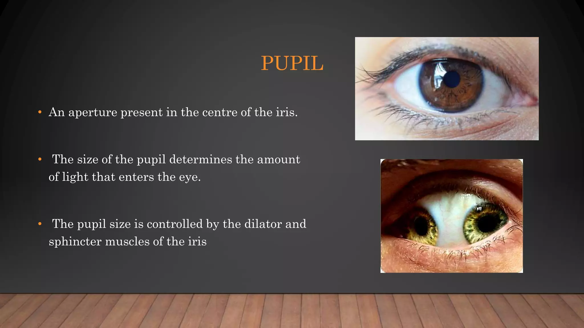
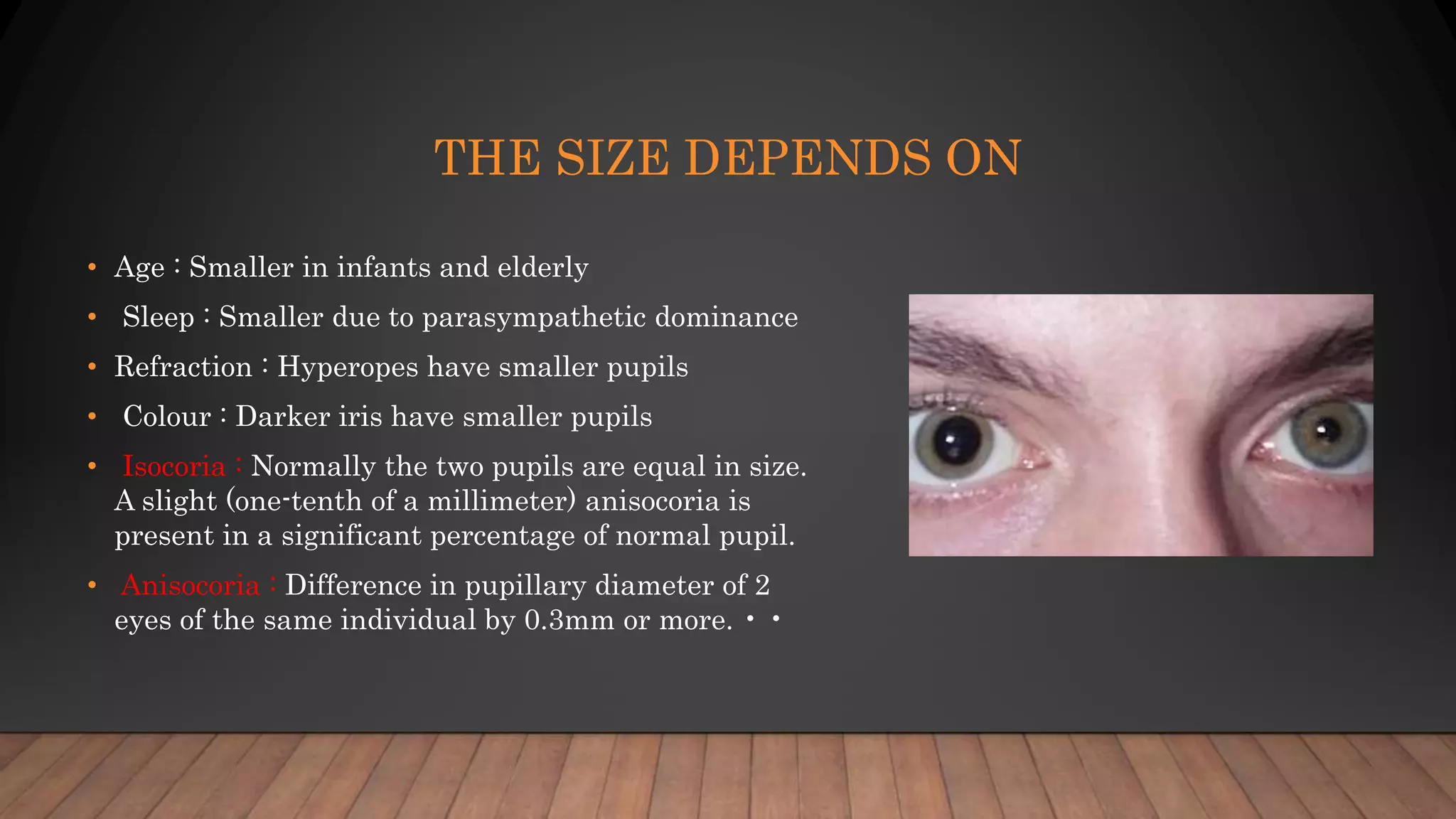
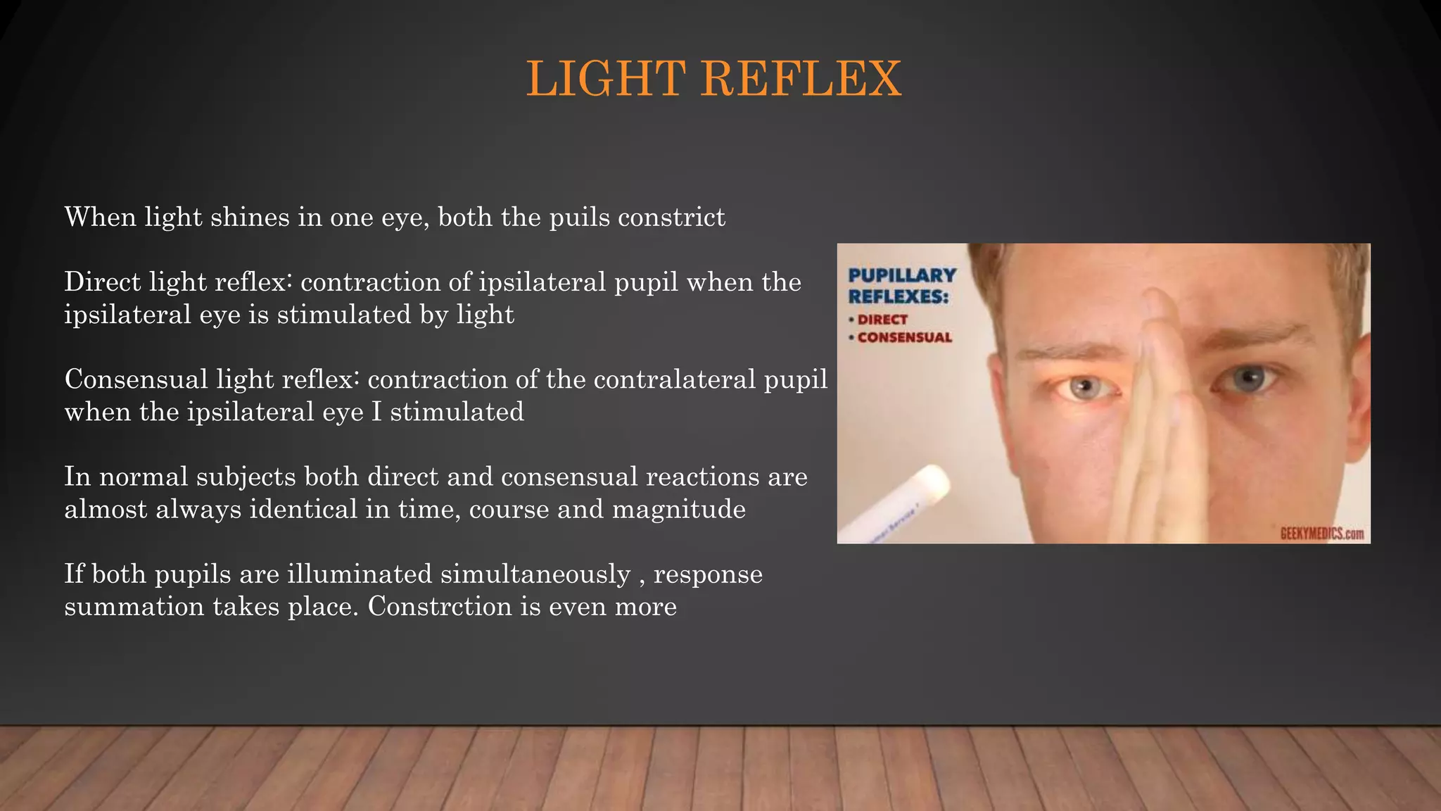
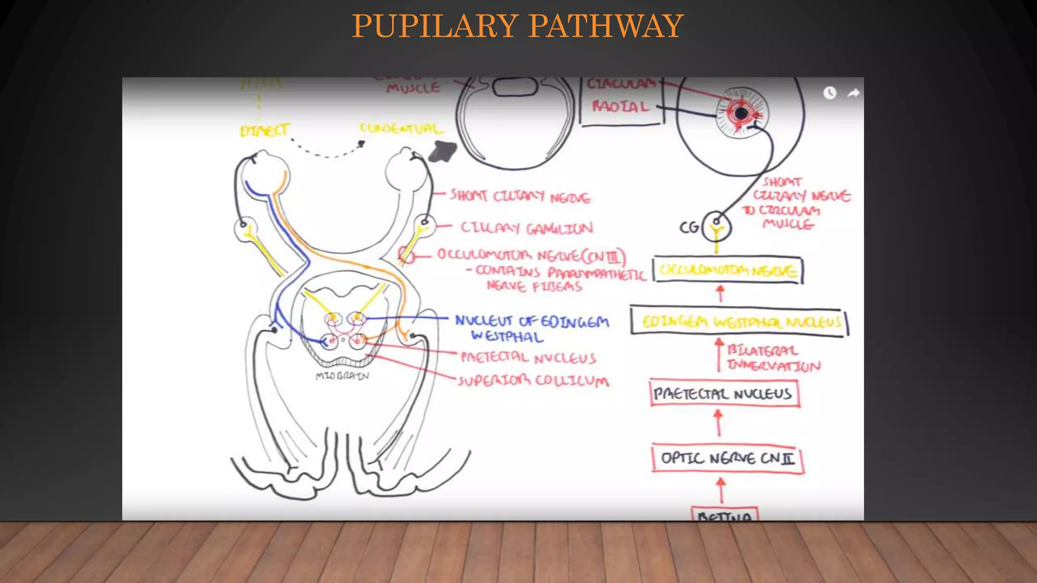
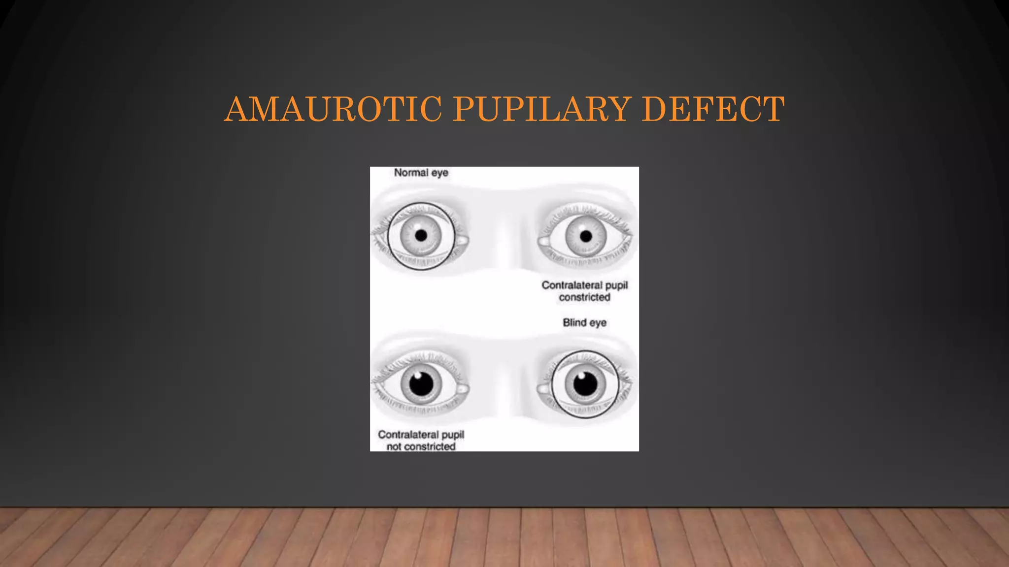
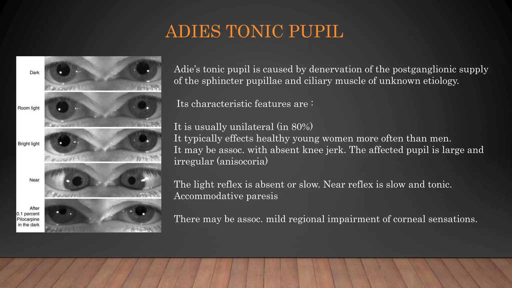
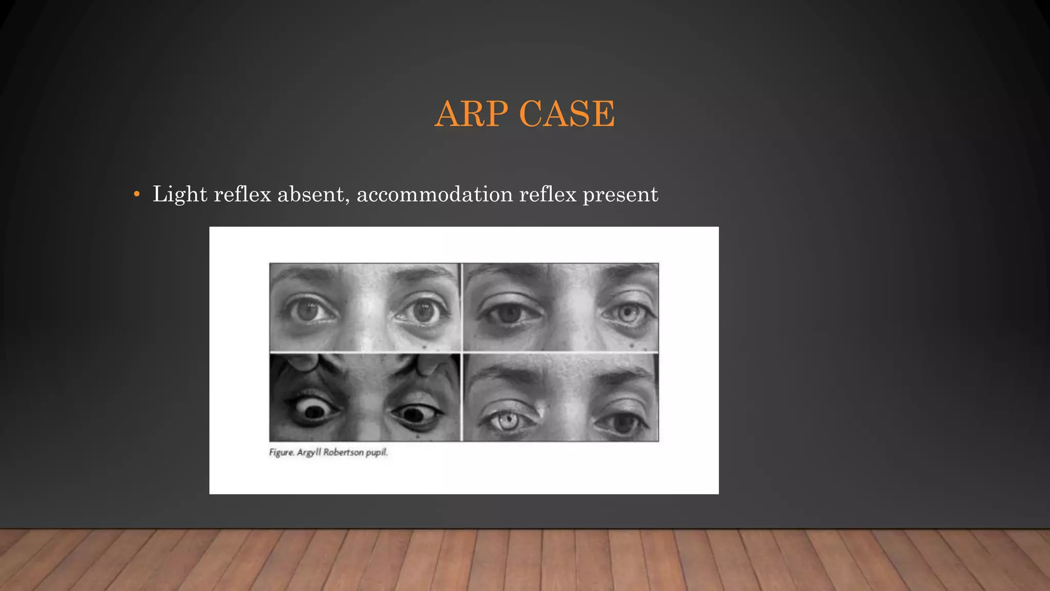
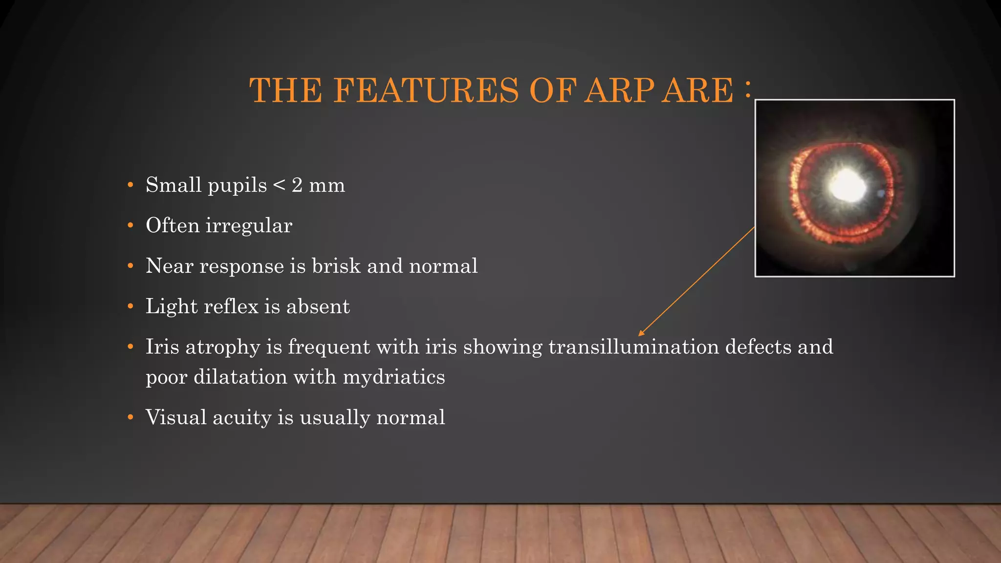
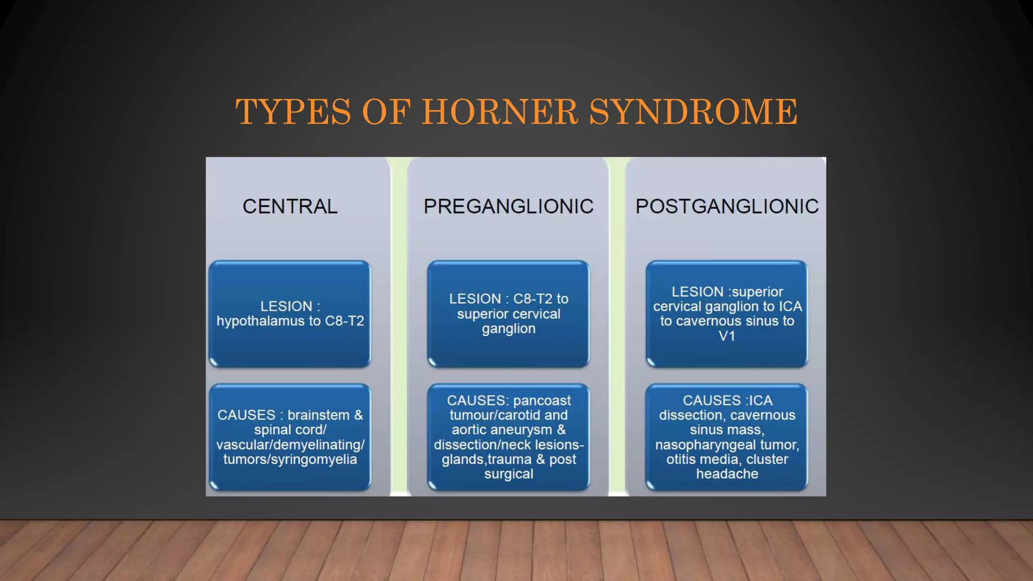
PUPILARY REFLEX
PATHWAYS AND
DISORDERS
DR. NAVEEN KUMAR CHALLA
PUPIL
• An aperture present in the centre of the iris.
• The size of the pupil determines the amount
of light that enters the eye.
• The pupil size is controlled by the dilator and
sphincter muscles of the iris
SIZE OF THE PUPIL
• • Normal pupil size varies from 2.5-4mm depending
upon the illumination. - Miotic pupils are less than
2mm - Mydriatic pupils are greater than 7mm
• • Diameter of the pupil size should be estimated in
light, using either a normal room light or a hand held
trans illuminator.
• • It should then be assessed in darkness using the
dimmest possible room light and then during near
stimulation using an accommodative target to
achieve maximum constriction of pupils.
SIZE OF THE PUPIL
• Owing to the refraction of the corneal surface the image of the pupil is
magnified by about 1/8 (an actual diameter of 3.5mm appears clinically to
be 4mm).
• It should then be assessed in darkness using the dimmest possible room
light and then during near stimulation using an accommodative target to
achieve maximum constriction of pupils.
THE SIZE DEPENDS ON
• Age : Smaller in infants and elderly
• Sleep : Smaller due to parasympathetic dominance
• Refraction : Hyperopes have smaller pupils
• Colour : Darker iris have smaller pupils
• Isocoria : Normally the two pupils are equal in size.
A slight (one-tenth of a millimeter) anisocoria is
present in a significant percentage of normal pupil.
• Anisocoria : Difference in pupillary diameter of 2
eyes of the same individual by 0.3mm or more. • •
FUNCTIONS OF THE PUPIL :
• It is the opening through which light enters the eye.
• It regulates the amount of light entering the eye by miosis in light and
mydriasis in darkness.
• It improves the visual acuity because it prevents the irregular refraction
by the periphery of the cornea and lens, and increases the depth of focus.
• It allows passage of aqueous humour from the posterior chamber to the
anterior chamber
SPHINCTER AND DILATOR PUPILLAE
• Muscle tissue of the iris consists of
• Sphincter pupillae
• Dilator pupillae
PUPILARY PATHWAYS
LIGHT REFLEX
When light shines in one eye, both the puils constrict
Direct light reflex: contraction of ipsilateral pupil when the
ipsilateral eye is stimulated by light
Consensual light reflex: contraction of the contralateral pupil
when the ipsilateral eye I stimulated
In normal subjects both direct and consensual reactions are
almost always identical in time, course and magnitude
If both pupils are illuminated simultaneously , response
summation takes place. Constrction is even more
PUPILARY PATHWAY
The light reflex is mediated by the retinal photoreceptors and subserved by four neurons
1. First (sensory): connects each retina with both pretectal nuclei in the midbrain at the level of the
superior colliculi. Impulses originating from the nasal retina are conducted by fibres which
decussate in the chiasm and pass up the opposite optic tract to terminate in the contralateral
pretectal nucleus. Impulses originating in the temporal retina are conducted by uncrossed fibres
(ipsilateral optic tract) which terminate in the ipsilateral pretectal nucleus.
2. Second (internuncial): connects each pretectal nucleus to both Edinger-Westphal nuclei.
3. Third (pre-ganglionic motor): connects the Edinger-Westphal nucleus to the ciliary ganglion. The
parasympathetic fibres pass through the oculomotor nerve, enter its inferior division and reach the
ciliary ganglion via the nerve to the inferior oblique muscle.
4. Fourth (post-ganglionic motor): leaves the ciliary ganglion and passes in the short ciliary nerves to
innervate the sphincter pupillae. The ciliary ganglion is located within the muscle cone, just
behind
the globe.
PUPILLARY REFLEXES
• LIGHT REFLEX
• CONVERGENCE REFLEX
• ACCOMMODATION REFLEX
Caused by a complete optic nerve or retinal lesion leading to total blindness on the affected side.
Characterized by the following :
The involved eye is completely blind (i.e no light perception) Absence of direct light reflex on the
affected side and absence of consenual light reflex on the normal side.
When the normal eye is stimulated, both pupils react normally. In diffuse illumination, both pupils
are equal in size. The near reflex is normal in both eyes
AFFERENT PATHWAY DEFECT
AMAUROTIC PUPILARY DEFECT
RELATIVE AFFERENT PATHWAY DEFECT
(RAPD) OR MARCUS GUNN PUPIL
• It is the response of a pupil to light in the presence of a relative afferent
pathway defect (RAPD).
• Caused by an incomplete optic nerve lesion or a severe retinal disease.
• It is best tested by ‘swinging flashlight test’
SWINGING FLASHLIGHT TEST
• To perform this test, a bright flashlight is shone on to one pupil and constriction is noted.
Then the flashlight is quickly moved to the contralateral pupil and response is noted.
• This swinging to and fro of flashlight is repeated several times while observing the pupillary
response.
• Normally, both pupils constrict equally and the pupil to which light is transferred remains
tightly constricted.
• In the presence of RAPD in one eye, the affected pupil will dilate (paradoxical response) when
the flashlight is moved from the normal eye to the abnormal eye.
• This response is called Marcus Gunn Pupil.
• It is the earliest indication of optic nerve disease even in the presence of a normal visual
acuity.
PUPILARY REFLEX CHARACTERSTERICS DUE TO
LESIONS
EFFERANT PUPILLARY DEFECTS
• Characterized by absence of both direct and consensual light reflex on the
affected side (say right eye) and presence of both direct and consensual
light reflex on the normal side (i.e left eye).
• On the affected side, near reflex is also absent and pupils remains fixed
and dilated.
COMMON CAUSES OF EFFERENT
PUPILARY DEFECTS
1. Brainstem lesions at the level of the superior colliculus and red nucleus. Lesions here
are often accompanied by long tract signs.
2. third nerve lesions. Compressive third nerve lesions classically have pupillary
involvement, although an efferent defect is not pathognomic of a compressive lesion, as
20% of microvascular palsies will affect the pupillary fibers.
3. Lesions of the ciliary ganglion or short ciliary nerves.
4. Iris damage secondary to previous surgery or grossly elevated IOP.
5. Drugs: inadvertent exposure to mydriatic agent such as atropine is a common cause of
fixed dilated pupil. Pilocarpine 1% will constrict a dilated pupil secondary to a neurological
lesion; it ll have no effect on dilated pupil caused by a mydriatic agent.
TONIC PUPIL
• Damage to the ciliary ganglion or short ciliary nerves produces tonic pupil
which is characterized by following :
• Reaction to light is absent and to near reflex is very slow and tonic.
• Accommodative paresis.
• The affected pupil is larger.
CAUSES OF TONIC PUPIL :
• Local tonic pupil • Viral ciliary ganglionitis (e.g Herpes zoster) • Orbital or
choroidal trauma or tumours • Blunt trauma to the globe may injure
branches of short ciliary nerves at the iris root.
• Neuropathic tonic pupil • Part of picture of peripheral neuropathy of
diabetes, alcoholism.
• Idiopathic tonic pupil with benign areflexia (Adie’s tonic pupil)
ADIES TONIC PUPIL
Adie’s tonic pupil is caused by denervation of the postganglionic supply
of the sphincter pupillae and ciliary muscle of unknown etiology.
Its characteristic features are :
It is usually unilateral (in 80%)
It typically effects healthy young women more often than men.
It may be assoc. with absent knee jerk. The affected pupil is large and
irregular (anisocoria)
The light reflex is absent or slow. Near reflex is slow and tonic.
Accommodative paresis
There may be assoc. mild regional impairment of corneal sensations.
ARGYLL ROBERTSON PUPIL ( ARP )
• Bilateral, miotic pupil with irregular margins and are asymmetrical.
• Light reflex is absent & accommodation reflex is retained.
• The lesion is at the pretectal area.
Commonly seen in tertiary neurosyphilis especially in Tabes.
Other causes include : - Diabetes - Wernicke’s encephalopathy - Encephalitis,
- Midbrain tumours - Hereditary neuropathies
ARP CASE
• Light reflex absent, accommodation reflex present
THE FEATURES OF ARP ARE :
• Small pupils < 2 mm
• Often irregular
• Near response is brisk and normal
• Light reflex is absent
• Iris atrophy is frequent with iris showing transillumination defects and
poor dilatation with mydriatics
• Visual acuity is usually normal
HORNER SYNDROME
HORNER SYNDROME
CLINICAL FEATURES OF HORNER’S
SYNDROME
• Ptosis – paralysis of muller muscle of upper eyelid ( usually 1-2mm )
• Upside down ptosis – weakness of the inferior tarsal muscle.
• Miosis – accentuated in dim light. Pupillary reactions are normal to light
and near Dilatation lag
• Heterochromia irides – seen in congenital Horner’s syndrome – the
pigment of iris stroma fail to develop
• Anhydrosis: Absence of sweat on the face
TYPES OF HORNER SYNDROME
CENTRAL HORNER’S SYNDROME • Assoc. with brain stem signs, signs of cervical cord
disease
PREGANGLIONIC HORNER’S SYNDROME • Lung or breast malignancies that has
spread to thoracic outlet • Facial anhydrosis • Brachial plexus palsies may be present
POSTGANGLIONIC HORNER’S SYNDROME • Ipsilateral vascular headache • h/o head
injury, or retroparotid trauma • Tumor of middle cranial fossa or cavernous sinus
• The near reflex, a synkinesis rather than a true reflex, is activated when
gaze is changed from a distant to a near target.
• It comprises Accommodation, Convergence And Miosis. Its called near triad
• Vision is not a prerequisite and there is no clinical condition in which the
light reflex is present but the near response absent.
• Although the final pathways for the near and light reflexes are identical
(i.e. 3rd nerve, ciliary ganglion, short ciliary nerves), the center for the near
reflex is ill defined
PUPILLARY REFLEX FOR NEAR
Fibers form Medial Rectus m.via III n
Mesencephalic n. of V n.
Convergence Center in Tectal
or Pre Tectal Region
EW Nucleus
Efferent fibers travel along III n.
Relay in Accessory Ganglion
Sphincter
Convergence reflex
PATHWAY OF ACCOMMODATION REFLEX
Retina
Optic nerve, Chaisma, Optic Tract
Lateral Geniculate Body
Striate Cortex
From the Para Striate Cortex
Occipito mesencephalic Tract and Pontine center
EW Nucleus
Via III n. to Sphincter Pupillae

PUPILARY REFLEX PATHWAYS.pptx

  • 1.
  • 2.
    PUPIL • An aperturepresent in the centre of the iris. • The size of the pupil determines the amount of light that enters the eye. • The pupil size is controlled by the dilator and sphincter muscles of the iris
  • 3.
    SIZE OF THEPUPIL • • Normal pupil size varies from 2.5-4mm depending upon the illumination. - Miotic pupils are less than 2mm - Mydriatic pupils are greater than 7mm • • Diameter of the pupil size should be estimated in light, using either a normal room light or a hand held trans illuminator. • • It should then be assessed in darkness using the dimmest possible room light and then during near stimulation using an accommodative target to achieve maximum constriction of pupils.
  • 4.
    SIZE OF THEPUPIL • Owing to the refraction of the corneal surface the image of the pupil is magnified by about 1/8 (an actual diameter of 3.5mm appears clinically to be 4mm). • It should then be assessed in darkness using the dimmest possible room light and then during near stimulation using an accommodative target to achieve maximum constriction of pupils.
  • 5.
    THE SIZE DEPENDSON • Age : Smaller in infants and elderly • Sleep : Smaller due to parasympathetic dominance • Refraction : Hyperopes have smaller pupils • Colour : Darker iris have smaller pupils • Isocoria : Normally the two pupils are equal in size. A slight (one-tenth of a millimeter) anisocoria is present in a significant percentage of normal pupil. • Anisocoria : Difference in pupillary diameter of 2 eyes of the same individual by 0.3mm or more. • •
  • 6.
    FUNCTIONS OF THEPUPIL : • It is the opening through which light enters the eye. • It regulates the amount of light entering the eye by miosis in light and mydriasis in darkness. • It improves the visual acuity because it prevents the irregular refraction by the periphery of the cornea and lens, and increases the depth of focus. • It allows passage of aqueous humour from the posterior chamber to the anterior chamber
  • 7.
    SPHINCTER AND DILATORPUPILLAE • Muscle tissue of the iris consists of • Sphincter pupillae • Dilator pupillae
  • 8.
  • 9.
    LIGHT REFLEX When lightshines in one eye, both the puils constrict Direct light reflex: contraction of ipsilateral pupil when the ipsilateral eye is stimulated by light Consensual light reflex: contraction of the contralateral pupil when the ipsilateral eye I stimulated In normal subjects both direct and consensual reactions are almost always identical in time, course and magnitude If both pupils are illuminated simultaneously , response summation takes place. Constrction is even more
  • 10.
  • 11.
    The light reflexis mediated by the retinal photoreceptors and subserved by four neurons 1. First (sensory): connects each retina with both pretectal nuclei in the midbrain at the level of the superior colliculi. Impulses originating from the nasal retina are conducted by fibres which decussate in the chiasm and pass up the opposite optic tract to terminate in the contralateral pretectal nucleus. Impulses originating in the temporal retina are conducted by uncrossed fibres (ipsilateral optic tract) which terminate in the ipsilateral pretectal nucleus. 2. Second (internuncial): connects each pretectal nucleus to both Edinger-Westphal nuclei. 3. Third (pre-ganglionic motor): connects the Edinger-Westphal nucleus to the ciliary ganglion. The parasympathetic fibres pass through the oculomotor nerve, enter its inferior division and reach the ciliary ganglion via the nerve to the inferior oblique muscle. 4. Fourth (post-ganglionic motor): leaves the ciliary ganglion and passes in the short ciliary nerves to innervate the sphincter pupillae. The ciliary ganglion is located within the muscle cone, just behind the globe.
  • 12.
    PUPILLARY REFLEXES • LIGHTREFLEX • CONVERGENCE REFLEX • ACCOMMODATION REFLEX
  • 13.
    Caused by acomplete optic nerve or retinal lesion leading to total blindness on the affected side. Characterized by the following : The involved eye is completely blind (i.e no light perception) Absence of direct light reflex on the affected side and absence of consenual light reflex on the normal side. When the normal eye is stimulated, both pupils react normally. In diffuse illumination, both pupils are equal in size. The near reflex is normal in both eyes AFFERENT PATHWAY DEFECT
  • 14.
  • 15.
    RELATIVE AFFERENT PATHWAYDEFECT (RAPD) OR MARCUS GUNN PUPIL • It is the response of a pupil to light in the presence of a relative afferent pathway defect (RAPD). • Caused by an incomplete optic nerve lesion or a severe retinal disease. • It is best tested by ‘swinging flashlight test’
  • 16.
    SWINGING FLASHLIGHT TEST •To perform this test, a bright flashlight is shone on to one pupil and constriction is noted. Then the flashlight is quickly moved to the contralateral pupil and response is noted. • This swinging to and fro of flashlight is repeated several times while observing the pupillary response. • Normally, both pupils constrict equally and the pupil to which light is transferred remains tightly constricted. • In the presence of RAPD in one eye, the affected pupil will dilate (paradoxical response) when the flashlight is moved from the normal eye to the abnormal eye. • This response is called Marcus Gunn Pupil. • It is the earliest indication of optic nerve disease even in the presence of a normal visual acuity.
  • 18.
  • 20.
    EFFERANT PUPILLARY DEFECTS •Characterized by absence of both direct and consensual light reflex on the affected side (say right eye) and presence of both direct and consensual light reflex on the normal side (i.e left eye). • On the affected side, near reflex is also absent and pupils remains fixed and dilated.
  • 21.
    COMMON CAUSES OFEFFERENT PUPILARY DEFECTS 1. Brainstem lesions at the level of the superior colliculus and red nucleus. Lesions here are often accompanied by long tract signs. 2. third nerve lesions. Compressive third nerve lesions classically have pupillary involvement, although an efferent defect is not pathognomic of a compressive lesion, as 20% of microvascular palsies will affect the pupillary fibers. 3. Lesions of the ciliary ganglion or short ciliary nerves. 4. Iris damage secondary to previous surgery or grossly elevated IOP. 5. Drugs: inadvertent exposure to mydriatic agent such as atropine is a common cause of fixed dilated pupil. Pilocarpine 1% will constrict a dilated pupil secondary to a neurological lesion; it ll have no effect on dilated pupil caused by a mydriatic agent.
  • 22.
    TONIC PUPIL • Damageto the ciliary ganglion or short ciliary nerves produces tonic pupil which is characterized by following : • Reaction to light is absent and to near reflex is very slow and tonic. • Accommodative paresis. • The affected pupil is larger.
  • 23.
    CAUSES OF TONICPUPIL : • Local tonic pupil • Viral ciliary ganglionitis (e.g Herpes zoster) • Orbital or choroidal trauma or tumours • Blunt trauma to the globe may injure branches of short ciliary nerves at the iris root. • Neuropathic tonic pupil • Part of picture of peripheral neuropathy of diabetes, alcoholism. • Idiopathic tonic pupil with benign areflexia (Adie’s tonic pupil)
  • 24.
    ADIES TONIC PUPIL Adie’stonic pupil is caused by denervation of the postganglionic supply of the sphincter pupillae and ciliary muscle of unknown etiology. Its characteristic features are : It is usually unilateral (in 80%) It typically effects healthy young women more often than men. It may be assoc. with absent knee jerk. The affected pupil is large and irregular (anisocoria) The light reflex is absent or slow. Near reflex is slow and tonic. Accommodative paresis There may be assoc. mild regional impairment of corneal sensations.
  • 25.
    ARGYLL ROBERTSON PUPIL( ARP ) • Bilateral, miotic pupil with irregular margins and are asymmetrical. • Light reflex is absent & accommodation reflex is retained. • The lesion is at the pretectal area. Commonly seen in tertiary neurosyphilis especially in Tabes. Other causes include : - Diabetes - Wernicke’s encephalopathy - Encephalitis, - Midbrain tumours - Hereditary neuropathies
  • 26.
    ARP CASE • Lightreflex absent, accommodation reflex present
  • 27.
    THE FEATURES OFARP ARE : • Small pupils < 2 mm • Often irregular • Near response is brisk and normal • Light reflex is absent • Iris atrophy is frequent with iris showing transillumination defects and poor dilatation with mydriatics • Visual acuity is usually normal
  • 28.
  • 29.
  • 30.
    CLINICAL FEATURES OFHORNER’S SYNDROME • Ptosis – paralysis of muller muscle of upper eyelid ( usually 1-2mm ) • Upside down ptosis – weakness of the inferior tarsal muscle. • Miosis – accentuated in dim light. Pupillary reactions are normal to light and near Dilatation lag • Heterochromia irides – seen in congenital Horner’s syndrome – the pigment of iris stroma fail to develop • Anhydrosis: Absence of sweat on the face
  • 31.
  • 32.
    CENTRAL HORNER’S SYNDROME• Assoc. with brain stem signs, signs of cervical cord disease PREGANGLIONIC HORNER’S SYNDROME • Lung or breast malignancies that has spread to thoracic outlet • Facial anhydrosis • Brachial plexus palsies may be present POSTGANGLIONIC HORNER’S SYNDROME • Ipsilateral vascular headache • h/o head injury, or retroparotid trauma • Tumor of middle cranial fossa or cavernous sinus
  • 33.
    • The nearreflex, a synkinesis rather than a true reflex, is activated when gaze is changed from a distant to a near target. • It comprises Accommodation, Convergence And Miosis. Its called near triad • Vision is not a prerequisite and there is no clinical condition in which the light reflex is present but the near response absent. • Although the final pathways for the near and light reflexes are identical (i.e. 3rd nerve, ciliary ganglion, short ciliary nerves), the center for the near reflex is ill defined PUPILLARY REFLEX FOR NEAR
  • 34.
    Fibers form MedialRectus m.via III n Mesencephalic n. of V n. Convergence Center in Tectal or Pre Tectal Region EW Nucleus Efferent fibers travel along III n. Relay in Accessory Ganglion Sphincter Convergence reflex
  • 35.
    PATHWAY OF ACCOMMODATIONREFLEX Retina Optic nerve, Chaisma, Optic Tract Lateral Geniculate Body Striate Cortex From the Para Striate Cortex Occipito mesencephalic Tract and Pontine center EW Nucleus Via III n. to Sphincter Pupillae