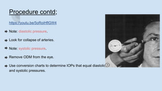
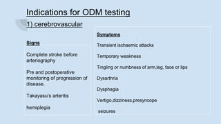
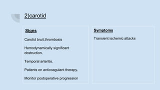
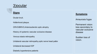
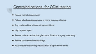
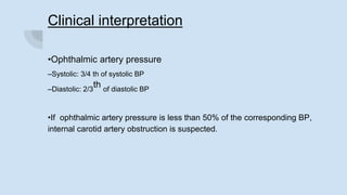
Ophthalmodynamometry.
ANUJA DHAKAL
BOVS PHASE II,NEH
layout
Introduction
History
Anatomy of vasculature
Principle
Methods and instrumentation
Procedure & comparison
precautions
Clinical significance
Indications & contraindications
Clinical interpretation
INTRODUCTION
Ophthalmodynamometry(ODM) is a clinical procedure which is
classically assumed to measure the pressure in the ipsilateral ophthalmic
artery so that the inferences can be derived about the patency of the
internal carotid artery.
Ophthalmodynamometry can be used as a screening procedure to detect
carotid artery occlusive diseases and,thus, can be important in the
prevention of cerebrovascular accidents.
HISTORY
Bailliart (1917)-introduced the first instrument for assessing the carotid
artery function(bailliart ophthalmodynamometer).
Baurmann (1936)-noted that the ODM reading was lowered in a patient
who had undergone ligation of the ipsilateral carotid artery.
Trotot (1944)-first diagnosis of thrombus of ICA ,based on ODM testing-
noted that the ODM reading on the ipsilateral side was significantly
lower than other side without disease.
Anatomy of carotid Artery.
In ophthalmodynamometry we are concerned
with internal carotid artery.
The internal carotid artery doesn’t give off any
branches in its cervical portion ,but several
small branches arise from the ICA in its
petrous portion.
The ophthalmic artery is the first major branch
of the intracranial carotid artery after it passes
through the cavernous sinus.
Caroticotympanic artery
Pterygoid artery
Cavernous artery
Hypophyseal artery
Meningeal artery
Ophthalmic artery
Anterior cerebral artery
Middle cerebral artery
Posterior communicating artery
Anterior choroidal artery
Central retinal artery
Short posterior ciliary artery
Long posterior ciliary artery
Anterior ciliary artery
Muscular arteries
Lacrimal artery
Supraorbital artery
Post & ant ethmoidal arteries
Medial palpebral artery
Supratrochlear artery
Dorsal nasal artery
Branches of ICA Branches of ophthalmic
artery
Ocular
branche
s
Orbital
branches
One of the smallest branches of the
ophthalmic artery, but its first,is the central
retinal artery.
while the clinician is viewing the
hemodynamics of the central retinal artery
during ophthalmodynamometry ,inferences
can be made about the physiologic
functioning of the ophthalmic artery ,even
though significant collateral formation may
occur with other vessels of the face and
head.
PRINCIPLE
Ophthalmodynamometry is based on the principle that due to direct anatomical
communication of internal carotid artery with ophthalmic artery ,if the blood flow through
the ICA is decreased then the blood flow through ophthalmic artery artery is also
decreased.Thus, measures the relative ophthalmic artery pressure.
More than 90 % of CA occlusion occur below the origin of ophthalmic artery, thus affecting
the blood flow into the ophthalmic artery and retinal arterioles.
Pressure is exerted on the sclera to increase IOP to the point where central retinal artery
collapses and it is supplied by greater blood through ophthalmic artery.
DR.BAILLIART OPHTHALMODYNAMOMETER
Methods and Instrumentation
There are two classic method used to raise the intraocular pressure to the
point at which it causes pulsation and subsequent collapse of the central
retinal artery .
1)Compression ophthalmodynamometry
2)Suction ophthalmodynamometry
1)Compression ophthalmodynamometry
It is the oldest non invasive method in use.
It uses an externally applied graded compression on the globe,the force being
created by a coiled spring .
As the external force is measured in grams of pressure,conversion scales is used
to transpose pressure into millimeters of mercury,or the intraocular pressure
equivalent to the external force.
The two most common method of compression ophthalmodynamometry are:
A) DIAL OPHTHALMODYNAMOMETRY
B) LINEAR OPHTHALMODYNAMOMETRY
a)DIAL ODM
The dial ODM consists of a scale on a dial with two arrows ,mounted on a
post containing the spring.
The drive arrow is connected to the spring,the second passenger arrow is
pushed by the notched drive arrow.
b)Linear ODM
The linear type of compression ophthalmodynamometry consists of a hollow
outer sleeve with a central post that has graduated markings.
Pressure on the central post causes the outer sleeve to move along the scale
so that the readings can be taken.
Procedure for compression ODM.
Procedure for compression ODM
Patients pupil must be dilated.(usually cycloplegic & mydriatics combination).
Explain procedure to the patient.
Anesthetize the eyes.
Clean and sterilize the ophthalmodynamometer.
Indicate appropriate fixation point to the patient.
Place footplate of the ODM temporally,close to the equator.
Visualize the optic nerve head with ophthalmoscope(either direct or indirect).
Gradually increase the pressure(steady rate).
Look for venous pulsations from the ophthalmoscope.
Procedure contd;
https://youtu.be/5oRoiHflGW4
Note: diastolic pressure.
Look for collapse of arteries.
Note: systolic pressure.
Remove ODM from the eye.
Use conversion charts to determine IOPs that equal diastolic
and systolic pressures.
2)suction ophthalmodynamometry
This is the second method in which negative pressure is applied in the form
of suction to increase intraocular pressure sufficiently to cause pulsation and
subsequent collapse of the central retinal artery.
This procedure also has the same principle of physical basis as compression
ODM,but here,a scleral vacuum suction cup is placed in a perilimbal position
and a negative pressure is applied.
Procedure for suction ophthalmodynamometry
Procedure contd;
Patient preparation is similar as compression technique.
The scleral cups are placed instead at the lateral side around the area of equator .
Central retinal artery is viewed through ophthalmoscopy be it direct or indirect.
Negative pressure is provided by using a foot pedal or the hand.
Noting of both diastolic and systolic pressure is similar to the earlier one.
Instrument should be removed immediately from the globe to reduce the length of
time and the effect of external pressure on fluids of eye.
Advantages of suction ODM over compression ODM
The suction method uses all of its force to compress the unlike compression ODM
which displaces the eyeball nasally.
Examiner doesn’t need to adjust the position unlike compression ODM.
Less painful and comfortable than compression method.
Sequelea of ODM:
Subconjuctival haemorrhage over the lateral rectus at the site of the footplate.
Some have noted central scotoma which persists till months.
(https://www.ncbi.nlm.nih.gov/pmc/articles/PMC2642107/bin/jnma00683-0064.tif)
Might result in retinal detachment due to excessive suction pressure.
Doppler ophthalmodynamometry
This technique described its usefulness in patients with
opaque media preventing visualization of the central retinal
artery.
Instead of an ODM, a miniature stethoscope is used but
instead of a diaphragm ,an ultrasound probe that transmits
and receives the signal,and then transforms it into audible
sound.
The probe is placed in the perilimbal area and the pressure
is applied until the pulsations of the central retinal artery is
heard.
The finding and interpretation of doppler
ophthalmodynamometry is not clear yet.
Precautions to be taken
A cooperative patient will make the test more easier and accurate therefore
adequate explanation is necessary.
The instrumentation should be correct( perpendicular).
The footplate can inadvertently slip,causing a corneal abrasion .
The fixation and view of blood vessels may be altered due to displaced globe.
Venous pulsation shouldn’t be constructed as a diastolic pressure.
Patient with bradycardia and cardiac arrhythmia have spurious determinations of
both systolic and diastolic end points.
Clinical significance:
It is significant for detecting presence of carotid artery insufficiency,which is the
major cause :
Cerebral vascular accident(stroke).
Central retinal artery occlusions.
In patient with reduced ophthalmic artery pressures,and arteriographically
significant carotid artery disease,16% will develop a stroke.
(Busuttil RW, Baker JD, Davidson RK, Machleder HI. Carotid artery stenosis - hemodynamic
significance and clinical course. JAMA. 1981 Apr 10;245(14):1438–1441)
contd;
Ophthalmodynamometry is mandatory in patients presenting with amaurosis fugax.
Results that indicate higher retinal arterial pressure in diabetes is at a high risk of
developing
retinopathy.(https://www.nejm.org/doi/full/10.1056/NEJM198003203021201)
About 2/3rd of patients with documented carotid occlusive disease present with
ocular symptoms and if patient have symptomatic carotid artery disease,as
many as 88% of them will complain of ocular
symptoms.(https://doi.org/10.1016/S0002-9394(14)76496-7)
Indications for ODM testing
1) cerebrovascular
Signs
Complete stroke before
arteriography
Pre and postoperative
monitoring of progression of
disease.
Takayasu’s arteritis
hemiplegia
Symptoms
Transient ischaemic attacks
Temporary weakness
Tingling or numbness of arm,leg, face or lips
Dysarthria
Dysphagia
Vertigo,dizziness,presyncope
seizures
2)carotid
Signs
Carotid bruit,thrombosis
Hemodynamically significant
obstruction.
Temporal arteritis.
Patients on anticoagulant therapy.
Monitor postoperative progression
Symptoms
Transient ischemic attacks
3)ocular
Signs
Ocular bruit.
Hollenhorst plaque.
CRVO/BRVO,Arteriosclerotic optic atrophy
History of systemic vascular occlusive disease
Venous stasis retinopathy
Unilateral vascular retinopathy,optic nerve head pallor
Unilateral decreased IOP
Diabetic,hypertensive patients
Symptoms
Amaurosis fugax
Permanent vision
loss secondary to
vascular occlusive
disease
Sudden loss of
vision.
Contraindications for ODM testing
Recent retinal detachment.
Patient who has glaucoma or is prone to acute attacks.
Any ocular,orbital inflammatory conditions.
High myopic eyes.
Recent cataract extraction,glaucoma filtration surgery,iridectomy.
Retinal or vitreous haemorrhage.
Hazy media obstructing visualization of optic nerve head
Clinical interpretation
•Ophthalmic artery pressure
–Systolic: 3/4 th of systolic BP
–Diastolic: 2/3
th of diastolic BP
•If ophthalmic artery pressure is less than 50% of the corresponding BP,
internal carotid artery obstruction is suspected.
A difference of 10% between diastolic findings or a difference of 15%
between systolic findings between two eyes could be suspected,whereas
15% difference in diastolic & 20% in systolic findings could be taken as
suspect.
(https://doi.org/10.1016/S0025-7125(16)34244-4)
The suspected patient’s medical history and other examinations must be
considered before testing for carotid artery occlusive disease.
Although interpretation may be a shade difficult in some patients,it yields
important clinical data that can be used as a screening test before the
invasive arteriography procedure.
Thank you !
Stay safe.

Ophthalmodynamometry.

  • 1.
  • 2.
    layout Introduction History Anatomy of vasculature Principle Methodsand instrumentation Procedure & comparison precautions Clinical significance Indications & contraindications Clinical interpretation
  • 3.
    INTRODUCTION Ophthalmodynamometry(ODM) is aclinical procedure which is classically assumed to measure the pressure in the ipsilateral ophthalmic artery so that the inferences can be derived about the patency of the internal carotid artery. Ophthalmodynamometry can be used as a screening procedure to detect carotid artery occlusive diseases and,thus, can be important in the prevention of cerebrovascular accidents.
  • 4.
    HISTORY Bailliart (1917)-introduced thefirst instrument for assessing the carotid artery function(bailliart ophthalmodynamometer). Baurmann (1936)-noted that the ODM reading was lowered in a patient who had undergone ligation of the ipsilateral carotid artery. Trotot (1944)-first diagnosis of thrombus of ICA ,based on ODM testing- noted that the ODM reading on the ipsilateral side was significantly lower than other side without disease.
  • 5.
    Anatomy of carotidArtery. In ophthalmodynamometry we are concerned with internal carotid artery. The internal carotid artery doesn’t give off any branches in its cervical portion ,but several small branches arise from the ICA in its petrous portion. The ophthalmic artery is the first major branch of the intracranial carotid artery after it passes through the cavernous sinus.
  • 6.
    Caroticotympanic artery Pterygoid artery Cavernousartery Hypophyseal artery Meningeal artery Ophthalmic artery Anterior cerebral artery Middle cerebral artery Posterior communicating artery Anterior choroidal artery Central retinal artery Short posterior ciliary artery Long posterior ciliary artery Anterior ciliary artery Muscular arteries Lacrimal artery Supraorbital artery Post & ant ethmoidal arteries Medial palpebral artery Supratrochlear artery Dorsal nasal artery Branches of ICA Branches of ophthalmic artery Ocular branche s Orbital branches
  • 7.
    One of thesmallest branches of the ophthalmic artery, but its first,is the central retinal artery. while the clinician is viewing the hemodynamics of the central retinal artery during ophthalmodynamometry ,inferences can be made about the physiologic functioning of the ophthalmic artery ,even though significant collateral formation may occur with other vessels of the face and head.
  • 8.
    PRINCIPLE Ophthalmodynamometry is basedon the principle that due to direct anatomical communication of internal carotid artery with ophthalmic artery ,if the blood flow through the ICA is decreased then the blood flow through ophthalmic artery artery is also decreased.Thus, measures the relative ophthalmic artery pressure. More than 90 % of CA occlusion occur below the origin of ophthalmic artery, thus affecting the blood flow into the ophthalmic artery and retinal arterioles. Pressure is exerted on the sclera to increase IOP to the point where central retinal artery collapses and it is supplied by greater blood through ophthalmic artery.
  • 9.
  • 10.
    Methods and Instrumentation Thereare two classic method used to raise the intraocular pressure to the point at which it causes pulsation and subsequent collapse of the central retinal artery . 1)Compression ophthalmodynamometry 2)Suction ophthalmodynamometry
  • 11.
    1)Compression ophthalmodynamometry It isthe oldest non invasive method in use. It uses an externally applied graded compression on the globe,the force being created by a coiled spring . As the external force is measured in grams of pressure,conversion scales is used to transpose pressure into millimeters of mercury,or the intraocular pressure equivalent to the external force. The two most common method of compression ophthalmodynamometry are: A) DIAL OPHTHALMODYNAMOMETRY B) LINEAR OPHTHALMODYNAMOMETRY
  • 12.
    a)DIAL ODM The dialODM consists of a scale on a dial with two arrows ,mounted on a post containing the spring. The drive arrow is connected to the spring,the second passenger arrow is pushed by the notched drive arrow.
  • 13.
    b)Linear ODM The lineartype of compression ophthalmodynamometry consists of a hollow outer sleeve with a central post that has graduated markings. Pressure on the central post causes the outer sleeve to move along the scale so that the readings can be taken.
  • 14.
  • 15.
    Procedure for compressionODM Patients pupil must be dilated.(usually cycloplegic & mydriatics combination). Explain procedure to the patient. Anesthetize the eyes. Clean and sterilize the ophthalmodynamometer. Indicate appropriate fixation point to the patient. Place footplate of the ODM temporally,close to the equator. Visualize the optic nerve head with ophthalmoscope(either direct or indirect). Gradually increase the pressure(steady rate). Look for venous pulsations from the ophthalmoscope.
  • 16.
    Procedure contd; https://youtu.be/5oRoiHflGW4 Note: diastolicpressure. Look for collapse of arteries. Note: systolic pressure. Remove ODM from the eye. Use conversion charts to determine IOPs that equal diastolic and systolic pressures.
  • 17.
    2)suction ophthalmodynamometry This isthe second method in which negative pressure is applied in the form of suction to increase intraocular pressure sufficiently to cause pulsation and subsequent collapse of the central retinal artery. This procedure also has the same principle of physical basis as compression ODM,but here,a scleral vacuum suction cup is placed in a perilimbal position and a negative pressure is applied.
  • 18.
    Procedure for suctionophthalmodynamometry
  • 19.
    Procedure contd; Patient preparationis similar as compression technique. The scleral cups are placed instead at the lateral side around the area of equator . Central retinal artery is viewed through ophthalmoscopy be it direct or indirect. Negative pressure is provided by using a foot pedal or the hand. Noting of both diastolic and systolic pressure is similar to the earlier one. Instrument should be removed immediately from the globe to reduce the length of time and the effect of external pressure on fluids of eye.
  • 20.
    Advantages of suctionODM over compression ODM The suction method uses all of its force to compress the unlike compression ODM which displaces the eyeball nasally. Examiner doesn’t need to adjust the position unlike compression ODM. Less painful and comfortable than compression method. Sequelea of ODM: Subconjuctival haemorrhage over the lateral rectus at the site of the footplate. Some have noted central scotoma which persists till months. (https://www.ncbi.nlm.nih.gov/pmc/articles/PMC2642107/bin/jnma00683-0064.tif) Might result in retinal detachment due to excessive suction pressure.
  • 21.
    Doppler ophthalmodynamometry This techniquedescribed its usefulness in patients with opaque media preventing visualization of the central retinal artery. Instead of an ODM, a miniature stethoscope is used but instead of a diaphragm ,an ultrasound probe that transmits and receives the signal,and then transforms it into audible sound. The probe is placed in the perilimbal area and the pressure is applied until the pulsations of the central retinal artery is heard. The finding and interpretation of doppler ophthalmodynamometry is not clear yet.
  • 22.
    Precautions to betaken A cooperative patient will make the test more easier and accurate therefore adequate explanation is necessary. The instrumentation should be correct( perpendicular). The footplate can inadvertently slip,causing a corneal abrasion . The fixation and view of blood vessels may be altered due to displaced globe. Venous pulsation shouldn’t be constructed as a diastolic pressure. Patient with bradycardia and cardiac arrhythmia have spurious determinations of both systolic and diastolic end points.
  • 23.
    Clinical significance: It issignificant for detecting presence of carotid artery insufficiency,which is the major cause : Cerebral vascular accident(stroke). Central retinal artery occlusions. In patient with reduced ophthalmic artery pressures,and arteriographically significant carotid artery disease,16% will develop a stroke. (Busuttil RW, Baker JD, Davidson RK, Machleder HI. Carotid artery stenosis - hemodynamic significance and clinical course. JAMA. 1981 Apr 10;245(14):1438–1441)
  • 24.
    contd; Ophthalmodynamometry is mandatoryin patients presenting with amaurosis fugax. Results that indicate higher retinal arterial pressure in diabetes is at a high risk of developing retinopathy.(https://www.nejm.org/doi/full/10.1056/NEJM198003203021201) About 2/3rd of patients with documented carotid occlusive disease present with ocular symptoms and if patient have symptomatic carotid artery disease,as many as 88% of them will complain of ocular symptoms.(https://doi.org/10.1016/S0002-9394(14)76496-7)
  • 25.
    Indications for ODMtesting 1) cerebrovascular Signs Complete stroke before arteriography Pre and postoperative monitoring of progression of disease. Takayasu’s arteritis hemiplegia Symptoms Transient ischaemic attacks Temporary weakness Tingling or numbness of arm,leg, face or lips Dysarthria Dysphagia Vertigo,dizziness,presyncope seizures
  • 26.
    2)carotid Signs Carotid bruit,thrombosis Hemodynamically significant obstruction. Temporalarteritis. Patients on anticoagulant therapy. Monitor postoperative progression Symptoms Transient ischemic attacks
  • 27.
    3)ocular Signs Ocular bruit. Hollenhorst plaque. CRVO/BRVO,Arterioscleroticoptic atrophy History of systemic vascular occlusive disease Venous stasis retinopathy Unilateral vascular retinopathy,optic nerve head pallor Unilateral decreased IOP Diabetic,hypertensive patients Symptoms Amaurosis fugax Permanent vision loss secondary to vascular occlusive disease Sudden loss of vision.
  • 28.
    Contraindications for ODMtesting Recent retinal detachment. Patient who has glaucoma or is prone to acute attacks. Any ocular,orbital inflammatory conditions. High myopic eyes. Recent cataract extraction,glaucoma filtration surgery,iridectomy. Retinal or vitreous haemorrhage. Hazy media obstructing visualization of optic nerve head
  • 29.
    Clinical interpretation •Ophthalmic arterypressure –Systolic: 3/4 th of systolic BP –Diastolic: 2/3 th of diastolic BP •If ophthalmic artery pressure is less than 50% of the corresponding BP, internal carotid artery obstruction is suspected.
  • 30.
    A difference of10% between diastolic findings or a difference of 15% between systolic findings between two eyes could be suspected,whereas 15% difference in diastolic & 20% in systolic findings could be taken as suspect. (https://doi.org/10.1016/S0025-7125(16)34244-4) The suspected patient’s medical history and other examinations must be considered before testing for carotid artery occlusive disease. Although interpretation may be a shade difficult in some patients,it yields important clinical data that can be used as a screening test before the invasive arteriography procedure.
  • 31.