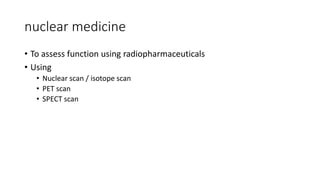
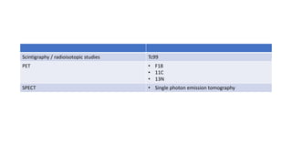
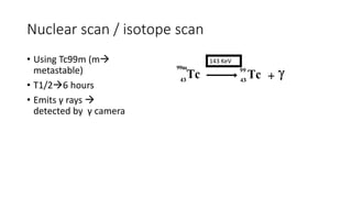
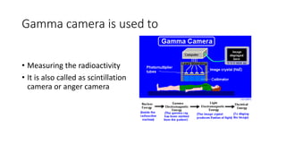
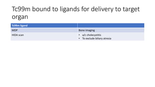
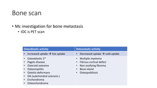
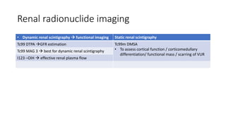
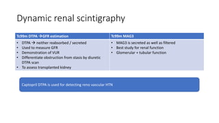
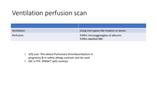
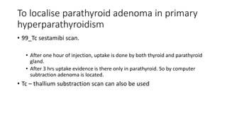
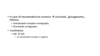
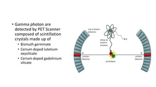
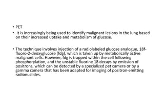
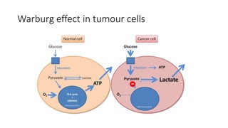
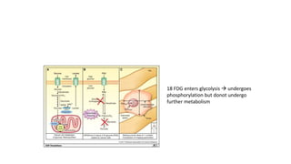
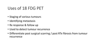
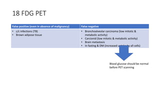
Nuclear medicine
nuclear medicine
• To assess function using radiopharmaceuticals
• Using
• Nuclear scan / isotope scan
• PET scan
• SPECT scan
Scintigraphy / radioisotopic studies Tc99
PET • F18
• 11C
• 13N
SPECT • Single photon emission tomography
Nuclear scan / isotope scan
• Using Tc99m (m
metastable)
• T1/26 hours
• Emits γ rays 
detected by γ camera
143 KeV
Gamma camera is used to
• Measuring the radioactivity
• It is also called as scintillation
camera or anger camera
Gamma camera is uded to detect
radioactivity
Tc99m bound to ligands for delivery to target
organ
Tc99m ligand
MDP Bone imaging
HIDA scan • a/c cholecystitis
• To exclude biliary atresia
Bone scan
• Mc investigation for bone metastasis
• IOC is PET scan
Osteoblastic activity Osteoclastic activity
• Increased uptake  hot uptake • Decreased uptake  cold uptake
• Osteoblastic 2*
• Pagets disease
• Osteroid osteoma
• Osteomyelitis
• Osteitis deformans
• OA (subchondral sclerosis )
• Enchondroma
• Osteochondroma
• Multiple myeloma
• Fibrous cortical defect
• Non ossifying fibroma
• Bone island
• Osteopoikilosis
Abnormal bone scan
Abnormal bone scane
Super scan • In diffuse metastatic ds of bone
• In diffuse metabolic ds of bone
• Renal failure
OM • Increased uptake in all 3 phases of bone scan
Mickey mouse sign • Pagets ds
Hot bone scan • Increased uptake in site where there is increased
uptake - neoplasm trauma infection avscular
necrosis
Cold scan • In multiple myeloma
• Histiocytosis
• Neuroblastoma
• RCC
• Thyroid carcinoma
Super scan
• Increased uptake in skeleton
• No background radiotracer activity
• But kidneys are not visualised
• In diffuse metastatic ds of bone
• In diffuse metabolic ds of bone
• Renal failure
Mickey mouse sign in pagets disease
• d/t increased uptake in
pedicles & spinous process
Big black beard / linoln sign
in pagets disease
• Increased condylar to condylar uptake in bone
scan
For infections of bone / osteomyelitis
• Gallium scan
• Indium labelled WBC scan (except in case of respiratory tract
infections )
• Increased uptake in all 3 phases
Nuclear medicine in heart
Myocardial perfusion studies • Thallium 201 (MC used)
• Tc99m tetrophosmin
• Tc99m sestamibi (MIBI scan)area of infarct
appears cold
Myocardial infarct imaging • Tc99 pyrophosphate (area of infarct appears hot)
MUGA scan • Tc99m labelled RBC are used
Myocardial perfusion scan
• SPECT scan is used To assess blood flow (perfusion )
• Using thallium 201 / Tc99 labelled tetrofosmin /Tc 99m labelled sestamibi
• Low perfused area appears cold (d/t decreased uptake of radioactive
material)
Stress thallium study
• Stress is given by physiologically / with dobutamine
• At peak of stress  inject thallium
• Uptake by normal myocardium with normal blood flow  hot areas
• Ischemic myocardium  cold areas
Reversible ischemia Ischemia on exercise but not on rest
Irreversible ischemia Ischemia both @ rest & execise
Myocardial infarct scintigraphy
• Using Tc pyrophosphate
• Uptake by infarcted myocardium
• Infarct  hot areas
• Blood flow is not assessed by this scan
• Gold standard for myocardial viability  PET scan
• Best investigation to differentiate b/w myocardial scar & hibernating
myocardium  PET
Cardiac PET uses
Myocardial metabolism FDG PET scan
Myocardial perfusion • radioactive ammonia (NH3)
• H20
• Rubidium 82
Rubidium 82  non cyclotron isotope used in PET scan
MUGA scan
• method for assessment of LV functionmeasure ejection fraction
• ECHO can also be used
• most accurate is MRI
• 99mTc-labelled albumin or red cells that are uniformly distributed
throughout the blood volume /
Tc 99m labelled RBC is used for
• Haemangioma
• GI bleeding
• Splenic disease
• Cardiac ventriculography
Nuclear medicine in GIT
Tc99m labelled RBC • Minimal lower GI Bleeding
• Sensitive test (0.1ml/min)
Tc99m pertechnate • Pertechnate is taken up by gastric mucosa  to
detect ectopic gastric mucosa
Tc99m sulphur colloid • Taken by RE cell
• To detect Focal nodular hyperaplasia (FNH) also
used in accessory spleen & abscess
• IOC for upper GI bleed  endoscopy
• IOC in lower GI bleed  mesenteric angiography
/ Tc99 RBC
Lower GI bleed
Mesenteric angiography Tc 99 RBC
Can detect 0.5 ml/min blood loss Can detect 0.1 ml/min blood loss
Uses of Tc99m labelled pertechnate
• Thyroid scintigraphy
• Salivary gland scintigraphy
• Meckels diverticulum
Renal radionuclide imaging
• Dynamic renal scintigraphy  functional imaging Static renal scintigraphy
Tc99 DTPA GFR estimation Tc99m DMSA
• To assess cortical function / corticomedullary
differentiation/ functional mass / scarring of VUR
Tc99 MAG 3  best for dynamic renal scintigraphy
I123 –OIH  effective renal plasma flow
Dynamic renal scintigraphy
Tc99m DTPA GFR estimation Tc99m MAG3
• DTPA  neither reabsorbed / secreted
• Used to measure GFR
• Demonstration of VUR
• Differentiate obstruction from stasis by diuretic
DTPA scan
• To assess transplanted kidney
• MAG3 is secreted as well as filtered
• Best study for renal function
• Glomerular + tubular function
Captopril DTPA is used for detecting reno vascular HTN
Ventilation perfusion scan
Ventilation Using inert gases like krypton or xenon
Perfusion Tc99m microaggreagtes of albumin
Tc99m labelled RBC
• V/Q scan to detect Pulmonary thromboembolism in
pregnancy & In iodine allergy contrast cant be used
• IOC in PTE MDCT with contrast
To localise parathyroid adenoma in primary
hyperparathyroidism
• 99_Tc sestamibi scan.
• After one hour of injection, uptake is done by both thyroid and parathyroid
gland.
• After 3 hrs uptake evidence is there only in parathyroid. So by computer
subtraction adenoma is located.
• Tc – thallium substraction scan can also be used
Radioactive iodine
T1/2 Uses
I131 8 days • Systemic therapy of thyroid metastasis
• Emits both β&γ rays
• β rays killing thyroid cells
I 125 60 days • Radioimmunoassay
• Brachytherapy of prostate
I124 4 days • Used in PET scanning
I 123 13 hrs • Functional thyroid scintigraphy
• Not available in india  done by Tc99
pertechnetiate
• Radioactive iodine uptake
Stable isotope of iodine I 127
• In case of neuroendocrine tumours  carcinoid , glucaganoma ,
VIPoma
• Somatostatin receptor scintigraphy
• Octreotide scintigraphy
• insulinoma
• IOC  EUS
• As somatostatin receptor is negative
HIDA scan
• Hepatic Iminodiacetic acid scan (HIDA Scan)is a biliary scintigraphic
scan.
• Applications & indications include:
• 1. Acute cholecystitis (investigation of choice)
• 2. Congenital biliary atresia
• 3. Biliary leak evaluation
• 4. Biliary – enteric fistula
• 5. Chronic GB dysfunction
Thyroid Scan
Hot nodule Cold thyroid nodule
Adenoma & thyroid Carcinoma
(extremely rare)
Inflammatory mass, Benign tumor &
Malignant tumor
SPECT scan
• Single positron emission computed tomography
• 360 * revolving γ scanner
• Also uses Tc99m
HMPAO SPECT • Lipophilic compound
• cross BBB
• For cerebral perfusion
• Alzheimers d/s  decrease perfusion in
temporoparietal
• Picks ds  decrease perfusion in
frontotemporal
Ictal SPECT with
HMPAO
• Hypermetabolic foci  localise
epileptogenic foci
• For lesions not localised
Interictal SPECT with
FDG
• Hypometabolic seizure
PET scan
PET
• This technique has been used in the
• evaluation of solitary pulmonary nodules and as
• aid to staging lung cancer,
• identification of mediastinal lymph node involvement by malignancy.
• Radiation induced necrosis
Uses gamma rays
Based on principle of annhilation
Annhilation
Annihilation
• Positron emitted from photon
emitting radionucleitide 
combine with electron to form
gamma rays of energy 511 KeV
• Gamma rays Are detected by
PET scanners coupled to
photomultiplier tubes
• Gamma photon are
detected by PET Scanner
composed of scintillation
crystals made up of
• Bismuth germinate
• Cerium doped lutetium
oxysilicate
• Cerium doped gadolinium
silicate
• PET
• It is increasingly being used to identify malignant lesions in the lung based
on their increased uptake and metabolism of glucose.
• The technique involves injection of a radiolabeled glucose analogue, 18f-
fluoro-2-deoxyglucose (fdg), which is taken up by metabolically active
malignant cells. However, fdg is trapped within the cell following
phosphorylation, and the unstable fluorine 18 decays by emission of
positrons, which can be detected by a specialized pet camera or by a
gamma camera that has been adapted for imaging of positron-emitting
radionuclides.
Cyclotron
• Used to produce artificial
isotopes
• F18 is produced in a cyclotron
by bombarding O18 enriched
water with high energy
photons in a cyclotron
• Other positron emitting C11
N13 & O15 can also be
produced
• 18FDG is most commonly
used
T1/2
Oxygen 2 minutes
Nitrogen 10 minutes
Carbon 20 minutes
Flourine 110 minutes
Role of 18 FDG in PET Scan
• In oncology
• Warburg effect in malignant cells
• Aerobic glycolysis
• Even in presence of O2 no TCA cycle
• Malignant cells have increased glucose utilisation d/t upregulation of
hexokinase activity
• Increased glucose uptake  increased 18 FDG uptake
Warburg effect in tumour cells
18 FDG enters glycolysis  undergoes
phosphorylation but donot undergo
further metabolism
Uses of 18 FDG PET
• Staging of various tumours
• Identifying metastasis
• Rx response & follow up
• Used to detect tumour recurrence
• Differentiate post surgical scarring / post RTx fibrosis from tumour
recurrence
18 FDG PET
False positive (even in absence of malignancy) False negative
• c/c infections (TB)
• Brown adipose tissue
• Bronchoalveolar carcinoma (low mitotic &
metabolic activity)
• Carcionid (low mitotic & metabolic activity)
• Brain metastasis
• In fasting & DM (increased uptake by all cells)
Blood glucose should be normal
before PET scanning
18 FDG PET in brain
• Greater uptake in brain & liver
• Not useful to detect brain metastasis
• 11C methinonine PET is better than 18 FDG PET to detect brain
metastasis
• Investigation of choice for brain metastasis is
MRI
• Investigation of choice for recurrent brain
tumour  PET scan (recurrence is seen against
post surgical or post RTx scarring)
IOC for solitary pulmonary nodule on CT scan
 PET scan
Bone metastasis
• IOC is PET
• PET scan >>> whole body MRI >>>bone scan
• Na + F- PET scan is better than 18 FDG
• 18 FDG cannot detect brain & liver metastasis
PET scanning in heart
• 2 types can be used
• N13 ammonia PET
• 18 FDG PET scan
N 13 ammonia PET to
detect myocardial perfusion
18 FDG PET  to detect
myocardial viability
/metabolism
Scar tissue -ve -ve
Hibernating myocardium -ve +ve (ischemic heart
preferentially uses glycolysis
rather than fatty acids)
• To differentiate hibernating myocardium
form scar tissue
• Only in hibernating myocardium stenting can
be done & it is useless in case of scar tissue
IOC in pheochromocytoma
Test
Adrenal pheochromocytoma Light bulb appearance in MRI
Extra adrenal pheochromocytoma 18F DOPA PET
Light bulb appearance on MRI of adrenal
pheochromocytoma
PET scan in carcinoid syndrome
• Carcinoid syndrome  low mitotic & low metabolic activity 18 FDG
will be negative
• Carcinoid syndrome  octreotide PET & DOTATOC PET are used

Nuclear medicine radiology revision notes

  • 1.
  • 2.
    nuclear medicine • Toassess function using radiopharmaceuticals • Using • Nuclear scan / isotope scan • PET scan • SPECT scan
  • 3.
    Scintigraphy / radioisotopicstudies Tc99 PET • F18 • 11C • 13N SPECT • Single photon emission tomography
  • 4.
    Nuclear scan /isotope scan • Using Tc99m (m metastable) • T1/26 hours • Emits γ rays  detected by γ camera 143 KeV
  • 5.
    Gamma camera isused to • Measuring the radioactivity • It is also called as scintillation camera or anger camera
  • 6.
    Gamma camera isuded to detect radioactivity
  • 7.
    Tc99m bound toligands for delivery to target organ Tc99m ligand MDP Bone imaging HIDA scan • a/c cholecystitis • To exclude biliary atresia
  • 8.
    Bone scan • Mcinvestigation for bone metastasis • IOC is PET scan Osteoblastic activity Osteoclastic activity • Increased uptake  hot uptake • Decreased uptake  cold uptake • Osteoblastic 2* • Pagets disease • Osteroid osteoma • Osteomyelitis • Osteitis deformans • OA (subchondral sclerosis ) • Enchondroma • Osteochondroma • Multiple myeloma • Fibrous cortical defect • Non ossifying fibroma • Bone island • Osteopoikilosis
  • 10.
  • 11.
    Abnormal bone scane Superscan • In diffuse metastatic ds of bone • In diffuse metabolic ds of bone • Renal failure OM • Increased uptake in all 3 phases of bone scan Mickey mouse sign • Pagets ds Hot bone scan • Increased uptake in site where there is increased uptake - neoplasm trauma infection avscular necrosis Cold scan • In multiple myeloma • Histiocytosis • Neuroblastoma • RCC • Thyroid carcinoma
  • 12.
    Super scan • Increaseduptake in skeleton • No background radiotracer activity • But kidneys are not visualised • In diffuse metastatic ds of bone • In diffuse metabolic ds of bone • Renal failure
  • 13.
    Mickey mouse signin pagets disease • d/t increased uptake in pedicles & spinous process
  • 15.
    Big black beard/ linoln sign in pagets disease • Increased condylar to condylar uptake in bone scan
  • 16.
    For infections ofbone / osteomyelitis • Gallium scan • Indium labelled WBC scan (except in case of respiratory tract infections ) • Increased uptake in all 3 phases
  • 17.
  • 18.
    Nuclear medicine inheart Myocardial perfusion studies • Thallium 201 (MC used) • Tc99m tetrophosmin • Tc99m sestamibi (MIBI scan)area of infarct appears cold Myocardial infarct imaging • Tc99 pyrophosphate (area of infarct appears hot) MUGA scan • Tc99m labelled RBC are used
  • 19.
    Myocardial perfusion scan •SPECT scan is used To assess blood flow (perfusion ) • Using thallium 201 / Tc99 labelled tetrofosmin /Tc 99m labelled sestamibi • Low perfused area appears cold (d/t decreased uptake of radioactive material)
  • 20.
    Stress thallium study •Stress is given by physiologically / with dobutamine • At peak of stress  inject thallium • Uptake by normal myocardium with normal blood flow  hot areas • Ischemic myocardium  cold areas Reversible ischemia Ischemia on exercise but not on rest Irreversible ischemia Ischemia both @ rest & execise
  • 21.
    Myocardial infarct scintigraphy •Using Tc pyrophosphate • Uptake by infarcted myocardium • Infarct  hot areas • Blood flow is not assessed by this scan
  • 22.
    • Gold standardfor myocardial viability  PET scan • Best investigation to differentiate b/w myocardial scar & hibernating myocardium  PET Cardiac PET uses Myocardial metabolism FDG PET scan Myocardial perfusion • radioactive ammonia (NH3) • H20 • Rubidium 82 Rubidium 82  non cyclotron isotope used in PET scan
  • 23.
    MUGA scan • methodfor assessment of LV functionmeasure ejection fraction • ECHO can also be used • most accurate is MRI • 99mTc-labelled albumin or red cells that are uniformly distributed throughout the blood volume /
  • 24.
    Tc 99m labelledRBC is used for • Haemangioma • GI bleeding • Splenic disease • Cardiac ventriculography
  • 25.
    Nuclear medicine inGIT Tc99m labelled RBC • Minimal lower GI Bleeding • Sensitive test (0.1ml/min) Tc99m pertechnate • Pertechnate is taken up by gastric mucosa  to detect ectopic gastric mucosa Tc99m sulphur colloid • Taken by RE cell • To detect Focal nodular hyperaplasia (FNH) also used in accessory spleen & abscess • IOC for upper GI bleed  endoscopy • IOC in lower GI bleed  mesenteric angiography / Tc99 RBC
  • 26.
    Lower GI bleed Mesentericangiography Tc 99 RBC Can detect 0.5 ml/min blood loss Can detect 0.1 ml/min blood loss
  • 27.
    Uses of Tc99mlabelled pertechnate • Thyroid scintigraphy • Salivary gland scintigraphy • Meckels diverticulum
  • 28.
    Renal radionuclide imaging •Dynamic renal scintigraphy  functional imaging Static renal scintigraphy Tc99 DTPA GFR estimation Tc99m DMSA • To assess cortical function / corticomedullary differentiation/ functional mass / scarring of VUR Tc99 MAG 3  best for dynamic renal scintigraphy I123 –OIH  effective renal plasma flow
  • 29.
    Dynamic renal scintigraphy Tc99mDTPA GFR estimation Tc99m MAG3 • DTPA  neither reabsorbed / secreted • Used to measure GFR • Demonstration of VUR • Differentiate obstruction from stasis by diuretic DTPA scan • To assess transplanted kidney • MAG3 is secreted as well as filtered • Best study for renal function • Glomerular + tubular function Captopril DTPA is used for detecting reno vascular HTN
  • 30.
    Ventilation perfusion scan VentilationUsing inert gases like krypton or xenon Perfusion Tc99m microaggreagtes of albumin Tc99m labelled RBC • V/Q scan to detect Pulmonary thromboembolism in pregnancy & In iodine allergy contrast cant be used • IOC in PTE MDCT with contrast
  • 31.
    To localise parathyroidadenoma in primary hyperparathyroidism • 99_Tc sestamibi scan. • After one hour of injection, uptake is done by both thyroid and parathyroid gland. • After 3 hrs uptake evidence is there only in parathyroid. So by computer subtraction adenoma is located. • Tc – thallium substraction scan can also be used
  • 32.
    Radioactive iodine T1/2 Uses I1318 days • Systemic therapy of thyroid metastasis • Emits both β&γ rays • β rays killing thyroid cells I 125 60 days • Radioimmunoassay • Brachytherapy of prostate I124 4 days • Used in PET scanning I 123 13 hrs • Functional thyroid scintigraphy • Not available in india  done by Tc99 pertechnetiate • Radioactive iodine uptake Stable isotope of iodine I 127
  • 33.
    • In caseof neuroendocrine tumours  carcinoid , glucaganoma , VIPoma • Somatostatin receptor scintigraphy • Octreotide scintigraphy • insulinoma • IOC  EUS • As somatostatin receptor is negative
  • 34.
    HIDA scan • HepaticIminodiacetic acid scan (HIDA Scan)is a biliary scintigraphic scan. • Applications & indications include: • 1. Acute cholecystitis (investigation of choice) • 2. Congenital biliary atresia • 3. Biliary leak evaluation • 4. Biliary – enteric fistula • 5. Chronic GB dysfunction
  • 35.
    Thyroid Scan Hot noduleCold thyroid nodule Adenoma & thyroid Carcinoma (extremely rare) Inflammatory mass, Benign tumor & Malignant tumor
  • 36.
    SPECT scan • Singlepositron emission computed tomography • 360 * revolving γ scanner • Also uses Tc99m
  • 37.
    HMPAO SPECT •Lipophilic compound • cross BBB • For cerebral perfusion • Alzheimers d/s  decrease perfusion in temporoparietal • Picks ds  decrease perfusion in frontotemporal Ictal SPECT with HMPAO • Hypermetabolic foci  localise epileptogenic foci • For lesions not localised Interictal SPECT with FDG • Hypometabolic seizure
  • 38.
  • 40.
    PET • This techniquehas been used in the • evaluation of solitary pulmonary nodules and as • aid to staging lung cancer, • identification of mediastinal lymph node involvement by malignancy. • Radiation induced necrosis
  • 41.
  • 42.
    Based on principleof annhilation
  • 44.
  • 45.
    Annihilation • Positron emittedfrom photon emitting radionucleitide  combine with electron to form gamma rays of energy 511 KeV • Gamma rays Are detected by PET scanners coupled to photomultiplier tubes
  • 46.
    • Gamma photonare detected by PET Scanner composed of scintillation crystals made up of • Bismuth germinate • Cerium doped lutetium oxysilicate • Cerium doped gadolinium silicate
  • 48.
    • PET • Itis increasingly being used to identify malignant lesions in the lung based on their increased uptake and metabolism of glucose. • The technique involves injection of a radiolabeled glucose analogue, 18f- fluoro-2-deoxyglucose (fdg), which is taken up by metabolically active malignant cells. However, fdg is trapped within the cell following phosphorylation, and the unstable fluorine 18 decays by emission of positrons, which can be detected by a specialized pet camera or by a gamma camera that has been adapted for imaging of positron-emitting radionuclides.
  • 49.
    Cyclotron • Used toproduce artificial isotopes • F18 is produced in a cyclotron by bombarding O18 enriched water with high energy photons in a cyclotron • Other positron emitting C11 N13 & O15 can also be produced • 18FDG is most commonly used T1/2 Oxygen 2 minutes Nitrogen 10 minutes Carbon 20 minutes Flourine 110 minutes
  • 50.
    Role of 18FDG in PET Scan • In oncology • Warburg effect in malignant cells • Aerobic glycolysis • Even in presence of O2 no TCA cycle • Malignant cells have increased glucose utilisation d/t upregulation of hexokinase activity • Increased glucose uptake  increased 18 FDG uptake
  • 51.
    Warburg effect intumour cells
  • 52.
    18 FDG entersglycolysis  undergoes phosphorylation but donot undergo further metabolism
  • 53.
    Uses of 18FDG PET • Staging of various tumours • Identifying metastasis • Rx response & follow up • Used to detect tumour recurrence • Differentiate post surgical scarring / post RTx fibrosis from tumour recurrence
  • 54.
    18 FDG PET Falsepositive (even in absence of malignancy) False negative • c/c infections (TB) • Brown adipose tissue • Bronchoalveolar carcinoma (low mitotic & metabolic activity) • Carcionid (low mitotic & metabolic activity) • Brain metastasis • In fasting & DM (increased uptake by all cells) Blood glucose should be normal before PET scanning
  • 55.
    18 FDG PETin brain • Greater uptake in brain & liver • Not useful to detect brain metastasis • 11C methinonine PET is better than 18 FDG PET to detect brain metastasis • Investigation of choice for brain metastasis is MRI • Investigation of choice for recurrent brain tumour  PET scan (recurrence is seen against post surgical or post RTx scarring)
  • 56.
    IOC for solitarypulmonary nodule on CT scan  PET scan
  • 57.
    Bone metastasis • IOCis PET • PET scan >>> whole body MRI >>>bone scan • Na + F- PET scan is better than 18 FDG • 18 FDG cannot detect brain & liver metastasis
  • 58.
    PET scanning inheart • 2 types can be used • N13 ammonia PET • 18 FDG PET scan N 13 ammonia PET to detect myocardial perfusion 18 FDG PET  to detect myocardial viability /metabolism Scar tissue -ve -ve Hibernating myocardium -ve +ve (ischemic heart preferentially uses glycolysis rather than fatty acids) • To differentiate hibernating myocardium form scar tissue • Only in hibernating myocardium stenting can be done & it is useless in case of scar tissue
  • 59.
    IOC in pheochromocytoma Test Adrenalpheochromocytoma Light bulb appearance in MRI Extra adrenal pheochromocytoma 18F DOPA PET
  • 60.
    Light bulb appearanceon MRI of adrenal pheochromocytoma
  • 61.
    PET scan incarcinoid syndrome • Carcinoid syndrome  low mitotic & low metabolic activity 18 FDG will be negative • Carcinoid syndrome  octreotide PET & DOTATOC PET are used