SlideShare a Scribd company logo
Radioisotope Imaging
A Bharath
Final year PG
• Introduction
• Historical perspective
• Components in Imaging
• Uses in surgical field
• PET and Hybrid imaging
Introduction
• Nuclear medicine is a medical specialty involving the application of
radioactive substances in the diagnosis and treatment of disease.
• Nuclear imaging is a form of physiologic imaging.
• Produces images of physiologic processes utilizing radioactive
isotopes
History
• In mid 1920s George de Hevesy made experiments with radionuclides
administered to rats
• Discovery of artificially produced radionuclides by Frédéric Joliot-Curie
and Irène Joliot-Curie in 1934
• In 1936, John Lawrence, known as "the father of nuclear medicine",
his brother Ernest Lawrence made the first application in patients of
an artificial radionuclide - Phosphorus-32 to treat leukemia.
• December 7, 1946 - article was published in the Journal of the
American Medical Association by Sam Seidlin - successful treatment
of a patient with thyroid cancer metastases using I-131
• Technetium-99m was first discovered in 1937 by C. Perrier and E.
Segre, is the most utilized element in nuclear medicine and is
employed in a wide variety of nuclear medicine imaging studies.
Components of imaging
• Radionucleide
•eg: Tc99m, I 123, I 131
• Radiopharmaceutical / Tracer
•pertechnate, sestamibi, NaI
• Gamma camera
Radionucleide / Radioisotope
There are several properties of the ideal radioisotope for diagnostic
purposes:
•Half life - short enough to limit radiation dose, but long enough to
allow good signal during imaging (ideally 1.5 x length of imaging)
•Emits only gamma rays
•Mono-energetic - ideal energy range is 100 to 250 keV
•Decays to stable daughter isotopes
•Easy to bind to different pharmaceuticals
Technetium 99m
• First artificial element to be produced
• Generated from Mo99 in a cyclotron
• Half life - 6hrs
• Emits gamma rays 140KeV
• Used in more than 70% of nuclear medicine scans
Radiopharmaceutical / Tracer
There are several properties of the ideal pharmaceutical:
•High target:non-target uptake ratio
•Easy and cheap to produce
•Non-toxic
•Does not alter physiology
Gamma Camera
• The Gamma camera consists of a
large area NaI crystal with a lead
collimator which allows only
transmission of radiation from a
particular point.
• The detector device can be
moved to cover larger areas.
Types of Imaging
• Static planar scintigraphy - provides 2D representations of a three
dimensional object by measuring the spatial distribution of the
radioisotope in the body
• Dynamic planar scintigraphy - measures temporal changes in the
spatial distribution of the radioisotopes in the body by taking multiple
images
• Single photon emission tomography (SPECT) or positron emission
tomography (PET) which allows to form 3D static or dynamic
representations of the organ and organ functions by taking multiple
images from different directions.
Commonly used scans in surgical field
• Thyroid scintigraphy
• Parathyroid sestamibi scan
• MIBG scan
• Bone scan
• DTPA, DMSA, MAG3
• HIDA scan
• Labelled RBC for GI bleeding
• Meckels diverticulum
RAIU test
• Radioactive iodine uptake test is used to evaluate functional status of
thyroid gland or nodule
• I 123 is given in form of Sodium iodide (100-200 uCi)
• Tracer is given orally
• Images are taken 24hr later
• Normal uptake is around 25%
• Based on uptake, gland/nodule is labelled as hot or cold
Increased Uptake
•Hyperthyroidism
•Hashimoto’s thyroiditis
•Decreased renal clearance of
iodine
•Iodine deficiency
Decreased Uptake
•Hypothyroidism (primary or
secondary)
•TSH resistance
•Thyroid hormone replacement,
suppression
•Iodine excess (dietary, drugs)
• I 123 has lower half life
• Emits pure Gamma rays
• Emitted gamma rays are ideal
for imaging
• Has minimal radiation exposure
risk
• Does not cause stunning of
tissue
• Expensive
• I 131 has higher half life
• 90% is beta decay
• Gamma rays emitted are of
higher energy
• Radiation exposure is 100 times
more
• Stuns tissue that it accumulates
in
• Much cheaper than I 123
• Routine use of RAIU test is ill advised
• Main implication is in hyperthyroid patient with goitre
• Malignancy is sen in 20% of cold noduls and 5% of hot nodules
• Though suggestive, malignancy is neither confirmed nor excluded
using RAIU test
• Thyroid scintigraphy can also be done using Tc99m but has few
drawbacks
• Tc pertechnate is rapidly taken up by follicular cells
• Trapped but not organified
• Short half life helps in rapid evaluation of functioning and non
functioning areas of thyroid gland
• Taken up by other structures like salaivary glands and vascular
structures
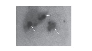
Parathyroid scintigraphy
• Sestamibi is monovalent, lipophilic cation which accumulates in
mitochondria
• Initially used for myocardial perfusion
• In 1989, its affinity for abnormal parathyroid tissue was identified
• Radionucleide used is Tc99m
• Sensitivity decreases in case of simultaneous thyroid pathology
3 types of imaging
•Subtraction imaging using both I 123 and Tc sestamibi
•Dual phase sestamibi scan
•Sestamibi SPECT
Pheochromocytoma
• Meta iodo benzyl guanine (I 123)
• Used for pre operative localization of Pheochromocytoma
• Tracer is given IV and images are taken at different time intervals
• F18 L DOPA scan is superior at identifying and localizing the tumor
• More costly and lesser availablity
MIBG is specifically done in patients who have
•equivocal CT/MRI
•suspected of having multifocal disease
•suspected of having malignant disease
•Highly specific for pheochromocytoma but sensitivity is only about 75-
90%
•F18 DOPA PET scans have higher sensitivity and specificity
Bone Scintigraphy
• Tc labelled methylene diphosphate
• Binds to hydroxyapatite crystals proportional to blood flow and
osteoblastic activity
• Hence is a marker of bone perfusion and turnover
• Mainly useful to detect bone secondaries
• Choice of radioisotope can vary depending on primary tumor
Renal cortex scintigraphy
• Di mercapto succinic acid
• Labelled with Tc99m
• Accumulates in Proximal tubular cells of renal cortex
• Static scan
• Position, size and morphology of renal cortex can be evaluated
• Any loss of renal cortex can be noted - renal scarring
• Pitfalls - UTI, HUN
Radioisotope Renogram
• Diethylene triamine penta acetate
• Mercapto acyltriglycine
• Both are Tc99m labelled
• DTPA is excreted through Glomerular filteration
• MAG3 is excrete through secretion in proximal tubules
Cholescintigraphy
• Hepatobiliary imido diacetic acid
• Tc99m labelled lidofenin
• Secreted into bile about 1hr after administration
• Can identify anomalies or obstructions in biliary tree
• Also used to diagnose gallbladder dysfunction
• Not the first line investigation and considered only if MRI is not
available
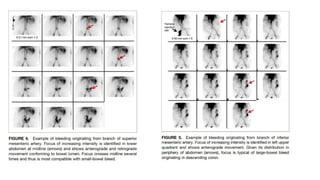
GIB scintigraphy
• Gastro intestinal bleeding scintigraphy
• initially Tc99m sulphur colloid was used
• Tc99m labelled RBC are used now
• Useful mainly in small bowel and lower gi bleeding
Meckels diverticulum
• 60% of diverticulae tend to have ectopic mucosa
• more than 70% have gastric mucosa
• Tc99m labelled Na pertechnate is used
• Taken up by mucin secreting cells of gastric mucosa
• Its sensitivity decreases after adolescence
• False positives can be seen with vasular malformations or other sites
of ectopic gastric mucosa
PET
• Positron emission tomography
• Positron emmitters are used as radionucleides
• Emitted positron travels short distance and interacts with electron
• Resulting in 2 gamma photons travelling at 180 degrees
• These are recognised by detectors and position is localised and a 3D
image is created
• PET scan is used in various fields but has important role in oncology
• F18 is the most commonly used isotope
• Used in form of Fluoro deoxy glucose
• Accumulated in cells that actively uptake glucose for metabolism
Hybrid Imaging
• PET-CT combines, in a single gantry, a PET scanner and a CT scanner,
to acquire sequential images from both devices in the same session,
which are combined into a single superposed image
• Functional imaging obtained by PET, can be more precisely correlated
with anatomic imaging obtained by CT scanning
Uses
• staging of carcinoma
• establish baseline staging before commencing treatment
• evaluation of an indeterminate/equivocal lesion on conventional
imaging
• assessing response to therapy
• evaluation of suspected disease recurrence or relapse
• to guide a biopsy (e.g. pleural biopsy for mesothelioma)
• In work up of an occult primary lesion
• differentiate between radiation-induced necrosis and tumour
recurrence
Take Home
• While some procedures have become obsolete owing to
developments in CT and MRI, still there are specific indications for
isotope scans
• Using in combination with CT is more informative
• Only contraindications are Pregnancy and Lactation
Thank You

More Related Content

What's hot

Gamma Camera
Gamma CameraGamma Camera
Gamma Camera
Muhammad Musaddiq
 
Computed tomography
Computed tomographyComputed tomography
Computed tomography
Rituraj Mishra
 
CT fluoroscopy1 (1).pptx
CT fluoroscopy1 (1).pptxCT fluoroscopy1 (1).pptx
CT fluoroscopy1 (1).pptx
VANI PUSHPA MUDAVATH
 
Basic principles of CT scanning
Basic principles of CT scanningBasic principles of CT scanning
CT artifact
CT artifact CT artifact
CT artifact
Ganesan Yogananthem
 
Interactions of ultrasound with matter
Interactions of ultrasound with matterInteractions of ultrasound with matter
Interactions of ultrasound with matter
Swapnil Shetty
 
Ct instrumentation and types of detector configuration
Ct instrumentation and types of detector configurationCt instrumentation and types of detector configuration
Ct instrumentation and types of detector configuration
SUJAN KARKI
 
Digital breast tomosynthesis
Digital breast tomosynthesisDigital breast tomosynthesis
Digital breast tomosynthesis
Yashawant Yadav
 
Fat suppression imaging
Fat suppression imagingFat suppression imaging
Fat suppression imaging
Roshan Shah
 
Everything Radionuclides - Nuclear Medicine
Everything Radionuclides - Nuclear MedicineEverything Radionuclides - Nuclear Medicine
Everything Radionuclides - Nuclear Medicine
Victor Ekpo
 
CT SCAN - 3 Marks - QUESTION AND ANSWERS
CT SCAN - 3 Marks - QUESTION AND ANSWERSCT SCAN - 3 Marks - QUESTION AND ANSWERS
CT SCAN - 3 Marks - QUESTION AND ANSWERS
Ganesan Yogananthem
 
Principles and application of PET CT & PET MR
Principles and application of PET CT & PET MRPrinciples and application of PET CT & PET MR
Principles and application of PET CT & PET MRcharusmita chaudhary
 
Gamma camera components
Gamma camera componentsGamma camera components
Gamma camera components
Syed Hammad .
 
CTDI (Computed Tomography Dose Index
CTDI (Computed Tomography Dose IndexCTDI (Computed Tomography Dose Index
CTDI (Computed Tomography Dose Index
Vivek Elangovan
 
Magnification(macro and micro radiography), distortion
Magnification(macro and micro radiography), distortionMagnification(macro and micro radiography), distortion
Magnification(macro and micro radiography), distortion
parthajyotidas11
 
CT Image reconstruction
CT Image reconstructionCT Image reconstruction
CT Image reconstruction
Santosh Ojha
 
Flat Panel Digital Fluoroscopy by Akash Das
Flat Panel Digital Fluoroscopy by Akash Das Flat Panel Digital Fluoroscopy by Akash Das
Flat Panel Digital Fluoroscopy by Akash Das
AkashDas169
 
MRI artifacts
MRI artifactsMRI artifacts
MRI artifacts
Sudil Paudyal
 
MRI 3-Marks QUESTIONS AND ANSWERS
MRI 3-Marks QUESTIONS AND ANSWERSMRI 3-Marks QUESTIONS AND ANSWERS
MRI 3-Marks QUESTIONS AND ANSWERS
Ganesan Yogananthem
 
Gas filled detectors
Gas filled detectorsGas filled detectors
Gas filled detectors
Amara Usman
 

What's hot (20)

Gamma Camera
Gamma CameraGamma Camera
Gamma Camera
 
Computed tomography
Computed tomographyComputed tomography
Computed tomography
 
CT fluoroscopy1 (1).pptx
CT fluoroscopy1 (1).pptxCT fluoroscopy1 (1).pptx
CT fluoroscopy1 (1).pptx
 
Basic principles of CT scanning
Basic principles of CT scanningBasic principles of CT scanning
Basic principles of CT scanning
 
CT artifact
CT artifact CT artifact
CT artifact
 
Interactions of ultrasound with matter
Interactions of ultrasound with matterInteractions of ultrasound with matter
Interactions of ultrasound with matter
 
Ct instrumentation and types of detector configuration
Ct instrumentation and types of detector configurationCt instrumentation and types of detector configuration
Ct instrumentation and types of detector configuration
 
Digital breast tomosynthesis
Digital breast tomosynthesisDigital breast tomosynthesis
Digital breast tomosynthesis
 
Fat suppression imaging
Fat suppression imagingFat suppression imaging
Fat suppression imaging
 
Everything Radionuclides - Nuclear Medicine
Everything Radionuclides - Nuclear MedicineEverything Radionuclides - Nuclear Medicine
Everything Radionuclides - Nuclear Medicine
 
CT SCAN - 3 Marks - QUESTION AND ANSWERS
CT SCAN - 3 Marks - QUESTION AND ANSWERSCT SCAN - 3 Marks - QUESTION AND ANSWERS
CT SCAN - 3 Marks - QUESTION AND ANSWERS
 
Principles and application of PET CT & PET MR
Principles and application of PET CT & PET MRPrinciples and application of PET CT & PET MR
Principles and application of PET CT & PET MR
 
Gamma camera components
Gamma camera componentsGamma camera components
Gamma camera components
 
CTDI (Computed Tomography Dose Index
CTDI (Computed Tomography Dose IndexCTDI (Computed Tomography Dose Index
CTDI (Computed Tomography Dose Index
 
Magnification(macro and micro radiography), distortion
Magnification(macro and micro radiography), distortionMagnification(macro and micro radiography), distortion
Magnification(macro and micro radiography), distortion
 
CT Image reconstruction
CT Image reconstructionCT Image reconstruction
CT Image reconstruction
 
Flat Panel Digital Fluoroscopy by Akash Das
Flat Panel Digital Fluoroscopy by Akash Das Flat Panel Digital Fluoroscopy by Akash Das
Flat Panel Digital Fluoroscopy by Akash Das
 
MRI artifacts
MRI artifactsMRI artifacts
MRI artifacts
 
MRI 3-Marks QUESTIONS AND ANSWERS
MRI 3-Marks QUESTIONS AND ANSWERSMRI 3-Marks QUESTIONS AND ANSWERS
MRI 3-Marks QUESTIONS AND ANSWERS
 
Gas filled detectors
Gas filled detectorsGas filled detectors
Gas filled detectors
 

Similar to Radioisotope imaging

Radionuclide imaging
Radionuclide imagingRadionuclide imaging
Radionuclide imaging
Sudarsan Agarwal
 
What is Nuclear Medicine and PET?
What is Nuclear Medicine and PET?What is Nuclear Medicine and PET?
What is Nuclear Medicine and PET?
todd_charge
 
Basics of Nuclear Medicine 2.pptxmmmmmmmmm
Basics of Nuclear Medicine 2.pptxmmmmmmmmmBasics of Nuclear Medicine 2.pptxmmmmmmmmm
Basics of Nuclear Medicine 2.pptxmmmmmmmmm
IbrahemIssacGaied
 
Pet ct and its benefit vs risk
Pet ct and its benefit vs riskPet ct and its benefit vs risk
Pet ct and its benefit vs risk
VibhaJoshi12
 
Common applications of nuclear medicine
Common applications of nuclear medicineCommon applications of nuclear medicine
Common applications of nuclear medicine
DR RML DELHI
 
Imaging in surgery
Imaging in surgeryImaging in surgery
Imaging in surgery
usifoh itaman
 
46_PET.pptx
46_PET.pptx46_PET.pptx
46_PET.pptx
DevPanda5
 
pet scan.ppt
 pet scan.ppt pet scan.ppt
pet scan.ppt
GauravDarekar3
 
Radiographic system clinical engineering
Radiographic system clinical engineeringRadiographic system clinical engineering
Radiographic system clinical engineering
HasibulHasanHredoy
 
nuclear medicine and its application in dentistry
nuclear medicine and its application in dentistrynuclear medicine and its application in dentistry
nuclear medicine and its application in dentistry
gaurav katheriya
 
OPHTHALMIC TUMORS
OPHTHALMIC TUMORSOPHTHALMIC TUMORS
OPHTHALMIC TUMORS
Kanhu Charan
 
NUCLEAR MEDICINE.pptx
NUCLEAR MEDICINE.pptxNUCLEAR MEDICINE.pptx
NUCLEAR MEDICINE.pptx
Aymanshahzad4
 
Renography
RenographyRenography
Renography
Victor Ekpo
 
RADIOTHERAPY FOR OPHTHALMOLOGISTS
RADIOTHERAPY FOR OPHTHALMOLOGISTSRADIOTHERAPY FOR OPHTHALMOLOGISTS
RADIOTHERAPY FOR OPHTHALMOLOGISTS
Kanhu Charan
 
Nuclear medicine radiology revision notes
Nuclear medicine radiology revision notesNuclear medicine radiology revision notes
Nuclear medicine radiology revision notes
TONY SCARIA
 
Nuclear Medicine in Thyroidology
Nuclear Medicine in ThyroidologyNuclear Medicine in Thyroidology
Nuclear Medicine in ThyroidologyLokender Yadav
 
Radioisotopes and their role in modern era imaging
Radioisotopes and their role in modern era imagingRadioisotopes and their role in modern era imaging
Radioisotopes and their role in modern era imaging
KhanAsma6
 
PET scan in Cardilogy
PET scan in CardilogyPET scan in Cardilogy
PET scan in Cardilogy
Raghu Kishore Galla
 
Radiotherapy in ENT
Radiotherapy in ENTRadiotherapy in ENT
Radiotherapy in ENT
Yousuf Choudhury
 
PET-CT
PET-CTPET-CT
PET-CT
Tushar singh
 

Similar to Radioisotope imaging (20)

Radionuclide imaging
Radionuclide imagingRadionuclide imaging
Radionuclide imaging
 
What is Nuclear Medicine and PET?
What is Nuclear Medicine and PET?What is Nuclear Medicine and PET?
What is Nuclear Medicine and PET?
 
Basics of Nuclear Medicine 2.pptxmmmmmmmmm
Basics of Nuclear Medicine 2.pptxmmmmmmmmmBasics of Nuclear Medicine 2.pptxmmmmmmmmm
Basics of Nuclear Medicine 2.pptxmmmmmmmmm
 
Pet ct and its benefit vs risk
Pet ct and its benefit vs riskPet ct and its benefit vs risk
Pet ct and its benefit vs risk
 
Common applications of nuclear medicine
Common applications of nuclear medicineCommon applications of nuclear medicine
Common applications of nuclear medicine
 
Imaging in surgery
Imaging in surgeryImaging in surgery
Imaging in surgery
 
46_PET.pptx
46_PET.pptx46_PET.pptx
46_PET.pptx
 
pet scan.ppt
 pet scan.ppt pet scan.ppt
pet scan.ppt
 
Radiographic system clinical engineering
Radiographic system clinical engineeringRadiographic system clinical engineering
Radiographic system clinical engineering
 
nuclear medicine and its application in dentistry
nuclear medicine and its application in dentistrynuclear medicine and its application in dentistry
nuclear medicine and its application in dentistry
 
OPHTHALMIC TUMORS
OPHTHALMIC TUMORSOPHTHALMIC TUMORS
OPHTHALMIC TUMORS
 
NUCLEAR MEDICINE.pptx
NUCLEAR MEDICINE.pptxNUCLEAR MEDICINE.pptx
NUCLEAR MEDICINE.pptx
 
Renography
RenographyRenography
Renography
 
RADIOTHERAPY FOR OPHTHALMOLOGISTS
RADIOTHERAPY FOR OPHTHALMOLOGISTSRADIOTHERAPY FOR OPHTHALMOLOGISTS
RADIOTHERAPY FOR OPHTHALMOLOGISTS
 
Nuclear medicine radiology revision notes
Nuclear medicine radiology revision notesNuclear medicine radiology revision notes
Nuclear medicine radiology revision notes
 
Nuclear Medicine in Thyroidology
Nuclear Medicine in ThyroidologyNuclear Medicine in Thyroidology
Nuclear Medicine in Thyroidology
 
Radioisotopes and their role in modern era imaging
Radioisotopes and their role in modern era imagingRadioisotopes and their role in modern era imaging
Radioisotopes and their role in modern era imaging
 
PET scan in Cardilogy
PET scan in CardilogyPET scan in Cardilogy
PET scan in Cardilogy
 
Radiotherapy in ENT
Radiotherapy in ENTRadiotherapy in ENT
Radiotherapy in ENT
 
PET-CT
PET-CTPET-CT
PET-CT
 

Recently uploaded

Charaka Samhita Sutra Sthana 9 Chapter khuddakachatuspadadhyaya
Charaka Samhita Sutra Sthana 9 Chapter khuddakachatuspadadhyayaCharaka Samhita Sutra Sthana 9 Chapter khuddakachatuspadadhyaya
Charaka Samhita Sutra Sthana 9 Chapter khuddakachatuspadadhyaya
Dr KHALID B.M
 
Phone Us ❤85270-49040❤ #ℂall #gIRLS In Surat By Surat @ℂall @Girls Hotel With...
Phone Us ❤85270-49040❤ #ℂall #gIRLS In Surat By Surat @ℂall @Girls Hotel With...Phone Us ❤85270-49040❤ #ℂall #gIRLS In Surat By Surat @ℂall @Girls Hotel With...
Phone Us ❤85270-49040❤ #ℂall #gIRLS In Surat By Surat @ℂall @Girls Hotel With...
Savita Shen $i11
 
Are There Any Natural Remedies To Treat Syphilis.pdf
Are There Any Natural Remedies To Treat Syphilis.pdfAre There Any Natural Remedies To Treat Syphilis.pdf
Are There Any Natural Remedies To Treat Syphilis.pdf
Little Cross Family Clinic
 
KDIGO 2024 guidelines for diabetologists
KDIGO 2024 guidelines for diabetologistsKDIGO 2024 guidelines for diabetologists
KDIGO 2024 guidelines for diabetologists
د.محمود نجيب
 
Ozempic: Preoperative Management of Patients on GLP-1 Receptor Agonists
Ozempic: Preoperative Management of Patients on GLP-1 Receptor Agonists  Ozempic: Preoperative Management of Patients on GLP-1 Receptor Agonists
Ozempic: Preoperative Management of Patients on GLP-1 Receptor Agonists
Saeid Safari
 
basicmodesofventilation2022-220313203758.pdf
basicmodesofventilation2022-220313203758.pdfbasicmodesofventilation2022-220313203758.pdf
basicmodesofventilation2022-220313203758.pdf
aljamhori teaching hospital
 
Surat @ℂall @Girls ꧁❤8527049040❤꧂@ℂall @Girls Service Vip Top Model Safe
Surat @ℂall @Girls ꧁❤8527049040❤꧂@ℂall @Girls Service Vip Top Model SafeSurat @ℂall @Girls ꧁❤8527049040❤꧂@ℂall @Girls Service Vip Top Model Safe
Surat @ℂall @Girls ꧁❤8527049040❤꧂@ℂall @Girls Service Vip Top Model Safe
Savita Shen $i11
 
ARTIFICIAL INTELLIGENCE IN HEALTHCARE.pdf
ARTIFICIAL INTELLIGENCE IN  HEALTHCARE.pdfARTIFICIAL INTELLIGENCE IN  HEALTHCARE.pdf
ARTIFICIAL INTELLIGENCE IN HEALTHCARE.pdf
Anujkumaranit
 
For Better Surat #ℂall #Girl Service ❤85270-49040❤ Surat #ℂall #Girls
For Better Surat #ℂall #Girl Service ❤85270-49040❤ Surat #ℂall #GirlsFor Better Surat #ℂall #Girl Service ❤85270-49040❤ Surat #ℂall #Girls
For Better Surat #ℂall #Girl Service ❤85270-49040❤ Surat #ℂall #Girls
Savita Shen $i11
 
Physiology of Special Chemical Sensation of Taste
Physiology of Special Chemical Sensation of TastePhysiology of Special Chemical Sensation of Taste
Physiology of Special Chemical Sensation of Taste
MedicoseAcademics
 
Flu Vaccine Alert in Bangalore Karnataka
Flu Vaccine Alert in Bangalore KarnatakaFlu Vaccine Alert in Bangalore Karnataka
Flu Vaccine Alert in Bangalore Karnataka
addon Scans
 
Charaka Samhita Sutra sthana Chapter 15 Upakalpaniyaadhyaya
Charaka Samhita Sutra sthana Chapter 15 UpakalpaniyaadhyayaCharaka Samhita Sutra sthana Chapter 15 Upakalpaniyaadhyaya
Charaka Samhita Sutra sthana Chapter 15 Upakalpaniyaadhyaya
Dr KHALID B.M
 
ANATOMY AND PHYSIOLOGY OF URINARY SYSTEM.pptx
ANATOMY AND PHYSIOLOGY OF URINARY SYSTEM.pptxANATOMY AND PHYSIOLOGY OF URINARY SYSTEM.pptx
ANATOMY AND PHYSIOLOGY OF URINARY SYSTEM.pptx
Swetaba Besh
 
Physiology of Chemical Sensation of smell.pdf
Physiology of Chemical Sensation of smell.pdfPhysiology of Chemical Sensation of smell.pdf
Physiology of Chemical Sensation of smell.pdf
MedicoseAcademics
 
Cervical & Brachial Plexus By Dr. RIG.pptx
Cervical & Brachial Plexus By Dr. RIG.pptxCervical & Brachial Plexus By Dr. RIG.pptx
Cervical & Brachial Plexus By Dr. RIG.pptx
Dr. Rabia Inam Gandapore
 
How to Give Better Lectures: Some Tips for Doctors
How to Give Better Lectures: Some Tips for DoctorsHow to Give Better Lectures: Some Tips for Doctors
How to Give Better Lectures: Some Tips for Doctors
LanceCatedral
 
Superficial & Deep Fascia of the NECK.pptx
Superficial & Deep Fascia of the NECK.pptxSuperficial & Deep Fascia of the NECK.pptx
Superficial & Deep Fascia of the NECK.pptx
Dr. Rabia Inam Gandapore
 
The Normal Electrocardiogram - Part I of II
The Normal Electrocardiogram - Part I of IIThe Normal Electrocardiogram - Part I of II
The Normal Electrocardiogram - Part I of II
MedicoseAcademics
 
Non-respiratory Functions of the Lungs.pdf
Non-respiratory Functions of the Lungs.pdfNon-respiratory Functions of the Lungs.pdf
Non-respiratory Functions of the Lungs.pdf
MedicoseAcademics
 
Ocular injury ppt Upendra pal optometrist upums saifai etawah
Ocular injury  ppt  Upendra pal  optometrist upums saifai etawahOcular injury  ppt  Upendra pal  optometrist upums saifai etawah
Ocular injury ppt Upendra pal optometrist upums saifai etawah
pal078100
 

Recently uploaded (20)

Charaka Samhita Sutra Sthana 9 Chapter khuddakachatuspadadhyaya
Charaka Samhita Sutra Sthana 9 Chapter khuddakachatuspadadhyayaCharaka Samhita Sutra Sthana 9 Chapter khuddakachatuspadadhyaya
Charaka Samhita Sutra Sthana 9 Chapter khuddakachatuspadadhyaya
 
Phone Us ❤85270-49040❤ #ℂall #gIRLS In Surat By Surat @ℂall @Girls Hotel With...
Phone Us ❤85270-49040❤ #ℂall #gIRLS In Surat By Surat @ℂall @Girls Hotel With...Phone Us ❤85270-49040❤ #ℂall #gIRLS In Surat By Surat @ℂall @Girls Hotel With...
Phone Us ❤85270-49040❤ #ℂall #gIRLS In Surat By Surat @ℂall @Girls Hotel With...
 
Are There Any Natural Remedies To Treat Syphilis.pdf
Are There Any Natural Remedies To Treat Syphilis.pdfAre There Any Natural Remedies To Treat Syphilis.pdf
Are There Any Natural Remedies To Treat Syphilis.pdf
 
KDIGO 2024 guidelines for diabetologists
KDIGO 2024 guidelines for diabetologistsKDIGO 2024 guidelines for diabetologists
KDIGO 2024 guidelines for diabetologists
 
Ozempic: Preoperative Management of Patients on GLP-1 Receptor Agonists
Ozempic: Preoperative Management of Patients on GLP-1 Receptor Agonists  Ozempic: Preoperative Management of Patients on GLP-1 Receptor Agonists
Ozempic: Preoperative Management of Patients on GLP-1 Receptor Agonists
 
basicmodesofventilation2022-220313203758.pdf
basicmodesofventilation2022-220313203758.pdfbasicmodesofventilation2022-220313203758.pdf
basicmodesofventilation2022-220313203758.pdf
 
Surat @ℂall @Girls ꧁❤8527049040❤꧂@ℂall @Girls Service Vip Top Model Safe
Surat @ℂall @Girls ꧁❤8527049040❤꧂@ℂall @Girls Service Vip Top Model SafeSurat @ℂall @Girls ꧁❤8527049040❤꧂@ℂall @Girls Service Vip Top Model Safe
Surat @ℂall @Girls ꧁❤8527049040❤꧂@ℂall @Girls Service Vip Top Model Safe
 
ARTIFICIAL INTELLIGENCE IN HEALTHCARE.pdf
ARTIFICIAL INTELLIGENCE IN  HEALTHCARE.pdfARTIFICIAL INTELLIGENCE IN  HEALTHCARE.pdf
ARTIFICIAL INTELLIGENCE IN HEALTHCARE.pdf
 
For Better Surat #ℂall #Girl Service ❤85270-49040❤ Surat #ℂall #Girls
For Better Surat #ℂall #Girl Service ❤85270-49040❤ Surat #ℂall #GirlsFor Better Surat #ℂall #Girl Service ❤85270-49040❤ Surat #ℂall #Girls
For Better Surat #ℂall #Girl Service ❤85270-49040❤ Surat #ℂall #Girls
 
Physiology of Special Chemical Sensation of Taste
Physiology of Special Chemical Sensation of TastePhysiology of Special Chemical Sensation of Taste
Physiology of Special Chemical Sensation of Taste
 
Flu Vaccine Alert in Bangalore Karnataka
Flu Vaccine Alert in Bangalore KarnatakaFlu Vaccine Alert in Bangalore Karnataka
Flu Vaccine Alert in Bangalore Karnataka
 
Charaka Samhita Sutra sthana Chapter 15 Upakalpaniyaadhyaya
Charaka Samhita Sutra sthana Chapter 15 UpakalpaniyaadhyayaCharaka Samhita Sutra sthana Chapter 15 Upakalpaniyaadhyaya
Charaka Samhita Sutra sthana Chapter 15 Upakalpaniyaadhyaya
 
ANATOMY AND PHYSIOLOGY OF URINARY SYSTEM.pptx
ANATOMY AND PHYSIOLOGY OF URINARY SYSTEM.pptxANATOMY AND PHYSIOLOGY OF URINARY SYSTEM.pptx
ANATOMY AND PHYSIOLOGY OF URINARY SYSTEM.pptx
 
Physiology of Chemical Sensation of smell.pdf
Physiology of Chemical Sensation of smell.pdfPhysiology of Chemical Sensation of smell.pdf
Physiology of Chemical Sensation of smell.pdf
 
Cervical & Brachial Plexus By Dr. RIG.pptx
Cervical & Brachial Plexus By Dr. RIG.pptxCervical & Brachial Plexus By Dr. RIG.pptx
Cervical & Brachial Plexus By Dr. RIG.pptx
 
How to Give Better Lectures: Some Tips for Doctors
How to Give Better Lectures: Some Tips for DoctorsHow to Give Better Lectures: Some Tips for Doctors
How to Give Better Lectures: Some Tips for Doctors
 
Superficial & Deep Fascia of the NECK.pptx
Superficial & Deep Fascia of the NECK.pptxSuperficial & Deep Fascia of the NECK.pptx
Superficial & Deep Fascia of the NECK.pptx
 
The Normal Electrocardiogram - Part I of II
The Normal Electrocardiogram - Part I of IIThe Normal Electrocardiogram - Part I of II
The Normal Electrocardiogram - Part I of II
 
Non-respiratory Functions of the Lungs.pdf
Non-respiratory Functions of the Lungs.pdfNon-respiratory Functions of the Lungs.pdf
Non-respiratory Functions of the Lungs.pdf
 
Ocular injury ppt Upendra pal optometrist upums saifai etawah
Ocular injury  ppt  Upendra pal  optometrist upums saifai etawahOcular injury  ppt  Upendra pal  optometrist upums saifai etawah
Ocular injury ppt Upendra pal optometrist upums saifai etawah
 

Radioisotope imaging

  • 2. • Introduction • Historical perspective • Components in Imaging • Uses in surgical field • PET and Hybrid imaging
  • 3. Introduction • Nuclear medicine is a medical specialty involving the application of radioactive substances in the diagnosis and treatment of disease. • Nuclear imaging is a form of physiologic imaging. • Produces images of physiologic processes utilizing radioactive isotopes
  • 4. History • In mid 1920s George de Hevesy made experiments with radionuclides administered to rats • Discovery of artificially produced radionuclides by Frédéric Joliot-Curie and Irène Joliot-Curie in 1934 • In 1936, John Lawrence, known as "the father of nuclear medicine", his brother Ernest Lawrence made the first application in patients of an artificial radionuclide - Phosphorus-32 to treat leukemia.
  • 5. • December 7, 1946 - article was published in the Journal of the American Medical Association by Sam Seidlin - successful treatment of a patient with thyroid cancer metastases using I-131 • Technetium-99m was first discovered in 1937 by C. Perrier and E. Segre, is the most utilized element in nuclear medicine and is employed in a wide variety of nuclear medicine imaging studies.
  • 6. Components of imaging • Radionucleide •eg: Tc99m, I 123, I 131 • Radiopharmaceutical / Tracer •pertechnate, sestamibi, NaI • Gamma camera
  • 7. Radionucleide / Radioisotope There are several properties of the ideal radioisotope for diagnostic purposes: •Half life - short enough to limit radiation dose, but long enough to allow good signal during imaging (ideally 1.5 x length of imaging) •Emits only gamma rays •Mono-energetic - ideal energy range is 100 to 250 keV •Decays to stable daughter isotopes •Easy to bind to different pharmaceuticals
  • 8. Technetium 99m • First artificial element to be produced • Generated from Mo99 in a cyclotron • Half life - 6hrs • Emits gamma rays 140KeV • Used in more than 70% of nuclear medicine scans
  • 9. Radiopharmaceutical / Tracer There are several properties of the ideal pharmaceutical: •High target:non-target uptake ratio •Easy and cheap to produce •Non-toxic •Does not alter physiology
  • 10. Gamma Camera • The Gamma camera consists of a large area NaI crystal with a lead collimator which allows only transmission of radiation from a particular point. • The detector device can be moved to cover larger areas.
  • 11.
  • 12. Types of Imaging • Static planar scintigraphy - provides 2D representations of a three dimensional object by measuring the spatial distribution of the radioisotope in the body • Dynamic planar scintigraphy - measures temporal changes in the spatial distribution of the radioisotopes in the body by taking multiple images
  • 13. • Single photon emission tomography (SPECT) or positron emission tomography (PET) which allows to form 3D static or dynamic representations of the organ and organ functions by taking multiple images from different directions.
  • 14. Commonly used scans in surgical field • Thyroid scintigraphy • Parathyroid sestamibi scan • MIBG scan • Bone scan • DTPA, DMSA, MAG3 • HIDA scan • Labelled RBC for GI bleeding • Meckels diverticulum
  • 15. RAIU test • Radioactive iodine uptake test is used to evaluate functional status of thyroid gland or nodule • I 123 is given in form of Sodium iodide (100-200 uCi) • Tracer is given orally • Images are taken 24hr later • Normal uptake is around 25% • Based on uptake, gland/nodule is labelled as hot or cold
  • 16. Increased Uptake •Hyperthyroidism •Hashimoto’s thyroiditis •Decreased renal clearance of iodine •Iodine deficiency Decreased Uptake •Hypothyroidism (primary or secondary) •TSH resistance •Thyroid hormone replacement, suppression •Iodine excess (dietary, drugs)
  • 17.
  • 18. • I 123 has lower half life • Emits pure Gamma rays • Emitted gamma rays are ideal for imaging • Has minimal radiation exposure risk • Does not cause stunning of tissue • Expensive • I 131 has higher half life • 90% is beta decay • Gamma rays emitted are of higher energy • Radiation exposure is 100 times more • Stuns tissue that it accumulates in • Much cheaper than I 123
  • 19. • Routine use of RAIU test is ill advised • Main implication is in hyperthyroid patient with goitre • Malignancy is sen in 20% of cold noduls and 5% of hot nodules • Though suggestive, malignancy is neither confirmed nor excluded using RAIU test • Thyroid scintigraphy can also be done using Tc99m but has few drawbacks
  • 20. • Tc pertechnate is rapidly taken up by follicular cells • Trapped but not organified • Short half life helps in rapid evaluation of functioning and non functioning areas of thyroid gland • Taken up by other structures like salaivary glands and vascular structures
  • 21. Parathyroid scintigraphy • Sestamibi is monovalent, lipophilic cation which accumulates in mitochondria • Initially used for myocardial perfusion • In 1989, its affinity for abnormal parathyroid tissue was identified • Radionucleide used is Tc99m • Sensitivity decreases in case of simultaneous thyroid pathology
  • 22. 3 types of imaging •Subtraction imaging using both I 123 and Tc sestamibi •Dual phase sestamibi scan •Sestamibi SPECT
  • 23.
  • 24. Pheochromocytoma • Meta iodo benzyl guanine (I 123) • Used for pre operative localization of Pheochromocytoma • Tracer is given IV and images are taken at different time intervals • F18 L DOPA scan is superior at identifying and localizing the tumor • More costly and lesser availablity
  • 25. MIBG is specifically done in patients who have •equivocal CT/MRI •suspected of having multifocal disease •suspected of having malignant disease •Highly specific for pheochromocytoma but sensitivity is only about 75- 90% •F18 DOPA PET scans have higher sensitivity and specificity
  • 26.
  • 27.
  • 28.
  • 29.
  • 30. Bone Scintigraphy • Tc labelled methylene diphosphate • Binds to hydroxyapatite crystals proportional to blood flow and osteoblastic activity • Hence is a marker of bone perfusion and turnover • Mainly useful to detect bone secondaries • Choice of radioisotope can vary depending on primary tumor
  • 31.
  • 32. Renal cortex scintigraphy • Di mercapto succinic acid • Labelled with Tc99m • Accumulates in Proximal tubular cells of renal cortex • Static scan • Position, size and morphology of renal cortex can be evaluated • Any loss of renal cortex can be noted - renal scarring • Pitfalls - UTI, HUN
  • 33.
  • 34.
  • 35. Radioisotope Renogram • Diethylene triamine penta acetate • Mercapto acyltriglycine • Both are Tc99m labelled • DTPA is excreted through Glomerular filteration • MAG3 is excrete through secretion in proximal tubules
  • 36.
  • 37. Cholescintigraphy • Hepatobiliary imido diacetic acid • Tc99m labelled lidofenin • Secreted into bile about 1hr after administration • Can identify anomalies or obstructions in biliary tree • Also used to diagnose gallbladder dysfunction • Not the first line investigation and considered only if MRI is not available
  • 38.
  • 39. GIB scintigraphy • Gastro intestinal bleeding scintigraphy • initially Tc99m sulphur colloid was used • Tc99m labelled RBC are used now • Useful mainly in small bowel and lower gi bleeding
  • 40.
  • 41.
  • 42. Meckels diverticulum • 60% of diverticulae tend to have ectopic mucosa • more than 70% have gastric mucosa • Tc99m labelled Na pertechnate is used • Taken up by mucin secreting cells of gastric mucosa • Its sensitivity decreases after adolescence • False positives can be seen with vasular malformations or other sites of ectopic gastric mucosa
  • 43.
  • 44.
  • 45. PET • Positron emission tomography • Positron emmitters are used as radionucleides • Emitted positron travels short distance and interacts with electron • Resulting in 2 gamma photons travelling at 180 degrees • These are recognised by detectors and position is localised and a 3D image is created
  • 46. • PET scan is used in various fields but has important role in oncology • F18 is the most commonly used isotope • Used in form of Fluoro deoxy glucose • Accumulated in cells that actively uptake glucose for metabolism
  • 47. Hybrid Imaging • PET-CT combines, in a single gantry, a PET scanner and a CT scanner, to acquire sequential images from both devices in the same session, which are combined into a single superposed image • Functional imaging obtained by PET, can be more precisely correlated with anatomic imaging obtained by CT scanning
  • 48. Uses • staging of carcinoma • establish baseline staging before commencing treatment • evaluation of an indeterminate/equivocal lesion on conventional imaging • assessing response to therapy • evaluation of suspected disease recurrence or relapse • to guide a biopsy (e.g. pleural biopsy for mesothelioma)
  • 49. • In work up of an occult primary lesion • differentiate between radiation-induced necrosis and tumour recurrence
  • 50. Take Home • While some procedures have become obsolete owing to developments in CT and MRI, still there are specific indications for isotope scans • Using in combination with CT is more informative • Only contraindications are Pregnancy and Lactation