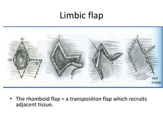
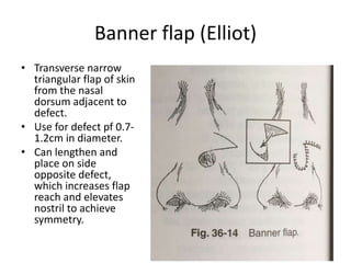
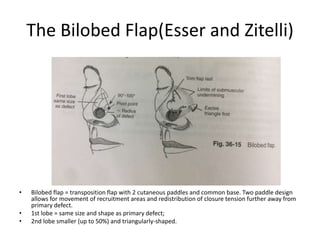
Nose Reconstruction
Anatomy of nose
 Divided into third based on underlying
skeletal structure
I. Proximal:- lies over nasal bone
II. Middle:- upper lateral cartilages.
III. Distal:- nasal tip with paired alae over
membraneous septum.
 Columella:- supported by medial crura of
alar cartilage.
Blood supply
• Angular artery:- lateral surface of caudal nose
• Superior labial artery:- nasal sill, nasal septum and base of columella.
• Dorsal branch of opthalmic artery:- axial arterial network for dorsal and lateral nasal skin.
• Infraorbital branch of internal maxillary artery:- dorsum and lateral sidewall of nose.
• Venous drainage parallels arterial supply.
Nerve supply
• Sensory
 Opthalmic division
(V1) of trigeminal
nerve:- radix, rhinion
and cephalic portion
of nasal sidewalls,
skin over dorsum to
tip.
 Maxillary division
(V2) of trigeminal
nerve:- lateral tissue
on lower half of
nose, columella and
lateral vestibule
• Motor
 Facial nerve VII:-
procerus, depressor
septi nasi and nasalis
History
• It began in ancient Egypt and ancient india.
• 800 BC: - Sushruta Samhita describes cheek
flap for nasal reconstruction.
• 16th century:- Gaspare Tagliacozzi used arm
pedicle tube flap.
• 1794: -Gentleman’s Magazine in London
describes Indian Method.
Principles of aesthetic nasal
reconstruction
Establish a goal.
Visualize the end result.
Create a plan.
Consider altering the wound in site, size, depth or
position.
Use the ideal or contralateral normal as a guide.
Replace missing tissue exactly to avoid overfilling
or underfilling of the defect.
Use ideal donor materials.
Ensure a stable platform.
Approach to reconstruction
The nasal base platform:- lip, cheek.
Combined, deep and extensive composite
defect repair on stages.
Nasal lining:- composite skin graft,
prelaminated skin graft and cartilage,
intranasal lining flap, skin graft, folded
forehead flap, microvascular lining
Nasal support
Nasal cover:-
Aesthetic subunit of the nose (Burget
and Menick)
• The Subunit Principle
Nasal subunits =
distinct topographic
regions defined by “
lighted ridges” and “
normal shadowed
valleys” of nasal
surface.
• Convex subunits:
Dorsum, Tip,
Columella, Paired
Alae.
• Concave subunits:
Sidewalls, Soft tissue
triangles.
• Subunit Principle:
Entire subunit must
be replaced if >50%
subunit lost.
The Subunit Principle: Controversies
• Scars placed within subunits can be well
camouflaged in nasal reconstruction.
• Excision of healthy tissue is unnecessary if one
can obtain a satisfactory scar within the borders
of a subunit.
• Rohrich et al presented a series of 1334 nasal
reconstruction cases in which a policy of
maximal conservation of native tissue.
Advocated reconstruction of the defect, not the
subunit
Good contour is the aesthetic endpoint.
Variable factors by skin zone = degrees of subcutaneous
fat, skin thickness, sebaceous content and mobility
• Zones of the Nose
• Zone I (upper dorsum and
sidewalls): Non-sebaceous,
thin, smooth, pliable and
mobile
• Zone II (supratip, tip and alar
lobules. Sebaceous glands,
thick, stiff, non-mobile.
• Zone III (alar margin, soft
tissue triangles, infratip
lobules and columella):- Thin ,
Fixed to underlying cartilage /
fibrofatty structures.
Analysis of defects
• Deficits in Surface Covering:-
Aesthetic subunit analysis.
Skin recruitmen.
• Structural Support:-
Establishes shape and contours of external nose.
Facilitates patency of nasal airway.
• Intranasal Lining:-
Prevents stenosis,
Promotes graft survival.
Maintain mucosal function (if possible)
Goals of Reconstruction
Maintain airway patency.
Replace missing layers with similar tissue.
Minimize morbidity.
Optimize aesthetics.
Surface Defect Repair Options
• Healing by Secondary Intent :-Medial Canthus,
Soft Tissue Triangle.
• Primary Closure.
• Skin Grafting.
• Composite Grafting
• Local Flaps:-Rhomboid, Bilobed (Zitelli), Rotation
Cheek Advancement, Axial Flaps / Interpolated
Flaps, Nasolabial, Paramedian Forehead.
• Microvascular Free Tissue Transfer:- Radial
Forearm Free Flap
Intranasal lining
“ Ideal” intranasal lining repair restores a thin ,
vascular and supple nasal lining.
Goals of reconstruction of intranasal lining
 Promote airway patency.
 Support structural cartilage grafts.
 Resist contracture.
 Preserve mucosal function.
Options for donor tissue include skin grafts,
septal mucoperichondrial flaps, turbinate flaps,
turn-over flaps (PMFF, glabellar), intra-oral
mucosal flaps and free flaps.
Intranasal lining flap
• Defect of the lateral midvault can be line with a dorsally based contralateral mucoperichondrial flapperfused by the
anterior ethmoid vessels.
• Defect on the nostril margin can be lined with a bipedicle vestibular flap or ipsilateral mucoperichondrial flap.
• The composite flap of entire septum based on the nasal spine can restore lining and basic support to the dorsum and
columella.
Nasal support
• Goals of structural
framework reconstruction:
• Facilitate nasal airway
patency (support nasal tip
and valves).
• Buttress reconstructed cover
layer so as to recreate
normal-appearing nasal
contour and external
landmarks.
• Central skeletal elements:
Midline support, Structural
integrity and projection of
pyramid.
• Tip and lower lateral skeletal
elements: Contour and
definition. Tip support and
orientation.
Graft sources
• Septum (bony / cartilaginous),
• Conchal cartilage,
• Osteochondral rib grafts,
• Calvarial bone.
• Graft Sources Cartilage grafts must be placed
on a well-vascularized bed to ensure viability.
(Nasal cover) Skin grafting
• Skin Grafting in Nasal Reconstruction:- FTSGs
well-suited to small defects of Zone I of the
nose and the infratip lobule.
• Donors site include: forehead, upper eyelid,
nasolabial fold, pre- or post-auricular areas or
supraclavicular fossa.
• Survive by plasmatic imbibition for first 48
hours; thus, cannot be placed over cartilage
grafts or avascular tissue.
Limbic flap
• The rhomboid flap = a transposition flap which recruits
adjacent tissue.
Banner flap (Elliot)
• Transverse narrow
triangular flap of skin
from the nasal
dorsum adjacent to
defect.
• Use for defect pf 0.7-
1.2cm in diameter.
• Can lengthen and
place on side
opposite defect,
which increases flap
reach and elevates
nostril to achieve
symmetry.
The Bilobed Flap(Esser and Zitelli)
• Bilobed flap = transposition flap with 2 cutaneous paddles and common base. Two paddle design
allows for movement of recruitment areas and redistribution of closure tension further away from
primary defect.
• 1st lobe = same size and shape as primary defect;
• 2nd lobe smaller (up to 50%) and triangularly-shaped.
Primary disadvantages of bilobed flap:
Multiple incisions resulting in extended scar
not amenable to strategic placement.
Curvilinear primary and secondary defects
prone to pin cushioning.
Extensive underlying scar bed impedes
lymphatic drainage.
Nasolabial Flap
 Random pattern flaps based on angular / facial artery perforators. May be based either superiorly
(alar or sidewall subunits) or inferiorly (columella).
 Utilize abundant skin of the medial cheek and nasolabial fold, which tends to be a good tissue match
for nasal cover.
 Suitable for reconstructing partial thickness defects of the ala.
Cheek Advancement Flap
• Cheek advancement flap well-suited for reconstruction of sidewall
defects > 1.5 cm. Medial cheek is an abundant skin reservoir with
good tissue match to the nasal sidewall. Incisions are strategically
placed within borders of aesthetic units of the face (nasolabial fold
and border of orbital and cheek units).
Paramedian forehead flap
CONCLUSION
Thank you

Nose reconstruction

  • 1.
  • 2.
    Anatomy of nose Divided into third based on underlying skeletal structure I. Proximal:- lies over nasal bone II. Middle:- upper lateral cartilages. III. Distal:- nasal tip with paired alae over membraneous septum.  Columella:- supported by medial crura of alar cartilage.
  • 5.
    Blood supply • Angularartery:- lateral surface of caudal nose • Superior labial artery:- nasal sill, nasal septum and base of columella. • Dorsal branch of opthalmic artery:- axial arterial network for dorsal and lateral nasal skin. • Infraorbital branch of internal maxillary artery:- dorsum and lateral sidewall of nose. • Venous drainage parallels arterial supply.
  • 6.
    Nerve supply • Sensory Opthalmic division (V1) of trigeminal nerve:- radix, rhinion and cephalic portion of nasal sidewalls, skin over dorsum to tip.  Maxillary division (V2) of trigeminal nerve:- lateral tissue on lower half of nose, columella and lateral vestibule • Motor  Facial nerve VII:- procerus, depressor septi nasi and nasalis
  • 7.
    History • It beganin ancient Egypt and ancient india. • 800 BC: - Sushruta Samhita describes cheek flap for nasal reconstruction. • 16th century:- Gaspare Tagliacozzi used arm pedicle tube flap. • 1794: -Gentleman’s Magazine in London describes Indian Method.
  • 8.
    Principles of aestheticnasal reconstruction Establish a goal. Visualize the end result. Create a plan. Consider altering the wound in site, size, depth or position. Use the ideal or contralateral normal as a guide. Replace missing tissue exactly to avoid overfilling or underfilling of the defect. Use ideal donor materials. Ensure a stable platform.
  • 9.
    Approach to reconstruction Thenasal base platform:- lip, cheek. Combined, deep and extensive composite defect repair on stages. Nasal lining:- composite skin graft, prelaminated skin graft and cartilage, intranasal lining flap, skin graft, folded forehead flap, microvascular lining Nasal support Nasal cover:-
  • 10.
    Aesthetic subunit ofthe nose (Burget and Menick) • The Subunit Principle Nasal subunits = distinct topographic regions defined by “ lighted ridges” and “ normal shadowed valleys” of nasal surface. • Convex subunits: Dorsum, Tip, Columella, Paired Alae. • Concave subunits: Sidewalls, Soft tissue triangles. • Subunit Principle: Entire subunit must be replaced if >50% subunit lost.
  • 11.
    The Subunit Principle:Controversies • Scars placed within subunits can be well camouflaged in nasal reconstruction. • Excision of healthy tissue is unnecessary if one can obtain a satisfactory scar within the borders of a subunit. • Rohrich et al presented a series of 1334 nasal reconstruction cases in which a policy of maximal conservation of native tissue. Advocated reconstruction of the defect, not the subunit Good contour is the aesthetic endpoint.
  • 12.
    Variable factors byskin zone = degrees of subcutaneous fat, skin thickness, sebaceous content and mobility • Zones of the Nose • Zone I (upper dorsum and sidewalls): Non-sebaceous, thin, smooth, pliable and mobile • Zone II (supratip, tip and alar lobules. Sebaceous glands, thick, stiff, non-mobile. • Zone III (alar margin, soft tissue triangles, infratip lobules and columella):- Thin , Fixed to underlying cartilage / fibrofatty structures.
  • 13.
    Analysis of defects •Deficits in Surface Covering:- Aesthetic subunit analysis. Skin recruitmen. • Structural Support:- Establishes shape and contours of external nose. Facilitates patency of nasal airway. • Intranasal Lining:- Prevents stenosis, Promotes graft survival. Maintain mucosal function (if possible)
  • 14.
    Goals of Reconstruction Maintainairway patency. Replace missing layers with similar tissue. Minimize morbidity. Optimize aesthetics.
  • 15.
    Surface Defect RepairOptions • Healing by Secondary Intent :-Medial Canthus, Soft Tissue Triangle. • Primary Closure. • Skin Grafting. • Composite Grafting • Local Flaps:-Rhomboid, Bilobed (Zitelli), Rotation Cheek Advancement, Axial Flaps / Interpolated Flaps, Nasolabial, Paramedian Forehead. • Microvascular Free Tissue Transfer:- Radial Forearm Free Flap
  • 16.
    Intranasal lining “ Ideal”intranasal lining repair restores a thin , vascular and supple nasal lining. Goals of reconstruction of intranasal lining  Promote airway patency.  Support structural cartilage grafts.  Resist contracture.  Preserve mucosal function. Options for donor tissue include skin grafts, septal mucoperichondrial flaps, turbinate flaps, turn-over flaps (PMFF, glabellar), intra-oral mucosal flaps and free flaps.
  • 17.
    Intranasal lining flap •Defect of the lateral midvault can be line with a dorsally based contralateral mucoperichondrial flapperfused by the anterior ethmoid vessels. • Defect on the nostril margin can be lined with a bipedicle vestibular flap or ipsilateral mucoperichondrial flap. • The composite flap of entire septum based on the nasal spine can restore lining and basic support to the dorsum and columella.
  • 22.
    Nasal support • Goalsof structural framework reconstruction: • Facilitate nasal airway patency (support nasal tip and valves). • Buttress reconstructed cover layer so as to recreate normal-appearing nasal contour and external landmarks. • Central skeletal elements: Midline support, Structural integrity and projection of pyramid. • Tip and lower lateral skeletal elements: Contour and definition. Tip support and orientation.
  • 23.
    Graft sources • Septum(bony / cartilaginous), • Conchal cartilage, • Osteochondral rib grafts, • Calvarial bone. • Graft Sources Cartilage grafts must be placed on a well-vascularized bed to ensure viability.
  • 25.
    (Nasal cover) Skingrafting • Skin Grafting in Nasal Reconstruction:- FTSGs well-suited to small defects of Zone I of the nose and the infratip lobule. • Donors site include: forehead, upper eyelid, nasolabial fold, pre- or post-auricular areas or supraclavicular fossa. • Survive by plasmatic imbibition for first 48 hours; thus, cannot be placed over cartilage grafts or avascular tissue.
  • 26.
    Limbic flap • Therhomboid flap = a transposition flap which recruits adjacent tissue.
  • 27.
    Banner flap (Elliot) •Transverse narrow triangular flap of skin from the nasal dorsum adjacent to defect. • Use for defect pf 0.7- 1.2cm in diameter. • Can lengthen and place on side opposite defect, which increases flap reach and elevates nostril to achieve symmetry.
  • 28.
    The Bilobed Flap(Esserand Zitelli) • Bilobed flap = transposition flap with 2 cutaneous paddles and common base. Two paddle design allows for movement of recruitment areas and redistribution of closure tension further away from primary defect. • 1st lobe = same size and shape as primary defect; • 2nd lobe smaller (up to 50%) and triangularly-shaped.
  • 29.
    Primary disadvantages ofbilobed flap: Multiple incisions resulting in extended scar not amenable to strategic placement. Curvilinear primary and secondary defects prone to pin cushioning. Extensive underlying scar bed impedes lymphatic drainage.
  • 30.
    Nasolabial Flap  Randompattern flaps based on angular / facial artery perforators. May be based either superiorly (alar or sidewall subunits) or inferiorly (columella).  Utilize abundant skin of the medial cheek and nasolabial fold, which tends to be a good tissue match for nasal cover.  Suitable for reconstructing partial thickness defects of the ala.
  • 31.
    Cheek Advancement Flap •Cheek advancement flap well-suited for reconstruction of sidewall defects > 1.5 cm. Medial cheek is an abundant skin reservoir with good tissue match to the nasal sidewall. Incisions are strategically placed within borders of aesthetic units of the face (nasolabial fold and border of orbital and cheek units).
  • 35.
  • 36.
  • 37.