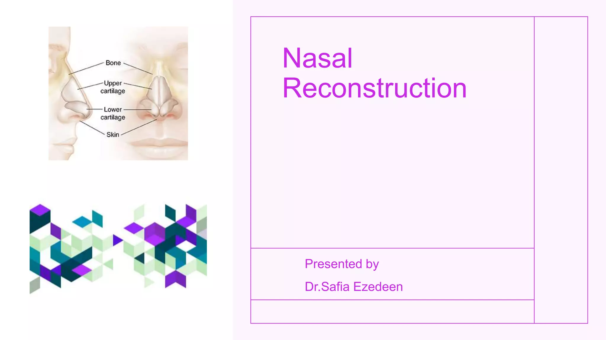
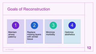
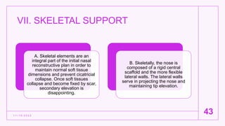
Nasal
Reconstruction
Presented by
Dr.Safia Ezedeen
Outline
1 1 / 1 5 / 2 0 2 2
2
Anatomy
Skin
coverage
Local
nasal flaps
Regional
flaps
Columella
Distant
transfers
Skeletal
support
Anatomy
A. components
1. Based on the underlying skeletal support, the nose can be divided into thirds.
The proximal third is supported by the bony nasal pyramid
The middle third is supported by the upper lateral cartilages
The distal third or lobule is supported by the alar (lower lateral) cartilages.
2. The septum is also critical to the support of the middle and distal third of the nose
1 1 / 1 5 / 2 0 2 2
3
Anatomy
1 1 / 1 5 / 2 0 2 2
4
B. Layers
The nose is composed of three layers:
skin, skeletal support, and mucosal lining
. Each layer must be taken into
consideration during reconstruction.
1. The skin is thick and sebaceous at the
tip and alar rims.
2. The skin is relatively thin over the
dorsum and sidewalls.
Aesthetic
subunits
1 1 / 1 5 / 2 0 2 2
5
Dorsum
Sidewalls
Tip
Soft tissue triangles
Columella
Alar-nostril
Aesthetic Subunits
1 1 / 1 5 / 2 0 2 2
6
Anatomy
1 1 / 1 5 / 2 0 2 2
7
Blood Supply
Arterial
• Angular artery (branch of facial artery): Lateral
surface of caudal nose
• Superior labial artery: Nasal sill, nasal septum,
and base of columella
• Dorsal nasal branch of ophthalmic artery: Axial
arterial network for dorsal and lateral nasal skin
• Infraorbital branch of internal maxillary artery:
Dorsum and lateral sidewalls of nose
Anatomy
Blood supply of
nose
1 1 / 1 5 / 2 0 2 2
8
Anatomy
1 1 / 1 5 / 2 0 2 2
9
Innervation
Sensory
• Ophthalmic division (V1)
of trigeminal nerve
Radix, rhinion, and cephalic
portion of nasal sidewalls,
skin over dorsum to tip
Anatomy
• Maxillary division (V2) of trigeminal nerve
Lateral tissue on lower half of nose, columella, and lateral vestibule
Motor
• Facial nerve VII
Procerus, depressor septi nasi, and nasalis
1 1 / 1 5 / 2 0 2 2
10
Assessment of
Defect
1 1 / 1 5 / 2 0 2 2
11
Location
Depth
• Skin and soft tissue coverage
• Cartilage and bone
Lining
Dimensions of defect
Goals of Reconstruction
1 1 / 1 5 / 2 0 2 2
12
Maintain
airway
patency
1
Replace
missing layers
with similar
tissue
2
Minimize
morbidity
3
Optimize
aesthetics
4
Rules for Reconstruction
1 1 / 1 5 / 2 0 2 2
13
If a defect occupies
more than 50% of a
subunit, enlarge defect
to incorporate entire
subunit and reconstruct
it as a whole.
Use undamaged
contralateral subunit as
the reconstructive
model.
Divide large defects into
multiple defects.
II. SKIN COVERAGE
1 1 / 1 5 / 2 0 2 2
14
A. Skin Grafts
1. Skin grafts are useful for partial-thickness defects on the dorsum or
sidewall. This may be suboptimal since texture, thickness, and color need
to match precisely or the reconstruction will look like a patch.
2. Common donor sites are the postauricular area for thin grafts and the
supraclavicular area for thicker grafts.
3. The tip is not a good area for a skin graft due to the relatively thick
sebaceous quality of the tissue in this location.
II. SKIN COVERAGE
1 1 / 1 5 / 2 0 2 2
15
5. The graft may be harvested from the helical margin or the root of the helix.
4. Composite grafts typically go through a characteristic color change from white to blue to pink over several days.
3. It may also be helpful to cool the grafts post-operatively.
2. It is helpful to allow the margins of the wound to granulate to increase the take of these grafts.
1. Full-thickness alar rim defects ≤1.5 cm in diameter may be reconstructed with auricular composite grafts consisting of skin and cartilage.
B. Composite Grafts
III. LOCAL NASAL FLAPS
1 1 / 1 5 / 2 0 2 2
16
A. Rhomboid
Flap
B. Bilobed
Flap
C. Dorsal
Nasal Flap
A. Rhomboid Flap
• 1. For small (<1.5 cm) dorsal and lateral sidewall defects, the
rhomboid flap may be used in elderly patients with excess skin.
• 2. The donor defect should be designed so that its long axis is a
line of minimal tension and along an aesthetic unit to camouflage
the scar.
1 1 / 1 5 / 2 0 2 2
17
A. Rhomboid Flap
1 1 / 1 5 / 2 0 2 2
18
B. Bilobed Flap
• 1. Used for dorsal midline, tip, and adjacent supratip defects up to
2 cm in its greatest diameter.
• 2. Some of the dorsal hump may be shaved off to facilitate closure.
• 3. The second flap is used to close the donor defect.
1 1 / 1 5 / 2 0 2 2
19
B. Bilobed Flap
1 1 / 1 5 / 2 0 2 2
20
C. Dorsal Nasal Flap
• 1. Used for dorsal defects <2 cm and small supratip defects.
• 2. This is a rotation advancement flap of the dorsal nasal skin
based laterally on the angular vessels and advanced caudally.
• 3. It may cause some cephalic tip rotation when used for larger
defects close to the tip.
1 1 / 1 5 / 2 0 2 2
21
Dorsal Nasal flap
1 1 / 1 5 / 2 0 2 2
22
IV. REGIONAL FLAPS
1 1 / 1 5 / 2 0 2 2
23
A.Advancement
Cheek Flap
B. Nasolabial
Flap
C. Glabellar
Flap
D. Paramedian
Forehead Flap
E. Gullwing
Flap
F. Scalping
(Converse)
Flap
A. Rotational/Advancement
Cheek Flap
1 1 / 1 5 / 2 0 2 2
24
1. Dorsolateral nasal defects may be
reconstructed with large rotational
advancement cheek flaps.
2. This flap is based on the subdermal
blood supply.
3. The inferior border of the incision is
placed along the alar crease. A
compensatory Burrow’s triangle may be
excised from the alar base and the
nasolabial area.
A. Rotational/Advancement Cheek Flap
1 1 / 1 5 / 2 0 2 2
25
B. Nasolabial
Flap
1 1 / 1 5 / 2 0 2 2
26
1. Used for larger defects (≤2.5 cm) of the
dorsum and sidewall.
2. This transposition flap can be based
either superiorly or inferiorly.
3. The flap is elevated in the
subcutaneous plane. This flap may also
be based only on a subcutaneous pedicle
to eliminate the dog-ear that results from
flap transposition.
B. Nasolabial Flap
1 1 / 1 5 / 2 0 2 2
27
C. Glabellar Flap
1 1 / 1 5 / 2 0 2 2
28
2. The glabellar skin can be transferred
as a rotation, midline transposition flap,
or an island flap. This flap is essentially
a dorsal nasal flap with a prominent
glabellar extension.
1. Used for defects of the upper
dorsum of the nose and medial
canthus.
C. Glabellar Flap
1 1 / 1 5 / 2 0 2 2
29
D. Paramedian
Forehead Flap
1 1 / 1 5 / 2 0 2 2
30
1. Used for large defects encroaching on the
columella, tip, or alar lobule and for subtotal
and total nasal reconstruction.
2. This is an axial flap based on the
supratrochlear artery, a terminal branch of the
ophthalmic artery. The flap may also be based
on the supraorbital or angular vessels.
3. The donor site may be closed primarily
when the defect is ≤3 cm in size.
4. Preliminary forehead skin expansion will
also help ease the close of the donor site.
D. Paramedian Forehead Flap
1 1 / 1 5 / 2 0 2 2
31
E. Gullwing Flap
1 1 / 1 5 / 2 0 2 2
32
2. This is useful for larger defects
involving the nasal tip, infratip region,
and lobule.
1. Millard modified the paramedian
forehead flap to include wide “wings”
or transverse extensions over the
natural horizontal furrows of the
forehead.
E. Gullwing Flap
1 1 / 1 5 / 2 0 2 2
33
F. Scalping
(Converse)
Flap
1 1 / 1 5 / 2 0 2 2
34
1. For large total and near-total defects of the nose,
the scalping flap may be used reliably.
2. It is elevated through a coronal incision behind
the superficial temporal artery, extending to a skin
paddle in the contralateral forehead. The frontalis
muscle is not carried in the distal end of the flap
3. The donor site on the forehead is closed with a
full-thickness skin graft.
4 .This flap is rarely used today as it has essentially
no advantages over the paramedian forehead flap.
F. Scalping (Converse) Flap
1 1 / 1 5 / 2 0 2 2
35
V. COLUMELLA
1 1 / 1 5 / 2 0 2 2
36
Columellar reconstruction can be
exceedingly difficult.
A. Nasolabial flaps—Best results
are achieved with a nasolabial
flap, preferably bilateral, and
transferred on a superior pedicle.
B. Upper lip forked flaps—Useful
in partial columellar loss or in the
elderly patient with a long upper
lip
V. COLUMELLA
1 1 / 1 5 / 2 0 2 2
37
. C. Forehead flap—A distal
extension to the paramedian
forehead flap may be rolled
inward or pinched and molded to
form the columella and to line the
vestibules.
D. Chondrocutaneous composite
graft—Auricular composite grafts
may be useful for isolated
columellar losses.
VI. DISTANT TRANSFERS
• Distant transfers are useful when the forehead is not available and
the defect is too large to repair by a graft or local flap.
1 1 / 1 5 / 2 0 2 2
38
1. Radial
forearm free flap
1 1 / 1 5 / 2 0 2 2
39
• First choice for free-tissue transfer
total nasal reconstruction.
• Thin, pliable tissue and easy to
transfer.
• Patients with normal Allen’s test can
provide an 8–10 cm vascular pedicle.
Recipient vessels are usually the facial
or superior labial arteries.
2. Dorsalis
pedis free
flap
1 1 / 1 5 / 2 0 2 2
40
• Has large, thin skin
paddle.
• Like the radial forearm
flap, can be harvested
as an osseocutaneous
transfer.
3. Postauricular
free flap
1 1 / 1 5 / 2 0 2 2
41
• A micro vascular modification
of the Washio technique.
• The dermis of the flap, which
is smooth and thin, may not
resemble the thick, sebaceous
nasal skin, but the donor site
is very inconspicuous.
4. Helical free
flap
1 1 / 1 5 / 2 0 2 2
42
• Limited to 3×3 cm surface area without distortion
of the anterior ear.
• Auricular tissue is carried on the anterior branch
of the temporal artery to bring the root of the helix
to the nose as a composite helical free flap.
• Particularly suited to nostril restoration.
VII. SKELETAL SUPPORT
1 1 / 1 5 / 2 0 2 2
43
A. Skeletal elements are an
integral part of the initial nasal
reconstructive plan in order to
maintain normal soft tissue
dimensions and prevent cicatricial
collapse. Once soft tissues
collapse and become fixed by scar,
secondary elevation is
disappointing.
B. Skeletally, the nose is
composed of a rigid central
scaffold and the more flexible
lateral walls. The lateral walls
serve in projecting the nose and
maintaining tip elevation.
VII. SKELETAL
SUPPORT
1 1 / 1 5 / 2 0 2 2
44
D. L strut
• A longitudinal piece of bone or cartilage is seated
on the nasal radix and extended along the dorsum
to the tip, where it is bent sharply to rest on the
anterior nasal spine.
• A costal osteochondral graft from the fifth rib can
be carved into appropriate hockeystick
configuration to project the tip and substitute for
the medial crura. Its disadvantage is side-to-side
instability and an excessively wide columella.
VII. SKELETAL
SUPPORT
1 1 / 1 5 / 2 0 2 2
45
E. Hinged septal flap—An L-shaped flap of septum is hinged
superiorly to provide nasal support.
F. Septal pivot flap—Simultaneous lining and some dorsal
skeletal support is provided with a composite flap of septum
pivoting anteriorly.
G. Cantilever graft
• Perhaps the most widely used method for restoring skeletal
support.
• A strong strut of bone is fixed to the nasal radix with screws or
wires and extends along the dorsum down to the tip. Absolute
rigid stability of the graft is important to prevent resorption.
• Cranium, ilium, and rib are acceptable sources for these
grafts.
VII. SKELETAL
SUPPORT
1 1 / 1 5 / 2 0 2 2
46
H. Lateral support
• Nonanatomically placed cartilage grafts
are necessary when reconstructing the
ala to prevent collapse. This is
particularly important close to the alar rim
. • Auricular cartilage is a good source for
these grafts.
Thank you
1 1 / 1 5 / 2 0 2 2
47
This Photo by Unknown author is licensed under CC BY-NC.

Nasal Reconstruction

  • 1.
  • 2.
    Outline 1 1 /1 5 / 2 0 2 2 2 Anatomy Skin coverage Local nasal flaps Regional flaps Columella Distant transfers Skeletal support
  • 3.
    Anatomy A. components 1. Basedon the underlying skeletal support, the nose can be divided into thirds. The proximal third is supported by the bony nasal pyramid The middle third is supported by the upper lateral cartilages The distal third or lobule is supported by the alar (lower lateral) cartilages. 2. The septum is also critical to the support of the middle and distal third of the nose 1 1 / 1 5 / 2 0 2 2 3
  • 4.
    Anatomy 1 1 /1 5 / 2 0 2 2 4 B. Layers The nose is composed of three layers: skin, skeletal support, and mucosal lining . Each layer must be taken into consideration during reconstruction. 1. The skin is thick and sebaceous at the tip and alar rims. 2. The skin is relatively thin over the dorsum and sidewalls.
  • 5.
    Aesthetic subunits 1 1 /1 5 / 2 0 2 2 5 Dorsum Sidewalls Tip Soft tissue triangles Columella Alar-nostril
  • 6.
    Aesthetic Subunits 1 1/ 1 5 / 2 0 2 2 6
  • 7.
    Anatomy 1 1 /1 5 / 2 0 2 2 7 Blood Supply Arterial • Angular artery (branch of facial artery): Lateral surface of caudal nose • Superior labial artery: Nasal sill, nasal septum, and base of columella • Dorsal nasal branch of ophthalmic artery: Axial arterial network for dorsal and lateral nasal skin • Infraorbital branch of internal maxillary artery: Dorsum and lateral sidewalls of nose
  • 8.
    Anatomy Blood supply of nose 11 / 1 5 / 2 0 2 2 8
  • 9.
    Anatomy 1 1 /1 5 / 2 0 2 2 9 Innervation Sensory • Ophthalmic division (V1) of trigeminal nerve Radix, rhinion, and cephalic portion of nasal sidewalls, skin over dorsum to tip
  • 10.
    Anatomy • Maxillary division(V2) of trigeminal nerve Lateral tissue on lower half of nose, columella, and lateral vestibule Motor • Facial nerve VII Procerus, depressor septi nasi, and nasalis 1 1 / 1 5 / 2 0 2 2 10
  • 11.
    Assessment of Defect 1 1/ 1 5 / 2 0 2 2 11 Location Depth • Skin and soft tissue coverage • Cartilage and bone Lining Dimensions of defect
  • 12.
    Goals of Reconstruction 11 / 1 5 / 2 0 2 2 12 Maintain airway patency 1 Replace missing layers with similar tissue 2 Minimize morbidity 3 Optimize aesthetics 4
  • 13.
    Rules for Reconstruction 11 / 1 5 / 2 0 2 2 13 If a defect occupies more than 50% of a subunit, enlarge defect to incorporate entire subunit and reconstruct it as a whole. Use undamaged contralateral subunit as the reconstructive model. Divide large defects into multiple defects.
  • 14.
    II. SKIN COVERAGE 11 / 1 5 / 2 0 2 2 14 A. Skin Grafts 1. Skin grafts are useful for partial-thickness defects on the dorsum or sidewall. This may be suboptimal since texture, thickness, and color need to match precisely or the reconstruction will look like a patch. 2. Common donor sites are the postauricular area for thin grafts and the supraclavicular area for thicker grafts. 3. The tip is not a good area for a skin graft due to the relatively thick sebaceous quality of the tissue in this location.
  • 15.
    II. SKIN COVERAGE 11 / 1 5 / 2 0 2 2 15 5. The graft may be harvested from the helical margin or the root of the helix. 4. Composite grafts typically go through a characteristic color change from white to blue to pink over several days. 3. It may also be helpful to cool the grafts post-operatively. 2. It is helpful to allow the margins of the wound to granulate to increase the take of these grafts. 1. Full-thickness alar rim defects ≤1.5 cm in diameter may be reconstructed with auricular composite grafts consisting of skin and cartilage. B. Composite Grafts
  • 16.
    III. LOCAL NASALFLAPS 1 1 / 1 5 / 2 0 2 2 16 A. Rhomboid Flap B. Bilobed Flap C. Dorsal Nasal Flap
  • 17.
    A. Rhomboid Flap •1. For small (<1.5 cm) dorsal and lateral sidewall defects, the rhomboid flap may be used in elderly patients with excess skin. • 2. The donor defect should be designed so that its long axis is a line of minimal tension and along an aesthetic unit to camouflage the scar. 1 1 / 1 5 / 2 0 2 2 17
  • 18.
    A. Rhomboid Flap 11 / 1 5 / 2 0 2 2 18
  • 19.
    B. Bilobed Flap •1. Used for dorsal midline, tip, and adjacent supratip defects up to 2 cm in its greatest diameter. • 2. Some of the dorsal hump may be shaved off to facilitate closure. • 3. The second flap is used to close the donor defect. 1 1 / 1 5 / 2 0 2 2 19
  • 20.
    B. Bilobed Flap 11 / 1 5 / 2 0 2 2 20
  • 21.
    C. Dorsal NasalFlap • 1. Used for dorsal defects <2 cm and small supratip defects. • 2. This is a rotation advancement flap of the dorsal nasal skin based laterally on the angular vessels and advanced caudally. • 3. It may cause some cephalic tip rotation when used for larger defects close to the tip. 1 1 / 1 5 / 2 0 2 2 21
  • 22.
    Dorsal Nasal flap 11 / 1 5 / 2 0 2 2 22
  • 23.
    IV. REGIONAL FLAPS 11 / 1 5 / 2 0 2 2 23 A.Advancement Cheek Flap B. Nasolabial Flap C. Glabellar Flap D. Paramedian Forehead Flap E. Gullwing Flap F. Scalping (Converse) Flap
  • 24.
    A. Rotational/Advancement Cheek Flap 11 / 1 5 / 2 0 2 2 24 1. Dorsolateral nasal defects may be reconstructed with large rotational advancement cheek flaps. 2. This flap is based on the subdermal blood supply. 3. The inferior border of the incision is placed along the alar crease. A compensatory Burrow’s triangle may be excised from the alar base and the nasolabial area.
  • 25.
    A. Rotational/Advancement CheekFlap 1 1 / 1 5 / 2 0 2 2 25
  • 26.
    B. Nasolabial Flap 1 1/ 1 5 / 2 0 2 2 26 1. Used for larger defects (≤2.5 cm) of the dorsum and sidewall. 2. This transposition flap can be based either superiorly or inferiorly. 3. The flap is elevated in the subcutaneous plane. This flap may also be based only on a subcutaneous pedicle to eliminate the dog-ear that results from flap transposition.
  • 27.
    B. Nasolabial Flap 11 / 1 5 / 2 0 2 2 27
  • 28.
    C. Glabellar Flap 11 / 1 5 / 2 0 2 2 28 2. The glabellar skin can be transferred as a rotation, midline transposition flap, or an island flap. This flap is essentially a dorsal nasal flap with a prominent glabellar extension. 1. Used for defects of the upper dorsum of the nose and medial canthus.
  • 29.
    C. Glabellar Flap 11 / 1 5 / 2 0 2 2 29
  • 30.
    D. Paramedian Forehead Flap 11 / 1 5 / 2 0 2 2 30 1. Used for large defects encroaching on the columella, tip, or alar lobule and for subtotal and total nasal reconstruction. 2. This is an axial flap based on the supratrochlear artery, a terminal branch of the ophthalmic artery. The flap may also be based on the supraorbital or angular vessels. 3. The donor site may be closed primarily when the defect is ≤3 cm in size. 4. Preliminary forehead skin expansion will also help ease the close of the donor site.
  • 31.
    D. Paramedian ForeheadFlap 1 1 / 1 5 / 2 0 2 2 31
  • 32.
    E. Gullwing Flap 11 / 1 5 / 2 0 2 2 32 2. This is useful for larger defects involving the nasal tip, infratip region, and lobule. 1. Millard modified the paramedian forehead flap to include wide “wings” or transverse extensions over the natural horizontal furrows of the forehead.
  • 33.
    E. Gullwing Flap 11 / 1 5 / 2 0 2 2 33
  • 34.
    F. Scalping (Converse) Flap 1 1/ 1 5 / 2 0 2 2 34 1. For large total and near-total defects of the nose, the scalping flap may be used reliably. 2. It is elevated through a coronal incision behind the superficial temporal artery, extending to a skin paddle in the contralateral forehead. The frontalis muscle is not carried in the distal end of the flap 3. The donor site on the forehead is closed with a full-thickness skin graft. 4 .This flap is rarely used today as it has essentially no advantages over the paramedian forehead flap.
  • 35.
    F. Scalping (Converse)Flap 1 1 / 1 5 / 2 0 2 2 35
  • 36.
    V. COLUMELLA 1 1/ 1 5 / 2 0 2 2 36 Columellar reconstruction can be exceedingly difficult. A. Nasolabial flaps—Best results are achieved with a nasolabial flap, preferably bilateral, and transferred on a superior pedicle. B. Upper lip forked flaps—Useful in partial columellar loss or in the elderly patient with a long upper lip
  • 37.
    V. COLUMELLA 1 1/ 1 5 / 2 0 2 2 37 . C. Forehead flap—A distal extension to the paramedian forehead flap may be rolled inward or pinched and molded to form the columella and to line the vestibules. D. Chondrocutaneous composite graft—Auricular composite grafts may be useful for isolated columellar losses.
  • 38.
    VI. DISTANT TRANSFERS •Distant transfers are useful when the forehead is not available and the defect is too large to repair by a graft or local flap. 1 1 / 1 5 / 2 0 2 2 38
  • 39.
    1. Radial forearm freeflap 1 1 / 1 5 / 2 0 2 2 39 • First choice for free-tissue transfer total nasal reconstruction. • Thin, pliable tissue and easy to transfer. • Patients with normal Allen’s test can provide an 8–10 cm vascular pedicle. Recipient vessels are usually the facial or superior labial arteries.
  • 40.
    2. Dorsalis pedis free flap 11 / 1 5 / 2 0 2 2 40 • Has large, thin skin paddle. • Like the radial forearm flap, can be harvested as an osseocutaneous transfer.
  • 41.
    3. Postauricular free flap 11 / 1 5 / 2 0 2 2 41 • A micro vascular modification of the Washio technique. • The dermis of the flap, which is smooth and thin, may not resemble the thick, sebaceous nasal skin, but the donor site is very inconspicuous.
  • 42.
    4. Helical free flap 11 / 1 5 / 2 0 2 2 42 • Limited to 3×3 cm surface area without distortion of the anterior ear. • Auricular tissue is carried on the anterior branch of the temporal artery to bring the root of the helix to the nose as a composite helical free flap. • Particularly suited to nostril restoration.
  • 43.
    VII. SKELETAL SUPPORT 11 / 1 5 / 2 0 2 2 43 A. Skeletal elements are an integral part of the initial nasal reconstructive plan in order to maintain normal soft tissue dimensions and prevent cicatricial collapse. Once soft tissues collapse and become fixed by scar, secondary elevation is disappointing. B. Skeletally, the nose is composed of a rigid central scaffold and the more flexible lateral walls. The lateral walls serve in projecting the nose and maintaining tip elevation.
  • 44.
    VII. SKELETAL SUPPORT 1 1/ 1 5 / 2 0 2 2 44 D. L strut • A longitudinal piece of bone or cartilage is seated on the nasal radix and extended along the dorsum to the tip, where it is bent sharply to rest on the anterior nasal spine. • A costal osteochondral graft from the fifth rib can be carved into appropriate hockeystick configuration to project the tip and substitute for the medial crura. Its disadvantage is side-to-side instability and an excessively wide columella.
  • 45.
    VII. SKELETAL SUPPORT 1 1/ 1 5 / 2 0 2 2 45 E. Hinged septal flap—An L-shaped flap of septum is hinged superiorly to provide nasal support. F. Septal pivot flap—Simultaneous lining and some dorsal skeletal support is provided with a composite flap of septum pivoting anteriorly. G. Cantilever graft • Perhaps the most widely used method for restoring skeletal support. • A strong strut of bone is fixed to the nasal radix with screws or wires and extends along the dorsum down to the tip. Absolute rigid stability of the graft is important to prevent resorption. • Cranium, ilium, and rib are acceptable sources for these grafts.
  • 46.
    VII. SKELETAL SUPPORT 1 1/ 1 5 / 2 0 2 2 46 H. Lateral support • Nonanatomically placed cartilage grafts are necessary when reconstructing the ala to prevent collapse. This is particularly important close to the alar rim . • Auricular cartilage is a good source for these grafts.
  • 47.
    Thank you 1 1/ 1 5 / 2 0 2 2 47 This Photo by Unknown author is licensed under CC BY-NC.