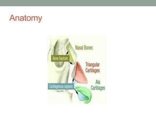
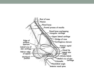
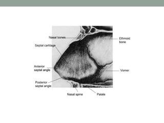
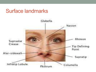
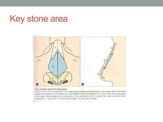
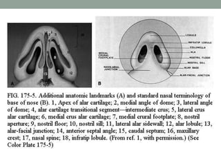
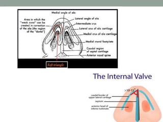
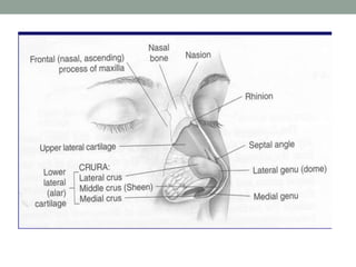
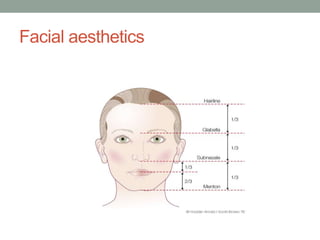
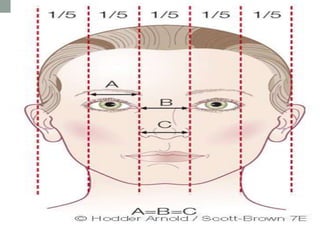
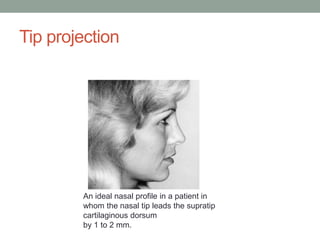
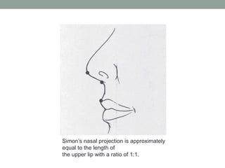
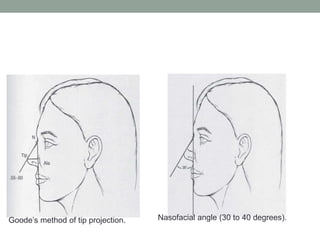
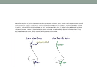
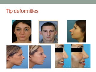
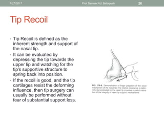
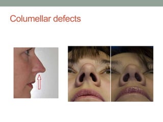
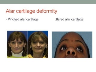
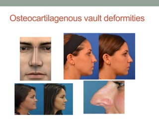
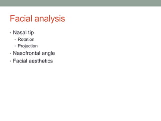
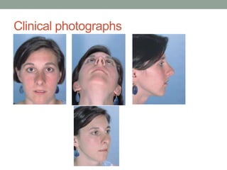
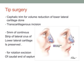
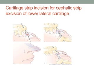
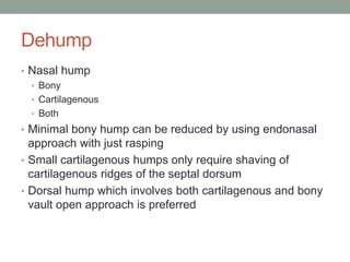
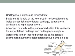
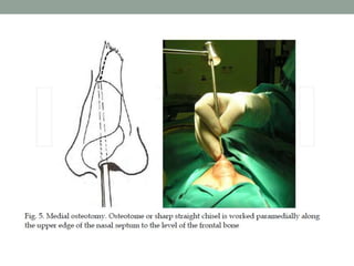
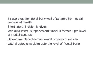
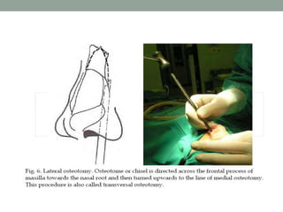
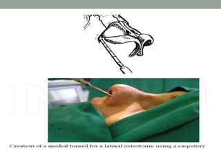
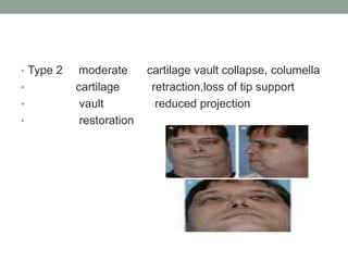
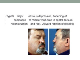
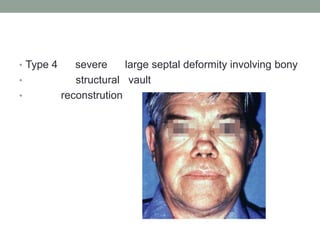
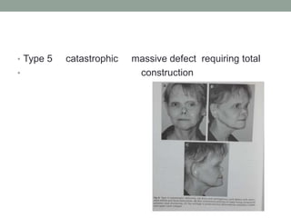
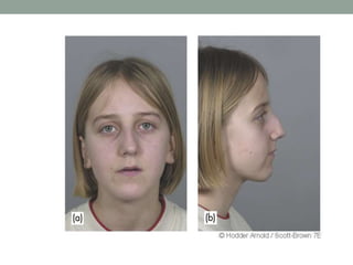
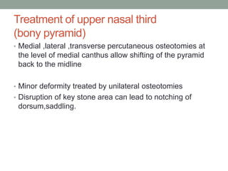
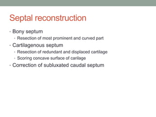
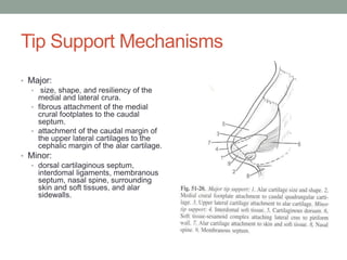
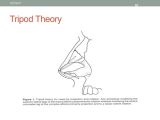
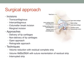
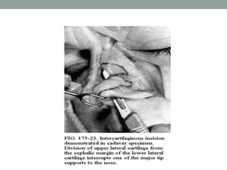
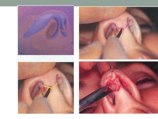
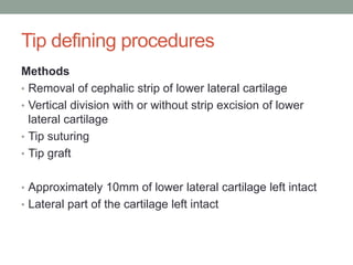
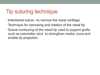
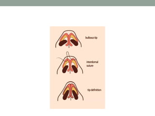
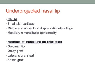
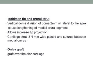
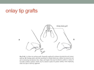
RHINOPLASTY
Dr.Disha sharma
Junior Resident
ENT-HNS
HISTORY
• Began - ancient egypt and India.
• Description of nasal reconstruction in Susruta samhita
(500 B.C.)
• 1887 - John Orlando Roe performed first intranasal
rhinoplasty.
• Jacques joseph – father of modern facial plastic surgery –
published his Treatise on rhinoplasty.
Anatomy
Surface landmarks
Key stone area
Weak zone
Facial aesthetics
Facial aesthetics
Facial analysis – tip rotation
Tip rotation generally occurs along an arc
produced by a
radius based at the external auditory canal.
Tip projection
An ideal nasal profile in a patient in
whom the nasal tip leads the supratip
cartilaginous dorsum
by 1 to 2 mm.
Simon’s nasal projection is approximately
equal to the length of
the upper lip with a ratio of 1:1.
Goode’s method of tip projection. Nasofacial angle (30 to 40 degrees).
Clinical Assessment
• Routine investigation
• Clinical assessment
Assess external nasal deformity
Quality of skin
Facial analysis
• Photographic documents
Basic principles to be taken care……
• Be conservative
• Should know where to stop
• Never promise miraculous results after surgery
• Beware of psychotic patients
• Consent
External nasal deformities
• Tip
• Bulbous
• Bifid
• Overprojected
• Underprojected
• Tip ptosis
• Columella
• Retracted
• Deviated
• Broadened
• Alar cartilage
• Pinching/flaring
• Osseocartilagenous vault
• Deviated
• Bony/cartilagenous/both
• Saddle
• Bony/cartilagenous/both
• Hump
• Bony/cartilagenous/both
• Tension nose
Tip deformities
Tip Recoil
• Tip Recoil is defined as the
inherent strength and support of
the nasal tip.
• It can be evaluated by
depressing the tip towards the
upper lip and watching for the
tip's supportive structure to
spring back into position.
• If the recoil is good, and the tip
cartilages resist the deforming
influence, then tip surgery can
usually be performed without
fear of substantial support loss.
1/27/2017 Prof Sameer ALI Bafaqeeh 26
Columellar defects
Alar cartilage deformity
• Pinched alar cartilage .flared alar cartilage
Osteocartilagenous vault deformities
Quality of skin
Thick skin
Masks refinement and
definition
Failure to contract – excess
soft tissue scar
Does not show small
irregularities
Thin skin
• Small irregularities
become visible
• Early healing
• Less oedema
• Ensure that all bony,
cartilageneous grafts or
implants are precisely
positioned and smoothly
contoured.
Facial analysis
• Nasal tip
• Rotation
• Projection
• Nasofrontal angle
• Facial aesthetics
Clinical photographs
Types of rhinoplasty
• Open approach Closed approach
• External rhinoplasty endonasal rhinoplasty
• Trans columellar incision incisions positioned inside
• the nostril
EXTERNAL RHINOPLASTY
Indications
• Extensive revision surgery
• Severe nasal trauma
• Congenital deformities: cleft lip nose
• Marked tip deformities
• Elaborate reduction and augmentation procedures
• Correction of extreme overprojection
Principles of external rhinoplasty
• Incision- mid-columella incision connected to bilateral
marginal incision
• Dissection in subperiosteal and subperichondrial planes
• Division of medial intercrural tissue offers access to
caudal septum and premaxillary spine
• Division of upper lateral cartilages from quadrilateral
cartilage offers acccessability to whole of septum
Advantages
• Extensive exposure for both septal and rhinoplasty
surgery
• Binocular vision
• Use of both hands
• Control of bleeding and diathermy
• precise placement and suturing of struts,battens and
shield grafts
• Valve area preserved
Techniques
Cartilage resections
Lateral /medial crura
Alar cartilage modifications and reorientation
Complete strip
Interrupted strip
Surgical technique
• Broken transcolumellar incision
• If columella short in case of cleft lip-V incision
• Mid-columella incision situated above medial crural foot
plates
• Vertical columellar incision made 1.5-2mm inside
vestibule
• Separate lateral incision given which is joined medially
over the domes
Surgical technique……
• Dissection carried in midline just cephalic to dome
subperichondrial plane
• Dissection of soft tissue of bony pyramid should start
above caudal end of nasal bone
• Nasal septum- between medial crurae of lower lateral
cartilage or hemitransfixion incision
• Strut used to
correct buckled medial crura
,strengthen weak medial crura
,correct tip asymmetries
,stable base for tip graft
Specific applications
The bony pyramid in external rhinoplasty
• Allows use of burr or reduction of the soft tissue envelope
at nasion to deepen the nasofrontal angle
• Application of soft tissue onlay grafts
• Bony dehumping together with lateral, medial, and
intermediate osteotomies
• The middle nasal vault
• Placement of cartilaginous strips or spreader grafts to
open up the nasal valve area and angles
• Middle nasal vault
Shaded areas showing placement of
spreader grafts
REDUCTION
RHINOPLASTY
Indications…
• Patients with ideal height and position of the nasion
associated with excess dorsal convexity
• Oversized alar cartilages producing increase tip and
lobule volume
Aims
o Aim-strong nasal dorsum in lateral profile-relates to ideal
nasion height
• Tip defining point-projecting just above dorsal line-to
create supratip break. In males may be on a straight line
with dorsum
Contraindications
Underprojected tip pseudohumps
Deep radix (deep root) of nose
Short over-rotated nose
Triad of thin skin
delicate alar side walls
bifidity
Techniques
• Nasal tip surgery
• Dehump
• Osteotomies
Tip surgery
Cephalic trim for volume reduction of lower lateral
cartilage done
Transcartilagenous incision
5mm of continous
Strip of lateral crus of
Lower lateral cartilage
Is preserved .
for rotation excision
Of caudal end of septum
Cartilage strip incision for cephalic strip
excision of lower lateral cartilage
Dehump
• Nasal hump
• Bony
• Cartilagenous
• Both
• Minimal bony hump can be reduced by using endonasal
approach with just rasping
• Small cartilagenous humps only require shaving of
cartilagenous ridges of the septal dorsum
• Dorsal hump which involves both cartilagenous and bony
vault open approach is preferred
• Cartilagenous dorsum is reduced first.
• Blade no.15 is held at the key area in horizontal plane to
incise across left upper lateral cartilage, quadrilateral
cartilage and right upper lateral .
• Advanced caudally in the plane of reduction this transects
the upper lateral cartilage and cartilagenous septum.
• Osteotome is then inserted under the cartilagenous
segment removing the osteocartilagenous hump en bloc
The Dorsal Hump
Osteotomies
• Medial osteotomy
• Lateral osteotomy
• Low to high
• Low to low
• Transverse osteotomy
• Intermediate osteotomy
Medial osteotomy
• It seperates the nasal bone from the septum
• Made on both side
• Nasal bone seperated at intranasal suture
• Short intercartilageneous given
• Outer peritosteun is pushed to the side
• Osteotome is placed at about 2mm paramedially
• Osteotome is worked through the bone slightly below the
level of frontal bone
Lateral osteotomy
• It seperates the lateral bony wall of pyramid from nasal
process of maxilla
• Short lateral incision is given
• Medial to lateral subperiosteal tunnel is formed upto level
of medial canthus
• Osteotome placed across frontal process of maxilla
• Lateral osteotomy done upto the level of frontal bone
Tranverse osteotomy
• Seperates the bony pyramid from frontal bone and nasal
spine of frontal bone
• Osteotomy made at a level just below nasion
Complications
• Polybeak appearance
• Inverted v deformity
• Flat nose
• Senstivity and pain
AUGMENTATION
RHINOPLASTY
Saddle nose
• Pug nose or boxer nose
Aetiology
1. Trauma
2. Nasal surgery
3. Familial/ethnic
4. Infections
5. Chronic Inflammatory conditions
6. tumour
Daniel and Brenner Classification for
saddle nose
• TYPE TERM DESCRIPTION
0 pseudosaddle relative depression of
cartilaginous dorsum relative
to bony septum
Type 1 minor minor decrease in septal support
cosmetic supratip depression and columella
concealment retraction
• Type 2 moderate cartilage vault collapse, columella
• cartilage retraction,loss of tip support
• vault reduced projection
• restoration
• Type3 major obvious depression, flattening of
• composite of middle vault,drop in septal dorsum
• reconstruction and roof. Upward rotation of nasal tip
• Type 4 severe large septal deformity involving bony
• structural vault
• reconstrution
• Type 5 catastrophic massive defect requiring total
• construction
Properties of implant materials
• Noncarcinogenic and nonimmunogenic
• Not toxic
• Nondestructive , should not impede healing
• Physical properties match the local tissues
• Nonresorbable
• Easily available
• Cost effective easy to sterilize
Graft Materials
• Autografts - cartilage, bone, dermis and fascia
• Homografts - Irradiated rib, pooled acellular dermis
• Xenografts - Leather, duck's sternum, bovine cartilage
• Precious metals - Titanium, gold, silver, metal alloys
• Inert bioimplants - Coral, ivory
• Synthetic compounds - Silicone, polytetrafluoroethylenes,
polyamide mesh
Synthetic biomaterials
• Silicone
• Teflon
• Proplast
• Gore-tex
• Medpore
• Mersiline
• Titanium
• ceramics
Type of autografts
• Septal cartilage
• Auricular cartilage
• Costal cartilage
• Bone graft
Septal cartilage graft
• Harvested using hemitransfixion/transfixion/external
approach
• Harvested posterior to an imaginary line drawn from
anterior nasal spine to osteocartilagenous junction
• Septal cartilage is more rigid and easier to carve and
shape
Conchal cartilage
• Conchal cartilage of 3.5cm in length ,antihelix fold to be
kept intact
• Can be harvested by anterior or posterior approach
• For anterior approach incision is given just anterior and
deep to antihelical fold
• Skin is elevated from underlying cartilage by blunt
dissection upto the posterior edge of external auditory
meatus
Costal cartilage
• Straight rib cartilage harvested from 6th to 8th rib through
3-4cm skin incision
• Rectus muscle is split vertically and retracted
Management ..
• Surgical approach
• External approach preferred
Suture fixation
Reduces risk of infection
Preparation of graft and implant
Use template to achieve the exact size
Avoid abrupt edges
Suture fixation in case layered cartilage graft
• Preparation of bed
• Level base to avoid rocking
• Rasped to remove irregularities
• Splinting
• Percutaneous securing sutures
• Taping the skin of the nose down
• External nasal splint
DEVIATED NOSE
Principles of surgical correction
• Deviation and a dorsal hump – reduction technique
• Deviated nose without dorsal hump- septal surgery
• Deviation with saddling- augmentation
Treatment of upper nasal third
(bony pyramid)
• Medial ,lateral ,transverse percutaneous osteotomies at
the level of medial canthus allow shifting of the pyramid
back to the midline
• Minor deformity treated by unilateral osteotomies
• Disruption of key stone area can lead to notching of
dorsum,saddling.
Septal reconstruction
• Bony septum
• Resection of most prominent and curved part
• Cartilagenous septum
• Resection of redundant and displaced cartilage
• Scoring concave surface of carilage
• Correction of subluxated caudal septum
Treatment of middle third
• Shaving of the convex part of the septum
• Use of bilateral and unilateral spreader graft
• Staggered cuts to weaken bowed cartilage
Treatment of lower third
If nasal spine central –post septal angle should be sutured
with figure of eight
Locking technique- untrimmed displaced caudal septum is
placed on contralateral side of thinned nasal spine
Absent caudal septum with deviated tip will require
replacement with graft.
NASAL TIP AND
NASOLABIALANGLE
Tip Support Mechanisms
• Major:
• size, shape, and resiliency of the
medial and lateral crura.
• fibrous attachment of the medial
crural footplates to the caudal
septum.
• attachment of the caudal margin of
the upper lateral cartilages to the
cephalic margin of the alar cartilage.
• Minor:
• dorsal cartilaginous septum,
interdomal ligaments, membranous
septum, nasal spine, surrounding
skin and soft tissues, and alar
sidewalls.
Tripod Theory
1/27/2017
97
Surgical approach
• Incisions
• Transcartilaginous
• Intercartilaginous
• Columellar break incision
• Marginal incision
• Approaches
• Delivery of tip cartilages
• Non-delivery of tip cartilages
• Open approach
• Retrograde approach
• Techniques
• Volume reduction with residual complete strip
• Volume reduction with suture reorientation of residual strip
• Interrupted strip
Transcartilagenous Incision
• Incise through vestibular skin
• Similar to intercartilaginous, but 3-5mm caudal to the
cephalic end of LLC
• This is caudal to the nasal valve
• Decreases risk of nasal obstruction (avoids scar
contracture of the valve)
Transcollumellar and marginal incision
• External approach
• Crosses collumella just above flared ends of the
medial crura
• Vertical columellar incision placed 1-2 mm inside
the vestibule
• Separate lateral incision given along the caudal
margin of lower lateral cartilage joined medially
over the domes.
Tip defining procedures
Methods
• Removal of cephalic strip of lower lateral cartilage
• Vertical division with or without strip excision of lower
lateral cartilage
• Tip suturing
• Tip graft
• Approximately 10mm of lower lateral cartilage left intact
• Lateral part of the cartilage left intact
Tip suturing technique
• Interdomal suture –to narrrow the nasal cartllage
• Technique for narrowing and rotation of the nasal tip
• Suture contouring of the nasal tip used to support grafts
such as columellar strut to strengthen media; crura and
enable tip projection
Underprojected nasal tip
• Cause
• Small alar cartilage
• Middle and upper third disproportionately large
• Maxillary n mandibular abnormality
• Methods of increasing tip projection
• Goldman tip
• Onlay graft
• Lateral crural steal
• Shield graft
• goldman tip and crural strut
• Vertical dome division of dome 2mm or lateral to the apex
• cause lengthening of medial crura segment
• Allows increase tip projection
• Cartilage strut 3-4 mm wide placed and sutured between
medial cruras
• Onlay graft
• graft over the alar cartilage
onlay tip grafts
• lateral crural steal
• Alar cartilage resected in intermediate crural area
alar cartilage may be delivered
lateral crura advanced to medial crura and
sutured
tip advancement and tip rotation
Shield grafts
In 1975 by Sheen
Indication-
Narrow middle nasal
Vault
Underprojected tip
Weak lower lateral
Cartilage
Incision- marginal
Or via external rhinoplasty
Columellar struts
Overprojecting tip
• Cause
• Overdevelopment of alar cartilage/ nasal spine/caudal
Septum/quadrangular cartilage dorsum
• Elongated columella
• Iatrogenic overprojection
• Methods of reduction
• transfixion excision
• Vertical dome division (goldman)
• Medial and lateral vertical segment excision
Vertical dome division –Irwing Goldman in 1957
Tip delivery approach
Vertical division of alar dome 1mm lateral to highest point
of dome
intermediate crura rotated anteriorly
intermediate crura sutured with medial crura
Medial crura stabilized their height trimmed
Tip Rotation Techniques
• Complete strip technique
• Interrupted strip technique
• Lateral interruption
• Medial interruption
• Lateral interruption with suture rotation
Complete Strip Techniques
1/27/2017 118
Interrupted Strip Techniques
REVISION RHINOPLASTY
common deformities….
• Polybeak
• Dropping tips
• Uncorrected broad nasal tip
• Irregularities on nasal dorsum
Polybeak deformity
• Causes
• Drooping of nasal tip
• Absolute or relative high septal angle
• Reconstruction
• Increase of tip projection
• Reduction of cartilagenous nasal dorsum
• Combination of both
Nasal Dorsal Irregularities
• More prominent in thin skinned patients- tension nose
• Most common site is K area
• Small bony irregularities can be easily rasped
• Cartilagenous are made smooth with knife or scissors
• Autogenous grafts-from radix to septal cartilage
• Crushed septal cartilage strut preferred
Columella deformities
• Acute nasolabial angle
• Retraction of columella
• Cause-
• Overresection of caudal septal end
• Resection of anterior nasal spine
• Creation of columella pocket between medial crura
Correction-
Distance between columellar skin and caudal septum by
inserting a cartilage strut
OVERSHORTENED
NOSE
Patients with this tend to take legal
Action
Too much cartilage or vestibular
Skin removed from lateral cartilage
• 1 septal-columella graft
• 2 spreaded graft between upper lateral cartilage
• 3interposition graft
B/w upper and
Lower lateral
cartilage
THANK YOU

Rhinoplasty

  • 1.
  • 2.
    HISTORY • Began -ancient egypt and India. • Description of nasal reconstruction in Susruta samhita (500 B.C.) • 1887 - John Orlando Roe performed first intranasal rhinoplasty. • Jacques joseph – father of modern facial plastic surgery – published his Treatise on rhinoplasty.
  • 3.
  • 6.
  • 7.
  • 9.
  • 11.
  • 12.
  • 15.
    Facial analysis –tip rotation Tip rotation generally occurs along an arc produced by a radius based at the external auditory canal.
  • 17.
    Tip projection An idealnasal profile in a patient in whom the nasal tip leads the supratip cartilaginous dorsum by 1 to 2 mm.
  • 18.
    Simon’s nasal projectionis approximately equal to the length of the upper lip with a ratio of 1:1.
  • 19.
    Goode’s method oftip projection. Nasofacial angle (30 to 40 degrees).
  • 21.
    Clinical Assessment • Routineinvestigation • Clinical assessment Assess external nasal deformity Quality of skin Facial analysis • Photographic documents
  • 22.
    Basic principles tobe taken care…… • Be conservative • Should know where to stop • Never promise miraculous results after surgery • Beware of psychotic patients • Consent
  • 23.
    External nasal deformities •Tip • Bulbous • Bifid • Overprojected • Underprojected • Tip ptosis • Columella • Retracted • Deviated • Broadened
  • 24.
    • Alar cartilage •Pinching/flaring • Osseocartilagenous vault • Deviated • Bony/cartilagenous/both • Saddle • Bony/cartilagenous/both • Hump • Bony/cartilagenous/both • Tension nose
  • 25.
  • 26.
    Tip Recoil • TipRecoil is defined as the inherent strength and support of the nasal tip. • It can be evaluated by depressing the tip towards the upper lip and watching for the tip's supportive structure to spring back into position. • If the recoil is good, and the tip cartilages resist the deforming influence, then tip surgery can usually be performed without fear of substantial support loss. 1/27/2017 Prof Sameer ALI Bafaqeeh 26
  • 27.
  • 28.
    Alar cartilage deformity •Pinched alar cartilage .flared alar cartilage
  • 29.
  • 30.
    Quality of skin Thickskin Masks refinement and definition Failure to contract – excess soft tissue scar Does not show small irregularities Thin skin • Small irregularities become visible • Early healing • Less oedema • Ensure that all bony, cartilageneous grafts or implants are precisely positioned and smoothly contoured.
  • 31.
    Facial analysis • Nasaltip • Rotation • Projection • Nasofrontal angle • Facial aesthetics
  • 32.
  • 33.
    Types of rhinoplasty •Open approach Closed approach • External rhinoplasty endonasal rhinoplasty • Trans columellar incision incisions positioned inside • the nostril
  • 34.
  • 35.
    Indications • Extensive revisionsurgery • Severe nasal trauma • Congenital deformities: cleft lip nose • Marked tip deformities • Elaborate reduction and augmentation procedures • Correction of extreme overprojection
  • 36.
    Principles of externalrhinoplasty • Incision- mid-columella incision connected to bilateral marginal incision • Dissection in subperiosteal and subperichondrial planes • Division of medial intercrural tissue offers access to caudal septum and premaxillary spine • Division of upper lateral cartilages from quadrilateral cartilage offers acccessability to whole of septum
  • 37.
    Advantages • Extensive exposurefor both septal and rhinoplasty surgery • Binocular vision • Use of both hands • Control of bleeding and diathermy • precise placement and suturing of struts,battens and shield grafts • Valve area preserved
  • 38.
    Techniques Cartilage resections Lateral /medialcrura Alar cartilage modifications and reorientation Complete strip Interrupted strip
  • 39.
    Surgical technique • Brokentranscolumellar incision • If columella short in case of cleft lip-V incision • Mid-columella incision situated above medial crural foot plates • Vertical columellar incision made 1.5-2mm inside vestibule • Separate lateral incision given which is joined medially over the domes
  • 40.
    Surgical technique…… • Dissectioncarried in midline just cephalic to dome subperichondrial plane • Dissection of soft tissue of bony pyramid should start above caudal end of nasal bone • Nasal septum- between medial crurae of lower lateral cartilage or hemitransfixion incision • Strut used to correct buckled medial crura ,strengthen weak medial crura ,correct tip asymmetries ,stable base for tip graft
  • 42.
    Specific applications The bonypyramid in external rhinoplasty • Allows use of burr or reduction of the soft tissue envelope at nasion to deepen the nasofrontal angle • Application of soft tissue onlay grafts • Bony dehumping together with lateral, medial, and intermediate osteotomies • The middle nasal vault • Placement of cartilaginous strips or spreader grafts to open up the nasal valve area and angles
  • 43.
    • Middle nasalvault Shaded areas showing placement of spreader grafts
  • 44.
  • 45.
    Indications… • Patients withideal height and position of the nasion associated with excess dorsal convexity • Oversized alar cartilages producing increase tip and lobule volume
  • 46.
    Aims o Aim-strong nasaldorsum in lateral profile-relates to ideal nasion height • Tip defining point-projecting just above dorsal line-to create supratip break. In males may be on a straight line with dorsum
  • 47.
    Contraindications Underprojected tip pseudohumps Deepradix (deep root) of nose Short over-rotated nose Triad of thin skin delicate alar side walls bifidity
  • 50.
    Techniques • Nasal tipsurgery • Dehump • Osteotomies
  • 51.
    Tip surgery Cephalic trimfor volume reduction of lower lateral cartilage done Transcartilagenous incision 5mm of continous Strip of lateral crus of Lower lateral cartilage Is preserved . for rotation excision Of caudal end of septum
  • 52.
    Cartilage strip incisionfor cephalic strip excision of lower lateral cartilage
  • 53.
    Dehump • Nasal hump •Bony • Cartilagenous • Both • Minimal bony hump can be reduced by using endonasal approach with just rasping • Small cartilagenous humps only require shaving of cartilagenous ridges of the septal dorsum • Dorsal hump which involves both cartilagenous and bony vault open approach is preferred
  • 54.
    • Cartilagenous dorsumis reduced first. • Blade no.15 is held at the key area in horizontal plane to incise across left upper lateral cartilage, quadrilateral cartilage and right upper lateral . • Advanced caudally in the plane of reduction this transects the upper lateral cartilage and cartilagenous septum. • Osteotome is then inserted under the cartilagenous segment removing the osteocartilagenous hump en bloc
  • 55.
  • 56.
    Osteotomies • Medial osteotomy •Lateral osteotomy • Low to high • Low to low • Transverse osteotomy • Intermediate osteotomy
  • 57.
    Medial osteotomy • Itseperates the nasal bone from the septum • Made on both side • Nasal bone seperated at intranasal suture • Short intercartilageneous given
  • 58.
    • Outer peritosteunis pushed to the side • Osteotome is placed at about 2mm paramedially • Osteotome is worked through the bone slightly below the level of frontal bone
  • 60.
  • 61.
    • It seperatesthe lateral bony wall of pyramid from nasal process of maxilla • Short lateral incision is given • Medial to lateral subperiosteal tunnel is formed upto level of medial canthus • Osteotome placed across frontal process of maxilla • Lateral osteotomy done upto the level of frontal bone
  • 65.
    Tranverse osteotomy • Seperatesthe bony pyramid from frontal bone and nasal spine of frontal bone • Osteotomy made at a level just below nasion
  • 66.
    Complications • Polybeak appearance •Inverted v deformity • Flat nose • Senstivity and pain
  • 67.
  • 68.
    Saddle nose • Pugnose or boxer nose
  • 69.
    Aetiology 1. Trauma 2. Nasalsurgery 3. Familial/ethnic 4. Infections 5. Chronic Inflammatory conditions 6. tumour
  • 70.
    Daniel and BrennerClassification for saddle nose • TYPE TERM DESCRIPTION 0 pseudosaddle relative depression of cartilaginous dorsum relative to bony septum
  • 71.
    Type 1 minorminor decrease in septal support cosmetic supratip depression and columella concealment retraction
  • 72.
    • Type 2moderate cartilage vault collapse, columella • cartilage retraction,loss of tip support • vault reduced projection • restoration
  • 73.
    • Type3 majorobvious depression, flattening of • composite of middle vault,drop in septal dorsum • reconstruction and roof. Upward rotation of nasal tip
  • 74.
    • Type 4severe large septal deformity involving bony • structural vault • reconstrution
  • 75.
    • Type 5catastrophic massive defect requiring total • construction
  • 76.
    Properties of implantmaterials • Noncarcinogenic and nonimmunogenic • Not toxic • Nondestructive , should not impede healing • Physical properties match the local tissues • Nonresorbable • Easily available • Cost effective easy to sterilize
  • 77.
    Graft Materials • Autografts- cartilage, bone, dermis and fascia • Homografts - Irradiated rib, pooled acellular dermis • Xenografts - Leather, duck's sternum, bovine cartilage • Precious metals - Titanium, gold, silver, metal alloys • Inert bioimplants - Coral, ivory • Synthetic compounds - Silicone, polytetrafluoroethylenes, polyamide mesh
  • 78.
    Synthetic biomaterials • Silicone •Teflon • Proplast • Gore-tex • Medpore • Mersiline • Titanium • ceramics
  • 79.
    Type of autografts •Septal cartilage • Auricular cartilage • Costal cartilage • Bone graft
  • 80.
    Septal cartilage graft •Harvested using hemitransfixion/transfixion/external approach • Harvested posterior to an imaginary line drawn from anterior nasal spine to osteocartilagenous junction • Septal cartilage is more rigid and easier to carve and shape
  • 81.
    Conchal cartilage • Conchalcartilage of 3.5cm in length ,antihelix fold to be kept intact • Can be harvested by anterior or posterior approach • For anterior approach incision is given just anterior and deep to antihelical fold • Skin is elevated from underlying cartilage by blunt dissection upto the posterior edge of external auditory meatus
  • 83.
    Costal cartilage • Straightrib cartilage harvested from 6th to 8th rib through 3-4cm skin incision • Rectus muscle is split vertically and retracted
  • 84.
    Management .. • Surgicalapproach • External approach preferred Suture fixation Reduces risk of infection Preparation of graft and implant Use template to achieve the exact size Avoid abrupt edges Suture fixation in case layered cartilage graft
  • 85.
    • Preparation ofbed • Level base to avoid rocking • Rasped to remove irregularities • Splinting • Percutaneous securing sutures • Taping the skin of the nose down • External nasal splint
  • 87.
  • 88.
    Principles of surgicalcorrection • Deviation and a dorsal hump – reduction technique • Deviated nose without dorsal hump- septal surgery • Deviation with saddling- augmentation
  • 90.
    Treatment of uppernasal third (bony pyramid) • Medial ,lateral ,transverse percutaneous osteotomies at the level of medial canthus allow shifting of the pyramid back to the midline • Minor deformity treated by unilateral osteotomies • Disruption of key stone area can lead to notching of dorsum,saddling.
  • 91.
    Septal reconstruction • Bonyseptum • Resection of most prominent and curved part • Cartilagenous septum • Resection of redundant and displaced cartilage • Scoring concave surface of carilage • Correction of subluxated caudal septum
  • 92.
    Treatment of middlethird • Shaving of the convex part of the septum • Use of bilateral and unilateral spreader graft • Staggered cuts to weaken bowed cartilage
  • 93.
    Treatment of lowerthird If nasal spine central –post septal angle should be sutured with figure of eight Locking technique- untrimmed displaced caudal septum is placed on contralateral side of thinned nasal spine Absent caudal septum with deviated tip will require replacement with graft.
  • 95.
  • 96.
    Tip Support Mechanisms •Major: • size, shape, and resiliency of the medial and lateral crura. • fibrous attachment of the medial crural footplates to the caudal septum. • attachment of the caudal margin of the upper lateral cartilages to the cephalic margin of the alar cartilage. • Minor: • dorsal cartilaginous septum, interdomal ligaments, membranous septum, nasal spine, surrounding skin and soft tissues, and alar sidewalls.
  • 97.
  • 98.
    Surgical approach • Incisions •Transcartilaginous • Intercartilaginous • Columellar break incision • Marginal incision • Approaches • Delivery of tip cartilages • Non-delivery of tip cartilages • Open approach • Retrograde approach • Techniques • Volume reduction with residual complete strip • Volume reduction with suture reorientation of residual strip • Interrupted strip
  • 101.
    Transcartilagenous Incision • Incisethrough vestibular skin • Similar to intercartilaginous, but 3-5mm caudal to the cephalic end of LLC • This is caudal to the nasal valve • Decreases risk of nasal obstruction (avoids scar contracture of the valve)
  • 103.
    Transcollumellar and marginalincision • External approach • Crosses collumella just above flared ends of the medial crura • Vertical columellar incision placed 1-2 mm inside the vestibule • Separate lateral incision given along the caudal margin of lower lateral cartilage joined medially over the domes.
  • 105.
    Tip defining procedures Methods •Removal of cephalic strip of lower lateral cartilage • Vertical division with or without strip excision of lower lateral cartilage • Tip suturing • Tip graft • Approximately 10mm of lower lateral cartilage left intact • Lateral part of the cartilage left intact
  • 106.
    Tip suturing technique •Interdomal suture –to narrrow the nasal cartllage • Technique for narrowing and rotation of the nasal tip • Suture contouring of the nasal tip used to support grafts such as columellar strut to strengthen media; crura and enable tip projection
  • 109.
    Underprojected nasal tip •Cause • Small alar cartilage • Middle and upper third disproportionately large • Maxillary n mandibular abnormality • Methods of increasing tip projection • Goldman tip • Onlay graft • Lateral crural steal • Shield graft
  • 110.
    • goldman tipand crural strut • Vertical dome division of dome 2mm or lateral to the apex • cause lengthening of medial crura segment • Allows increase tip projection • Cartilage strut 3-4 mm wide placed and sutured between medial cruras • Onlay graft • graft over the alar cartilage
  • 111.
  • 112.
    • lateral cruralsteal • Alar cartilage resected in intermediate crural area alar cartilage may be delivered lateral crura advanced to medial crura and sutured tip advancement and tip rotation
  • 113.
    Shield grafts In 1975by Sheen Indication- Narrow middle nasal Vault Underprojected tip Weak lower lateral Cartilage Incision- marginal Or via external rhinoplasty
  • 114.
  • 115.
    Overprojecting tip • Cause •Overdevelopment of alar cartilage/ nasal spine/caudal Septum/quadrangular cartilage dorsum • Elongated columella • Iatrogenic overprojection • Methods of reduction • transfixion excision • Vertical dome division (goldman) • Medial and lateral vertical segment excision
  • 116.
    Vertical dome division–Irwing Goldman in 1957 Tip delivery approach Vertical division of alar dome 1mm lateral to highest point of dome intermediate crura rotated anteriorly intermediate crura sutured with medial crura Medial crura stabilized their height trimmed
  • 117.
    Tip Rotation Techniques •Complete strip technique • Interrupted strip technique • Lateral interruption • Medial interruption • Lateral interruption with suture rotation
  • 118.
  • 119.
  • 120.
  • 121.
    common deformities…. • Polybeak •Dropping tips • Uncorrected broad nasal tip • Irregularities on nasal dorsum
  • 122.
    Polybeak deformity • Causes •Drooping of nasal tip • Absolute or relative high septal angle • Reconstruction • Increase of tip projection • Reduction of cartilagenous nasal dorsum • Combination of both
  • 124.
    Nasal Dorsal Irregularities •More prominent in thin skinned patients- tension nose • Most common site is K area • Small bony irregularities can be easily rasped • Cartilagenous are made smooth with knife or scissors • Autogenous grafts-from radix to septal cartilage • Crushed septal cartilage strut preferred
  • 125.
    Columella deformities • Acutenasolabial angle • Retraction of columella • Cause- • Overresection of caudal septal end • Resection of anterior nasal spine • Creation of columella pocket between medial crura Correction- Distance between columellar skin and caudal septum by inserting a cartilage strut
  • 126.
    OVERSHORTENED NOSE Patients with thistend to take legal Action Too much cartilage or vestibular Skin removed from lateral cartilage
  • 127.
    • 1 septal-columellagraft • 2 spreaded graft between upper lateral cartilage • 3interposition graft B/w upper and Lower lateral cartilage
  • 128.