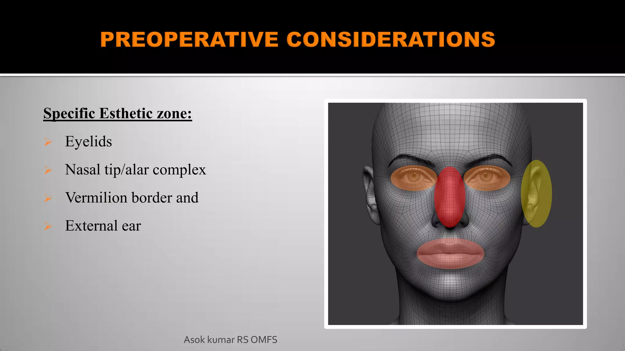
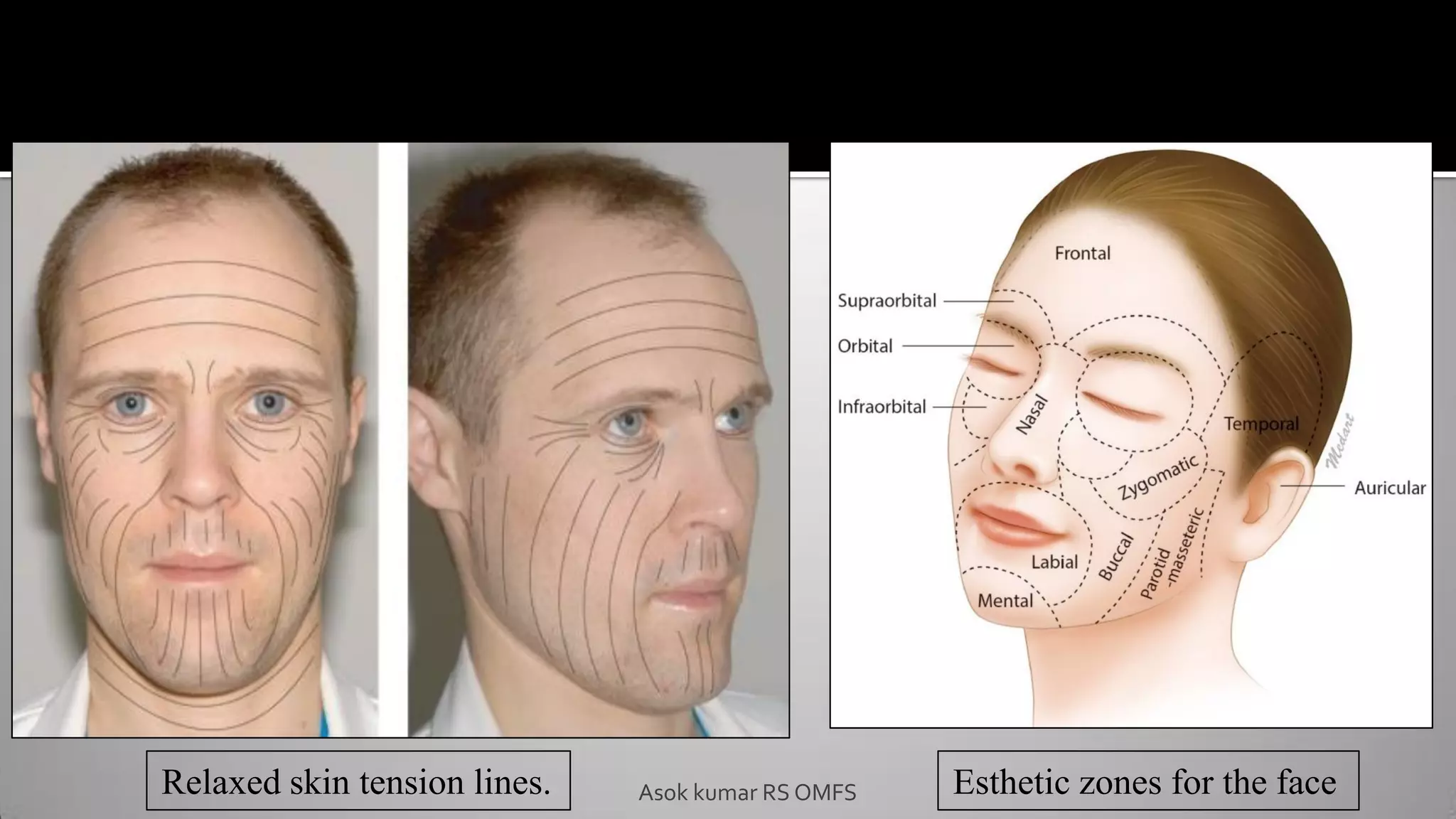
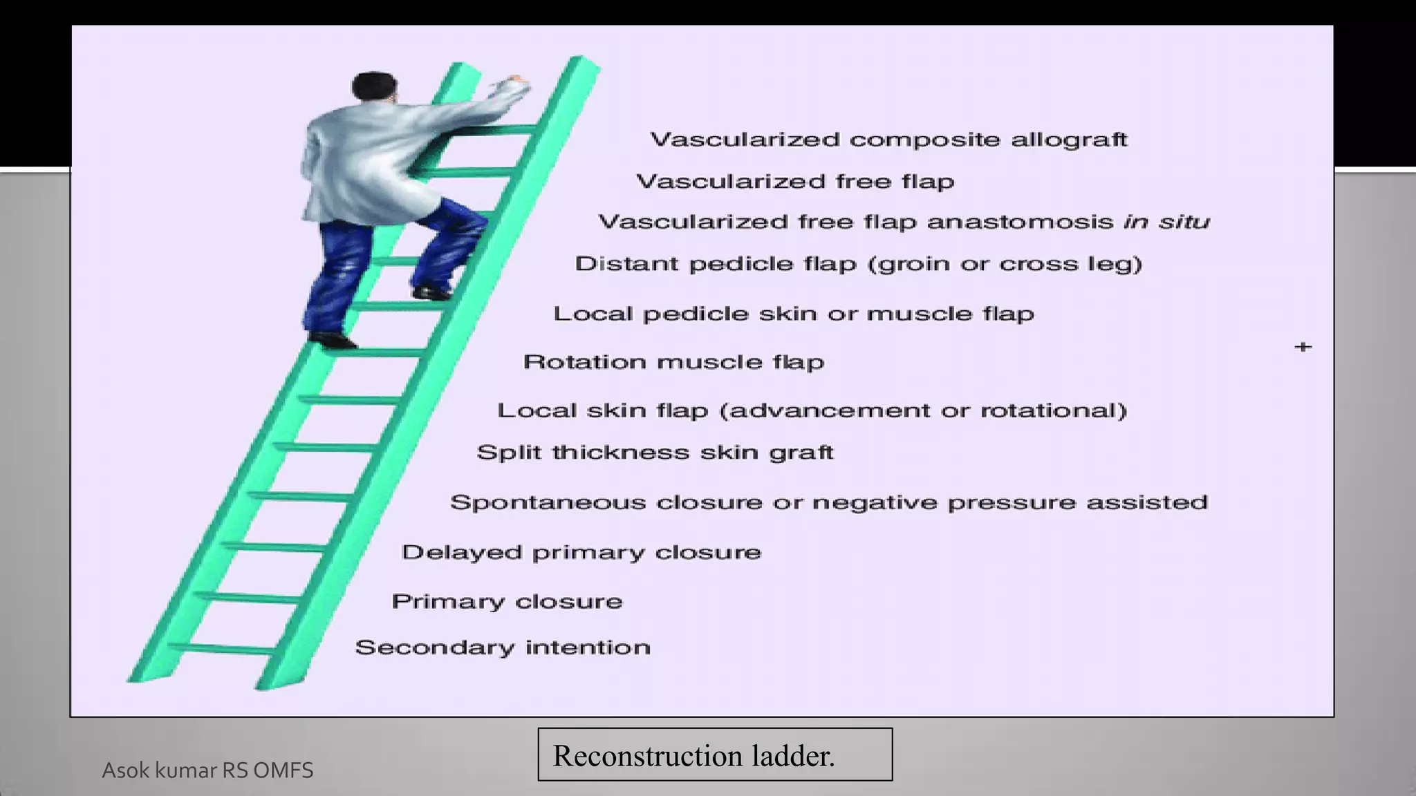
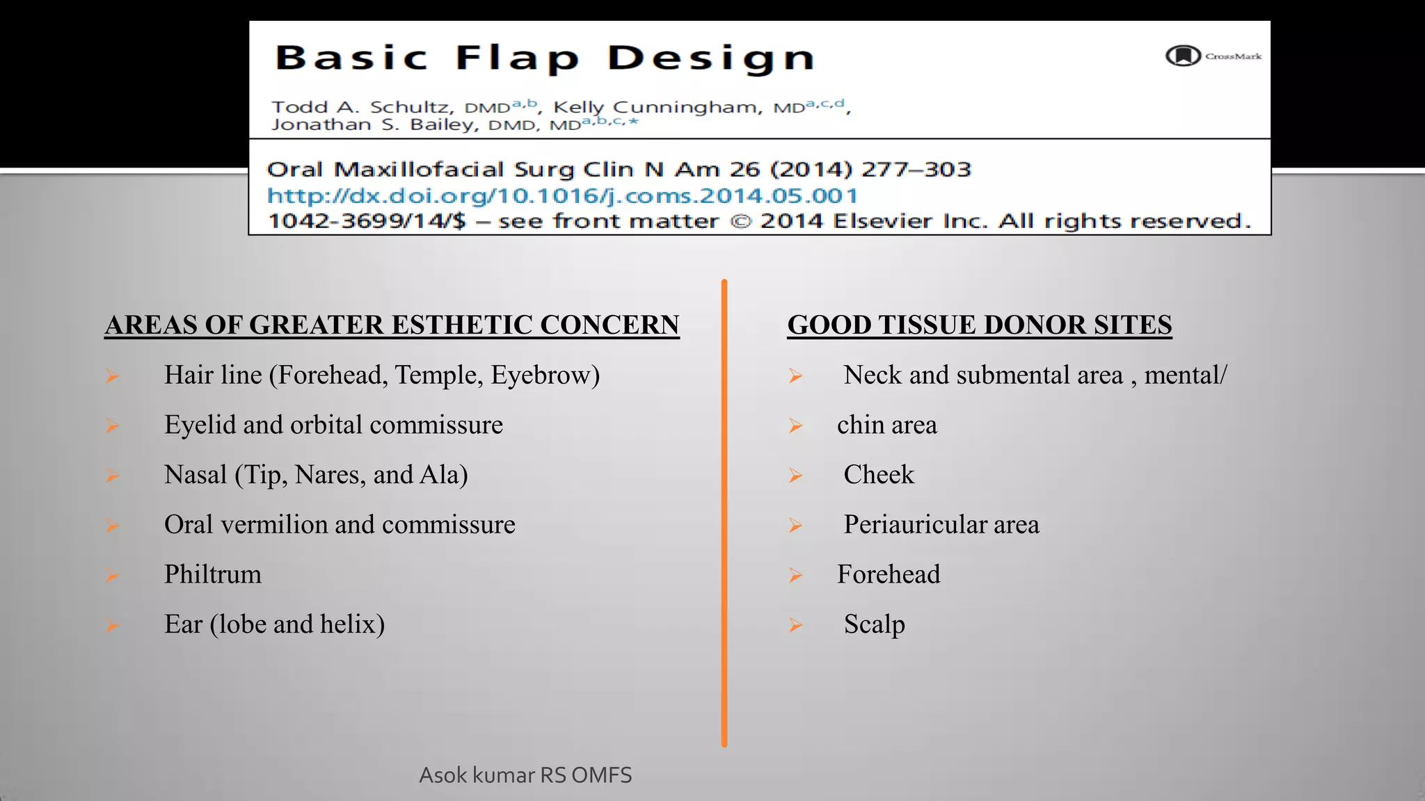
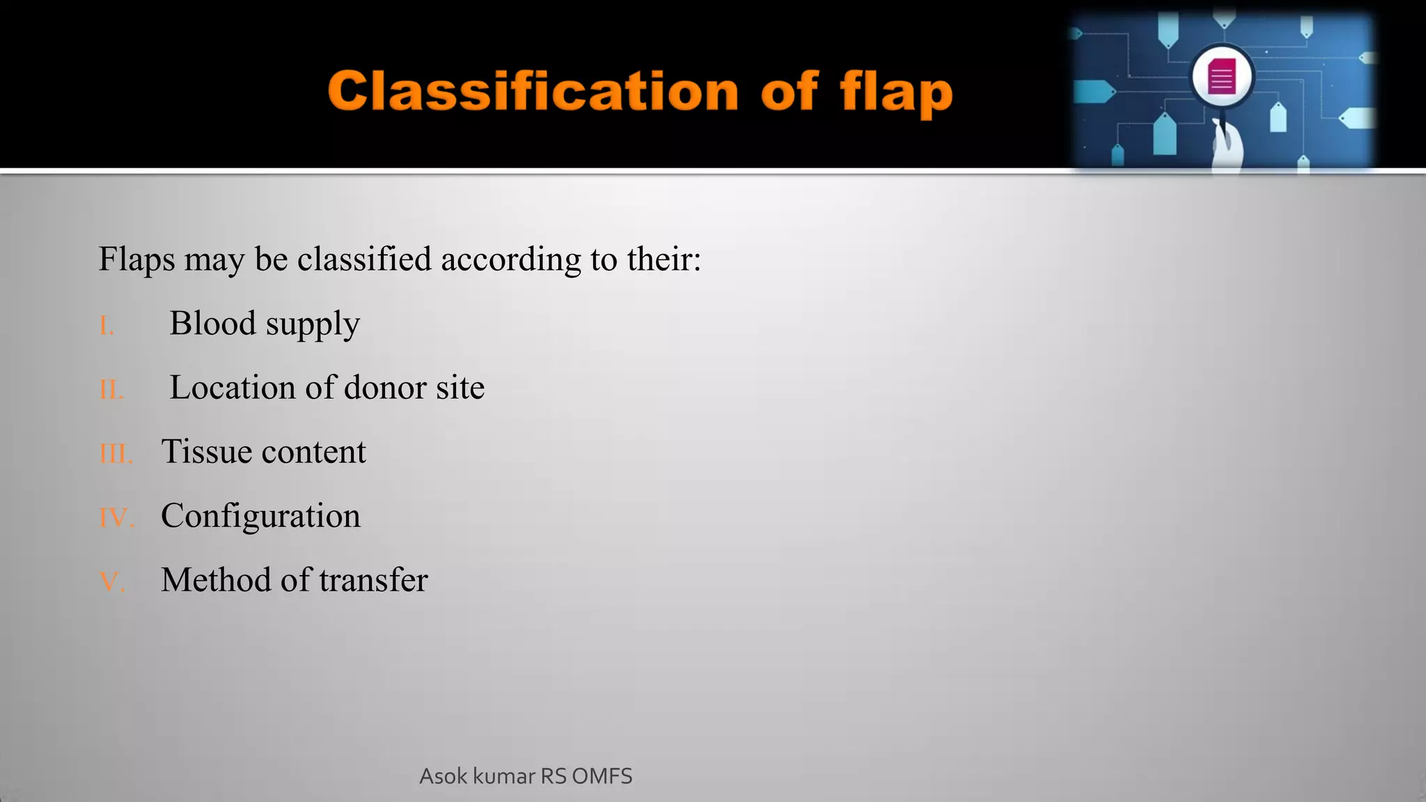
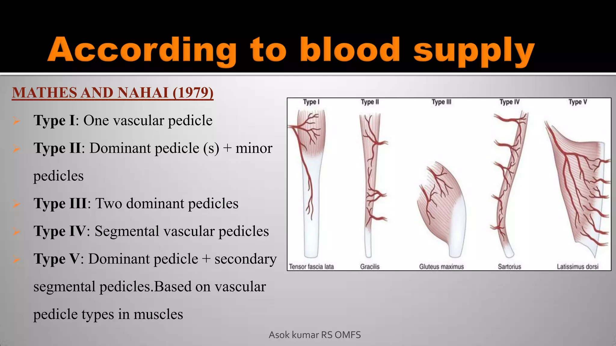
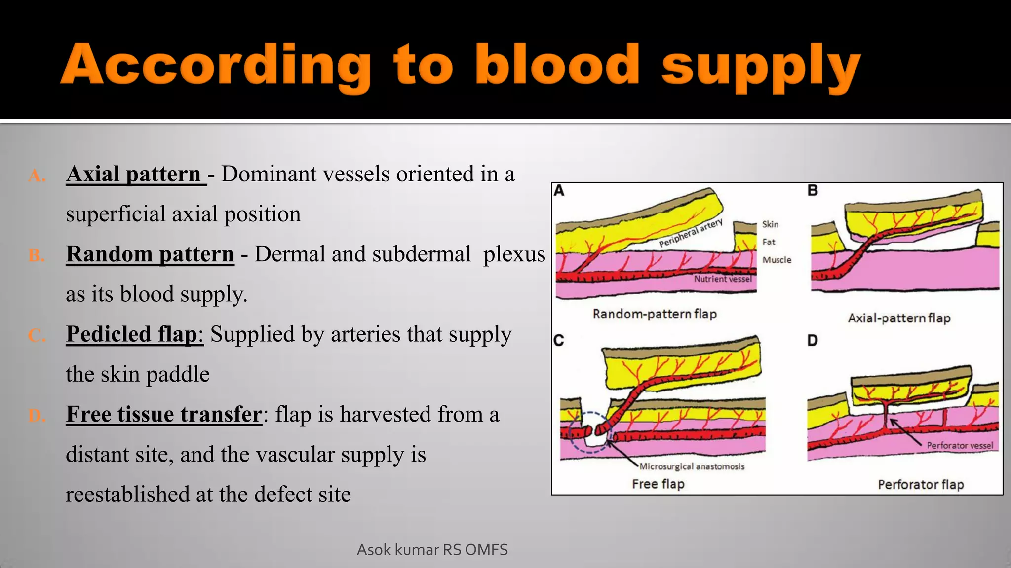
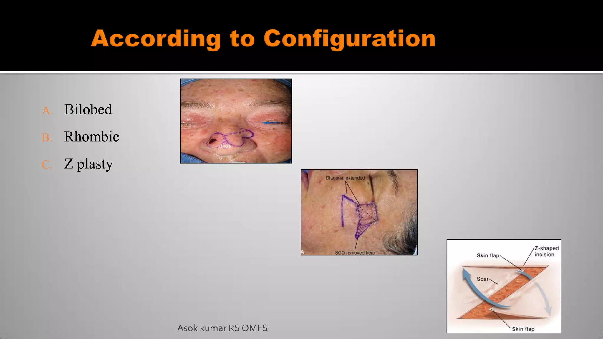
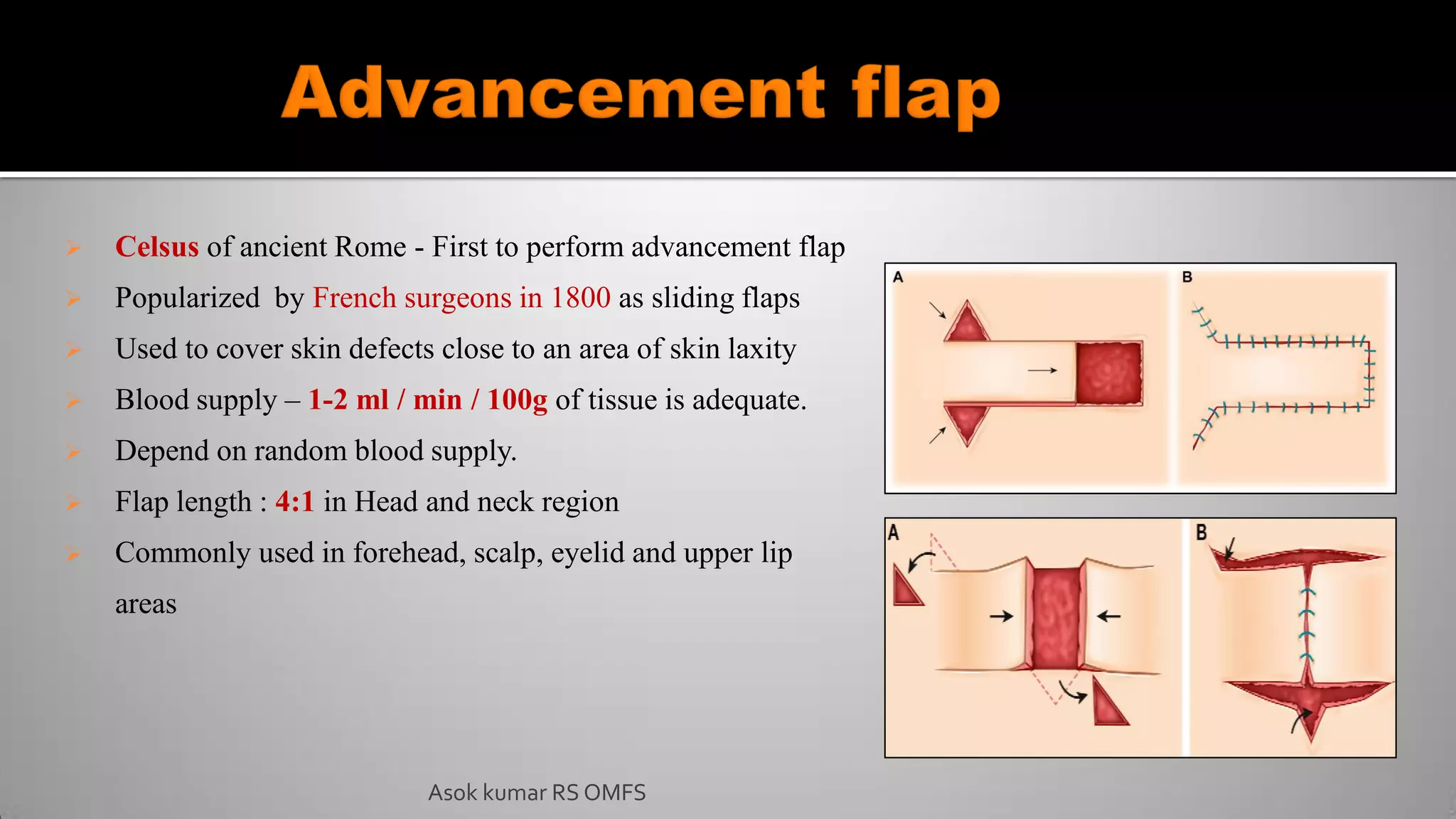
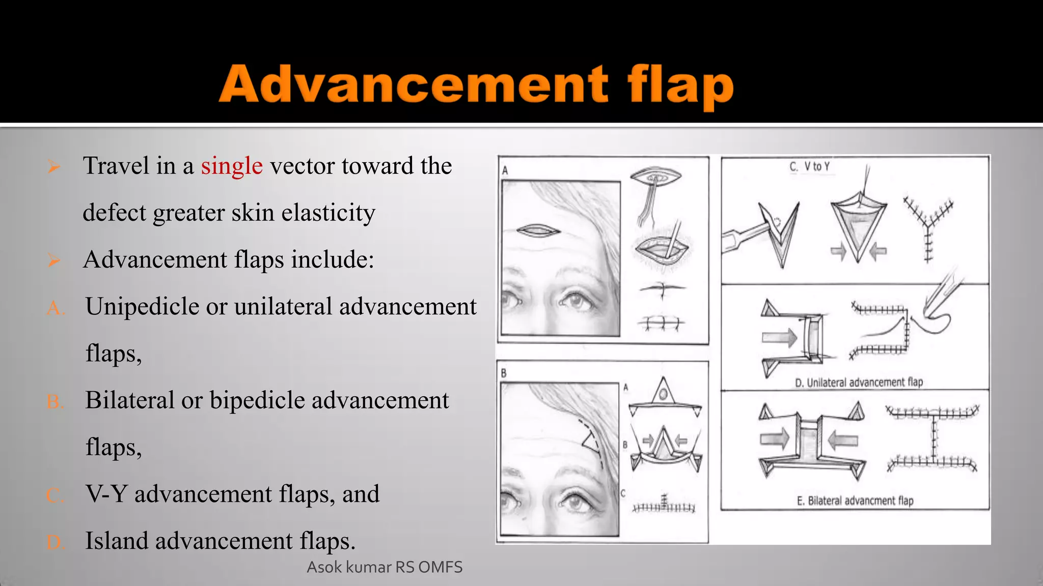
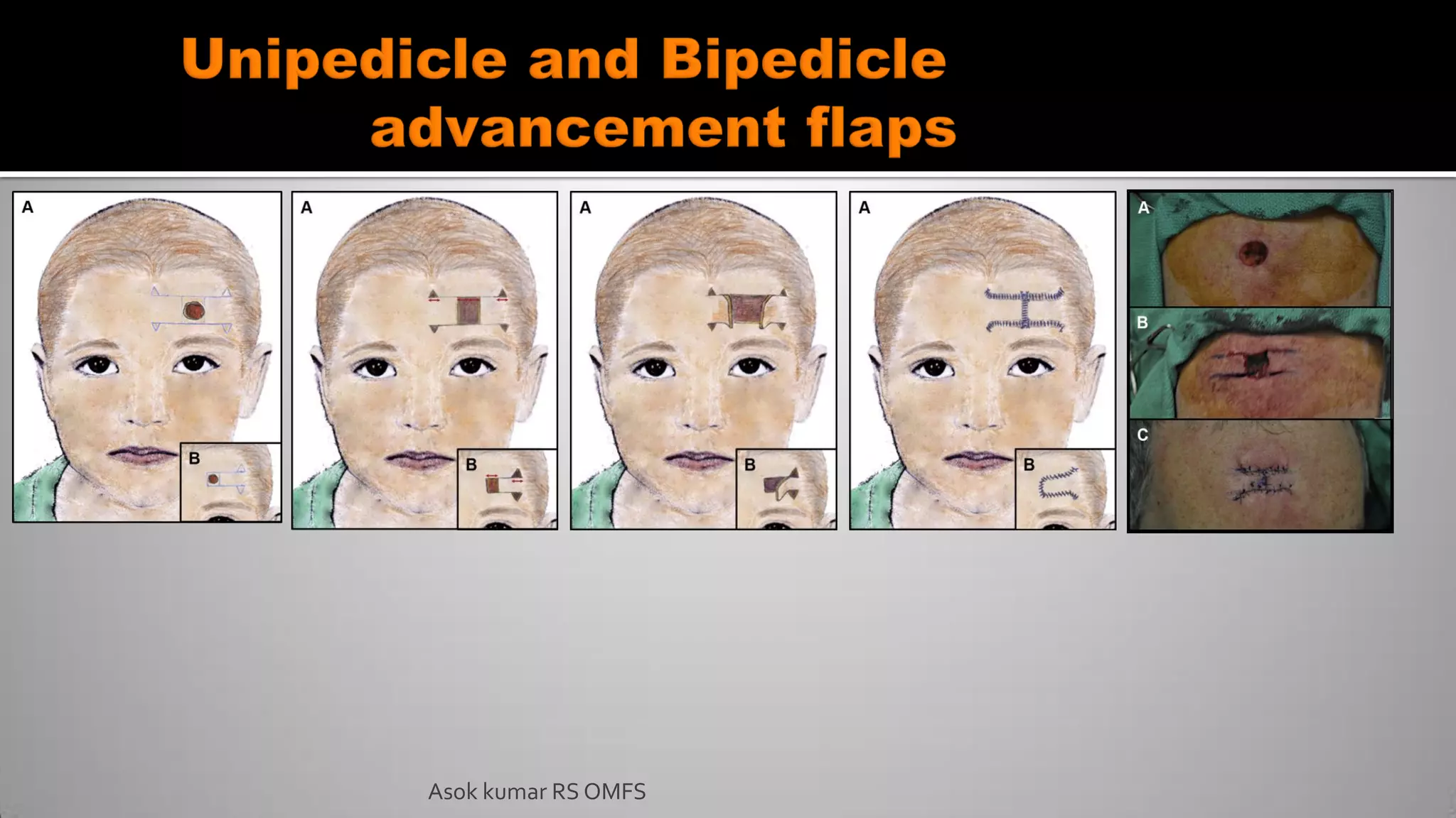
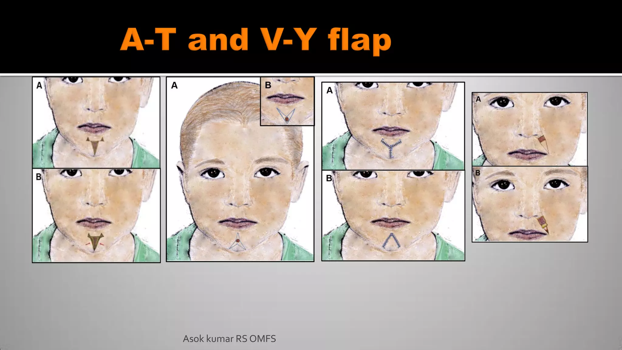
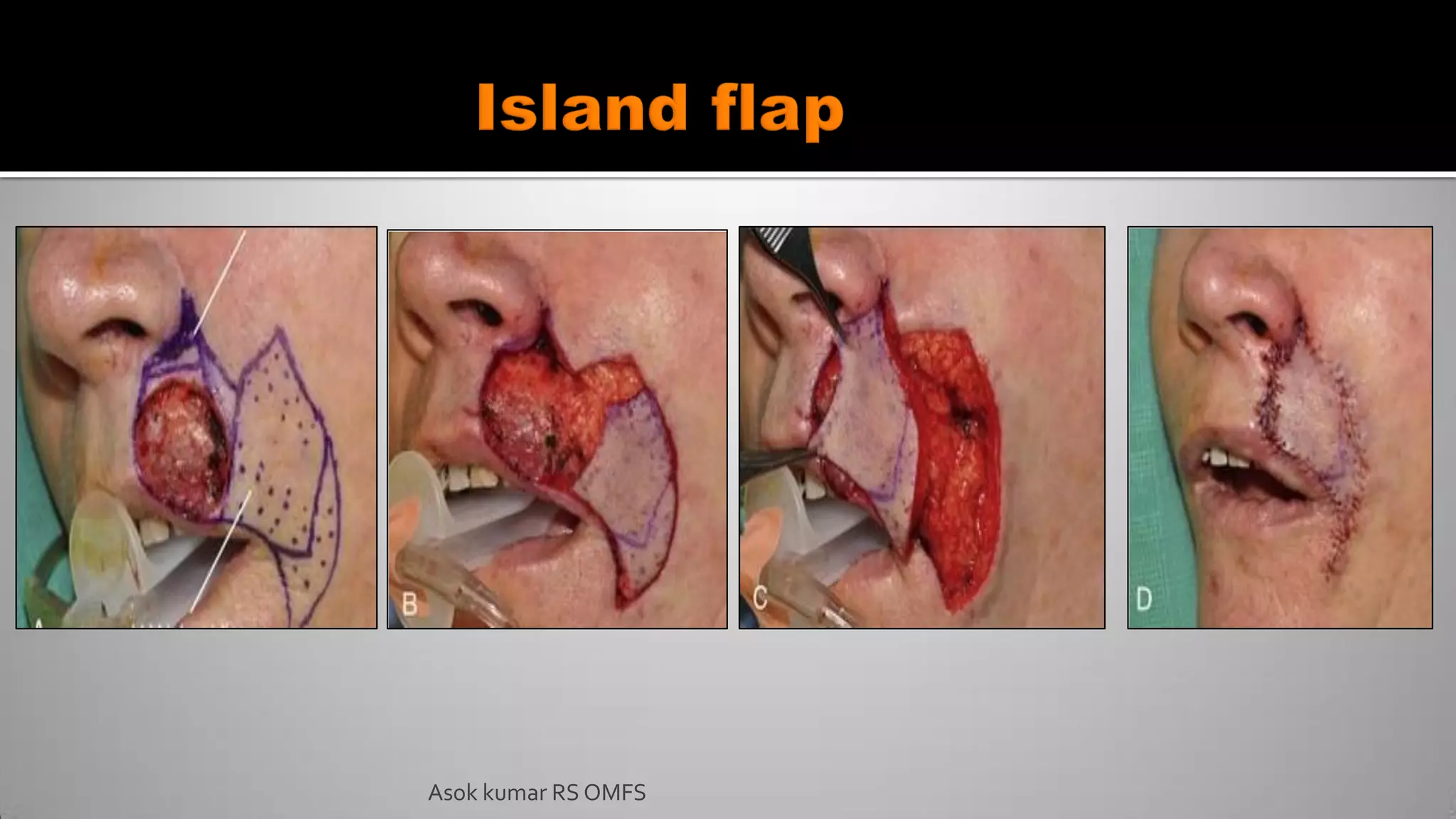
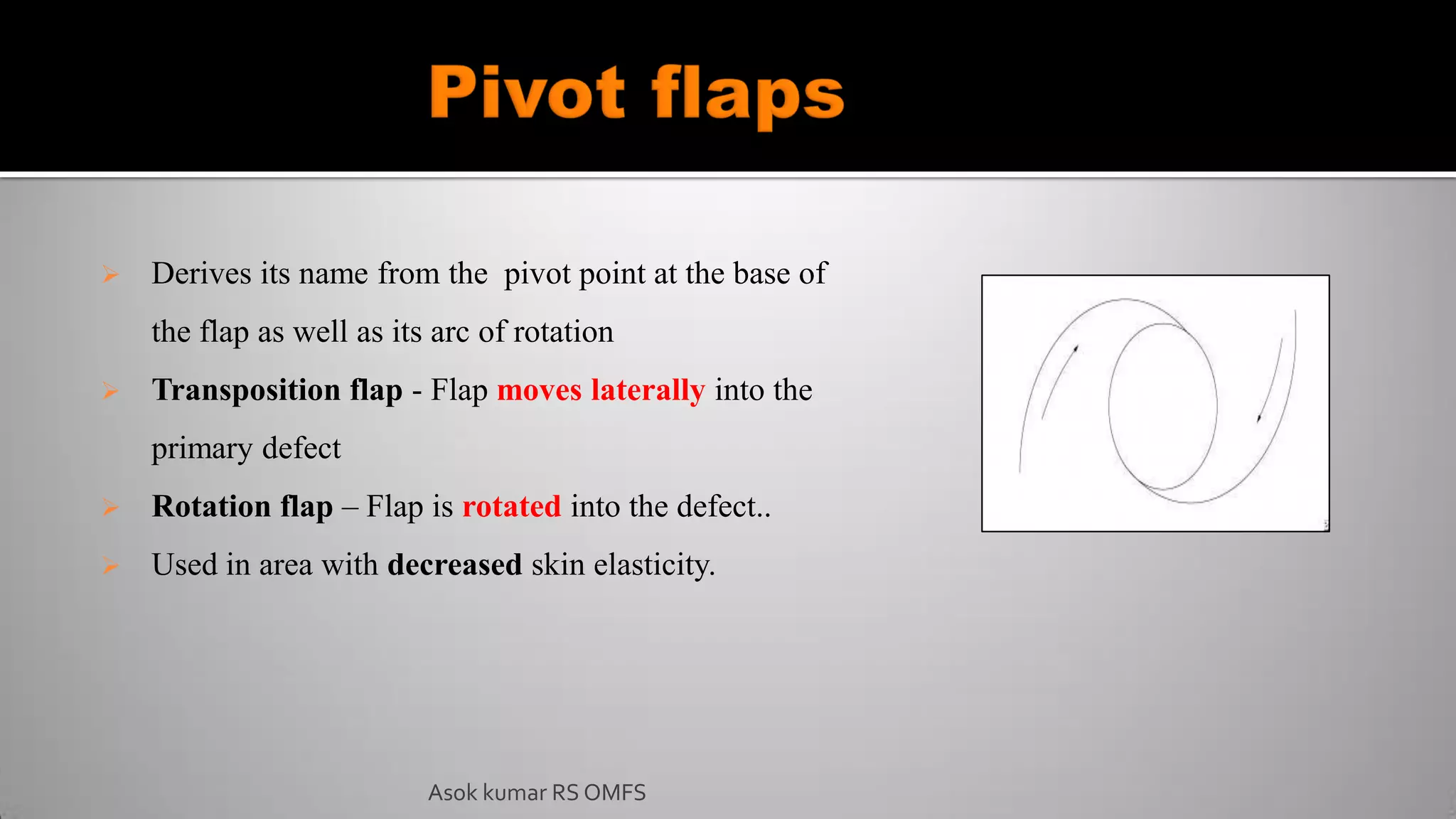
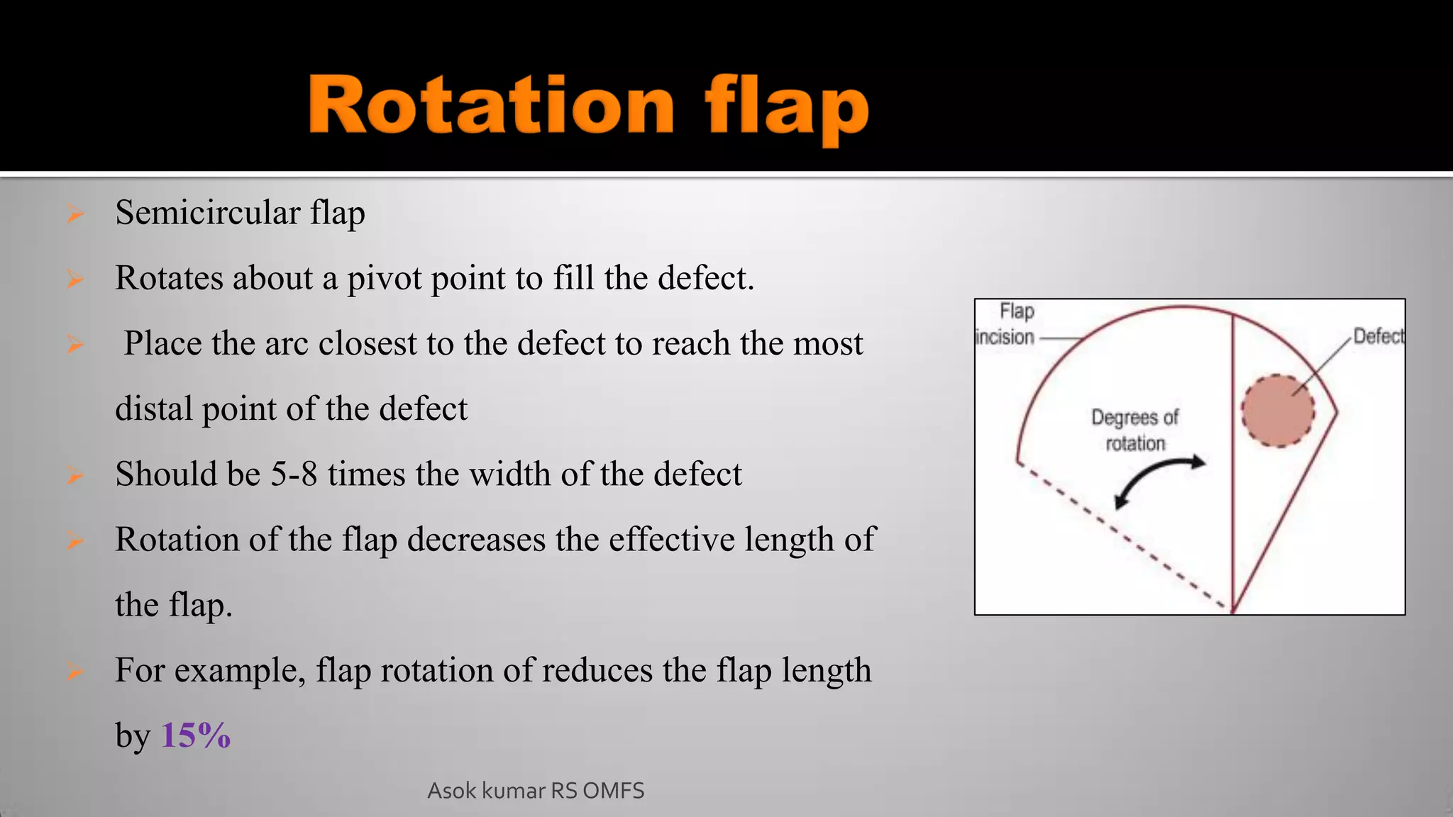
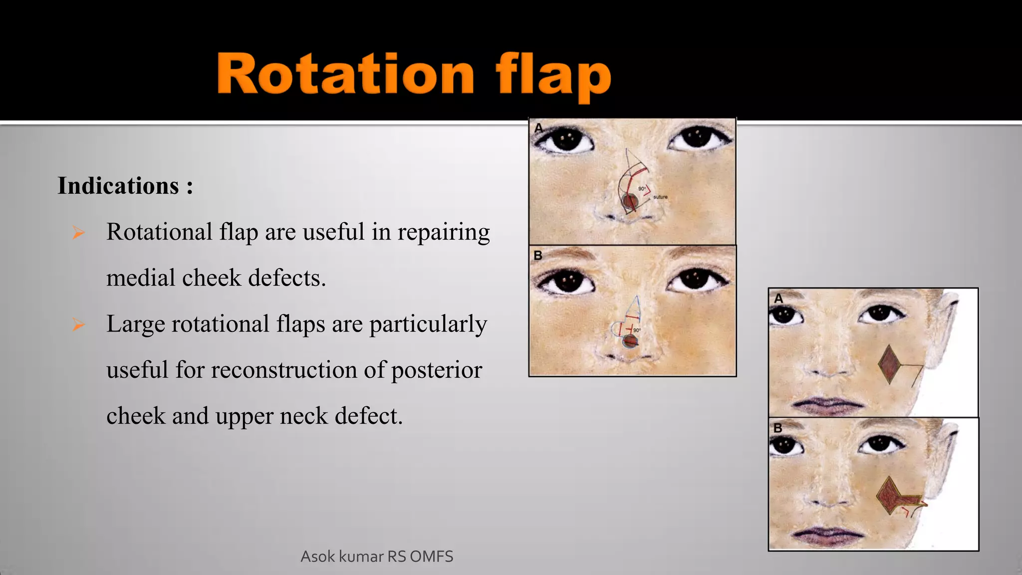
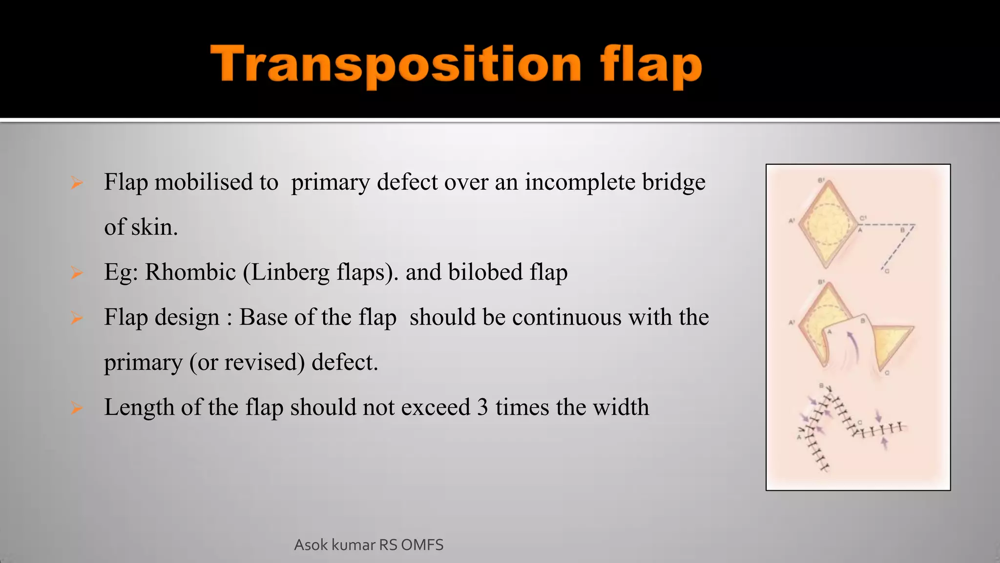
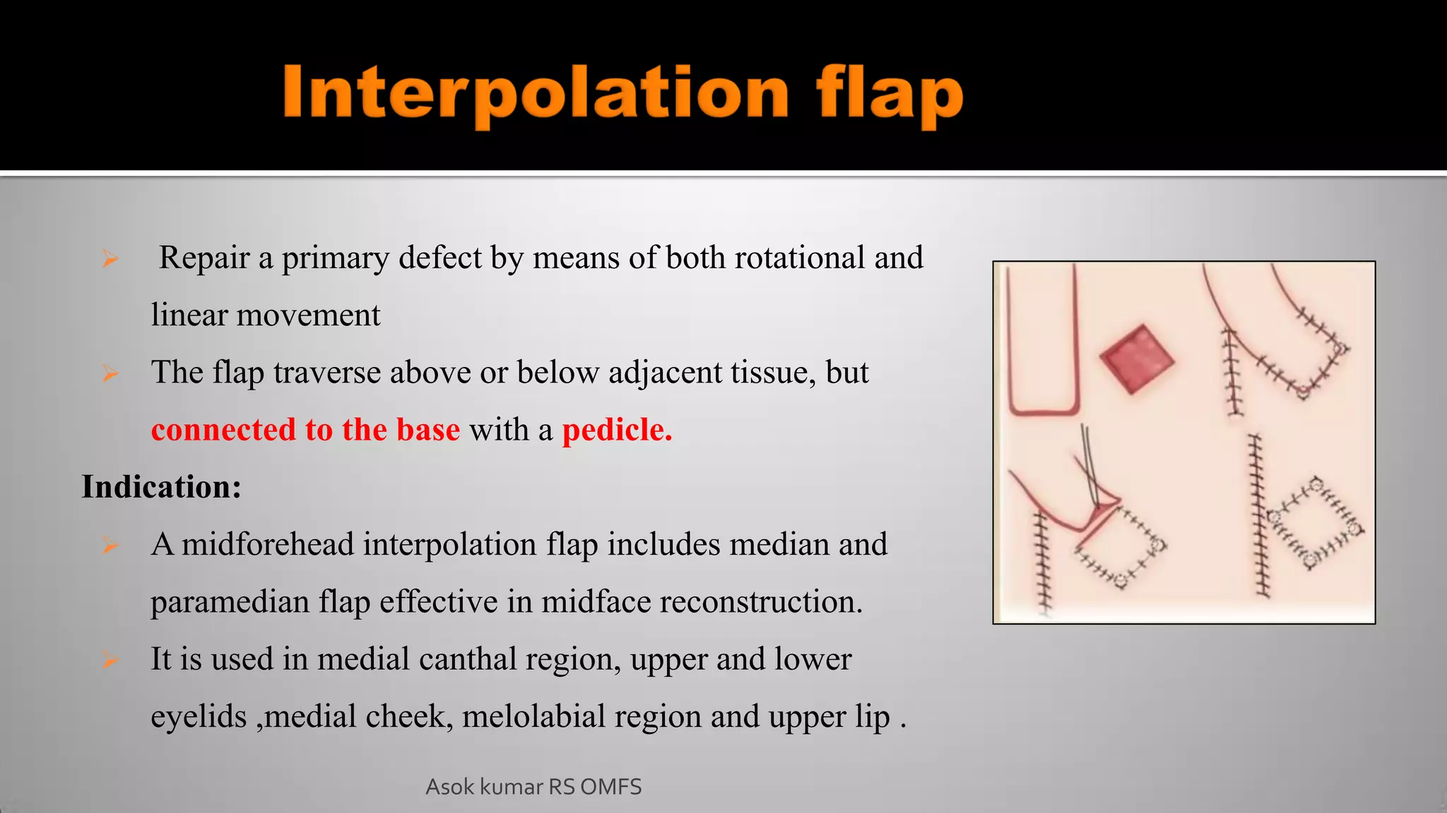
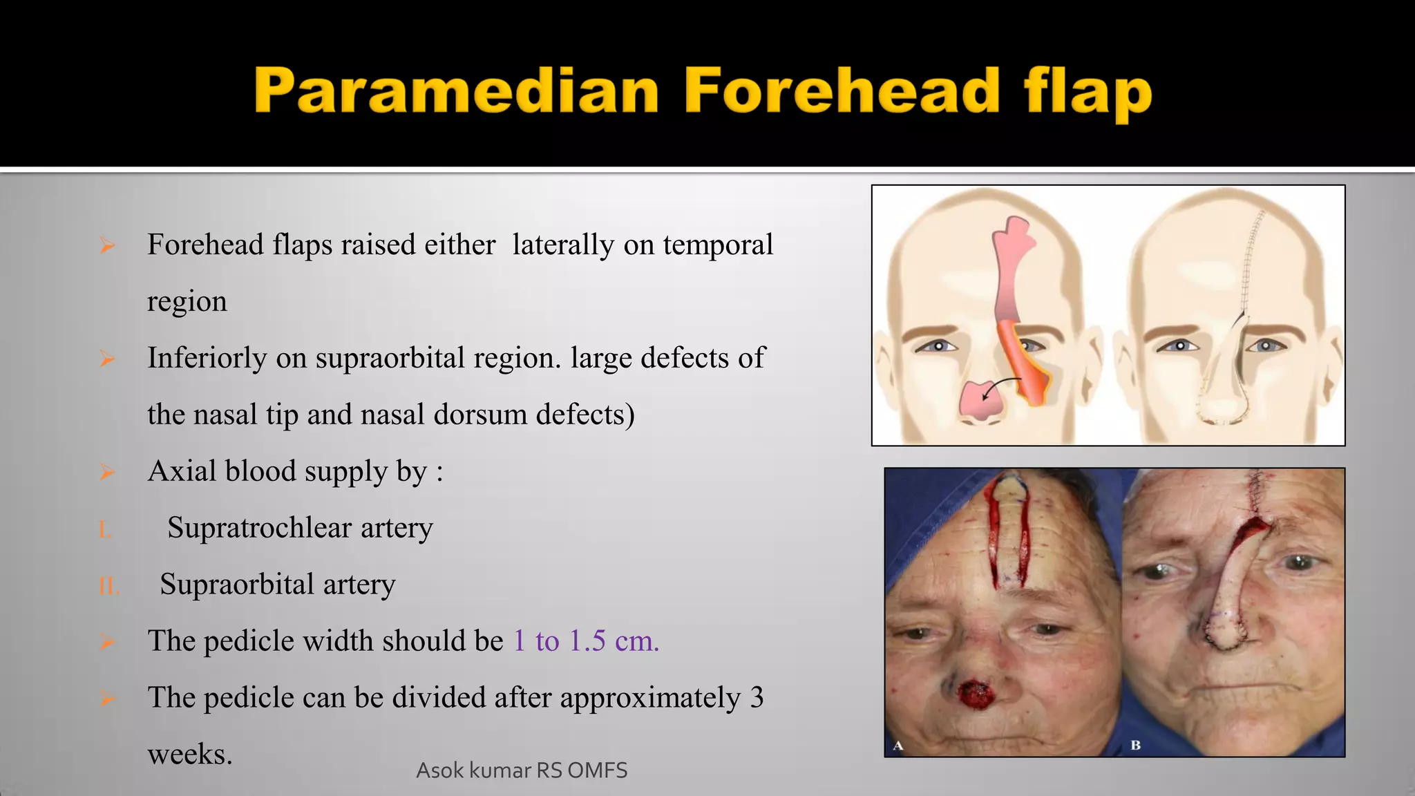
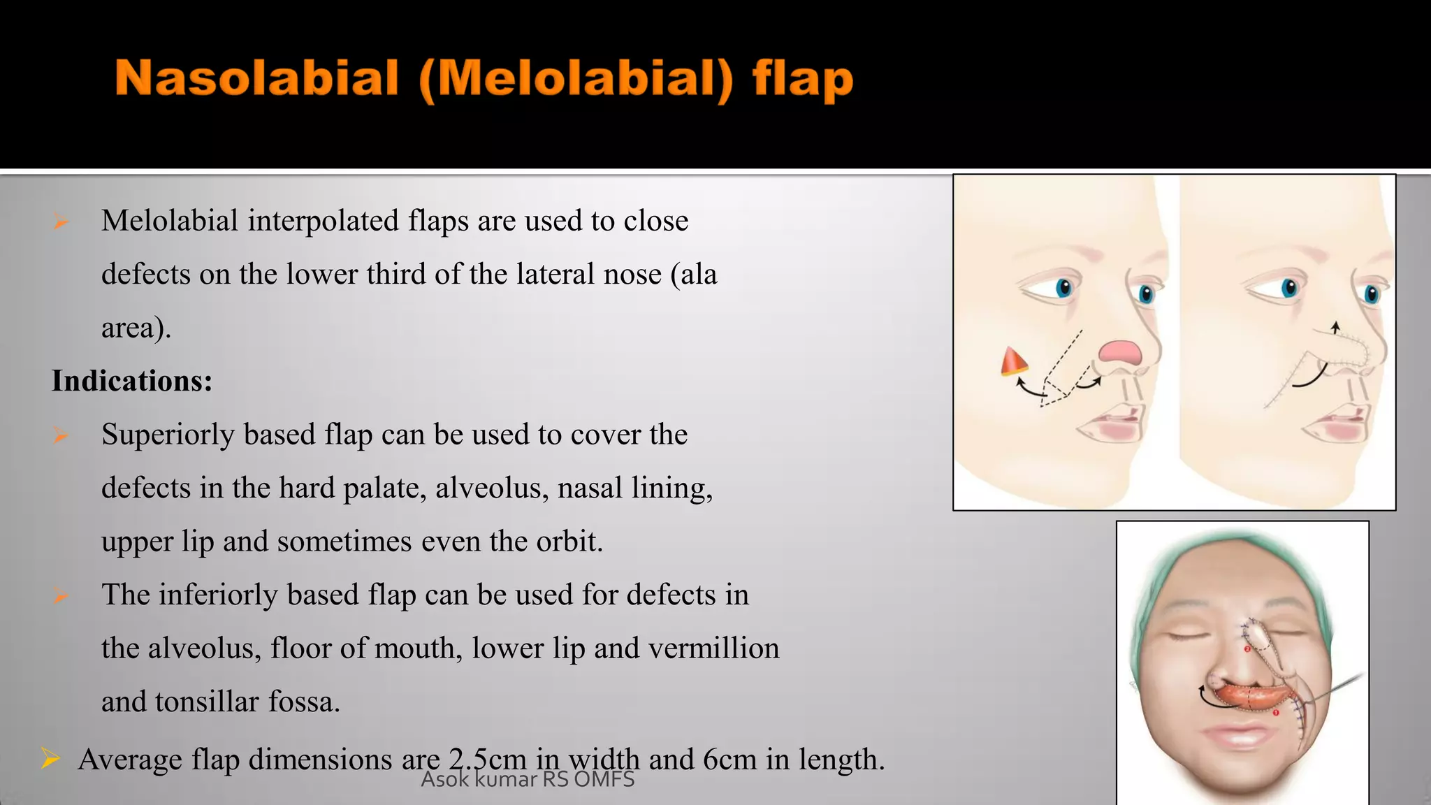
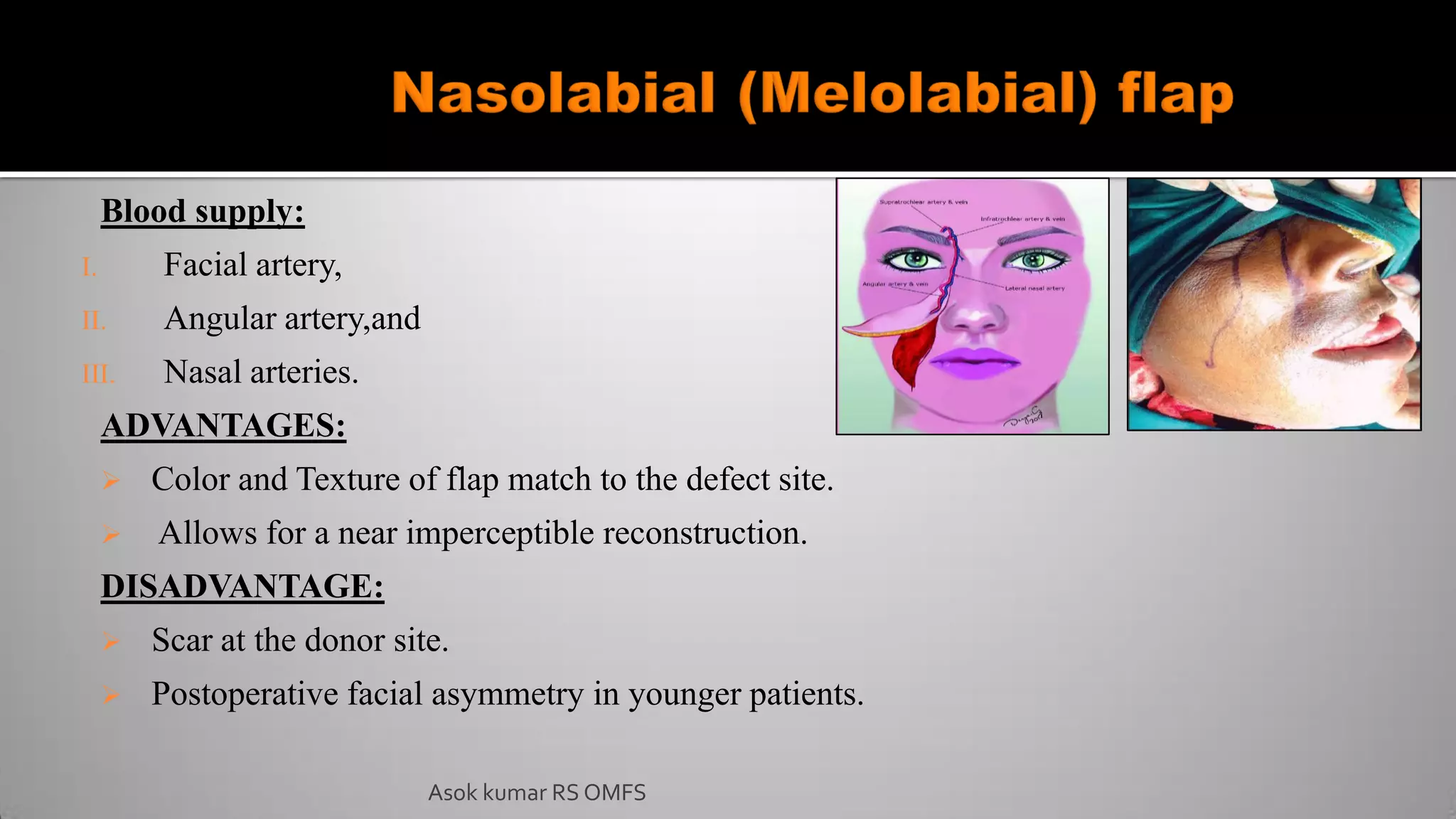
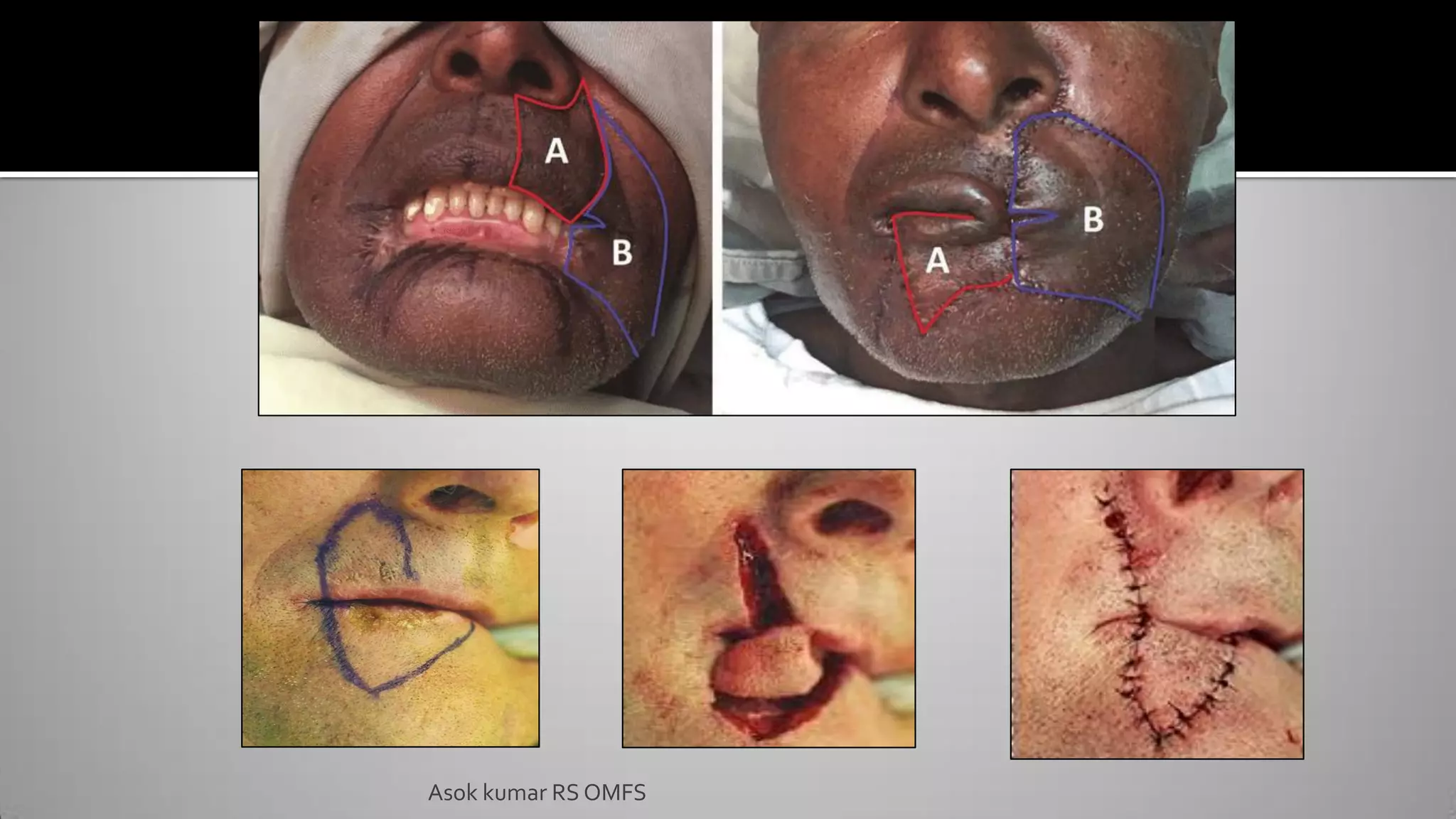
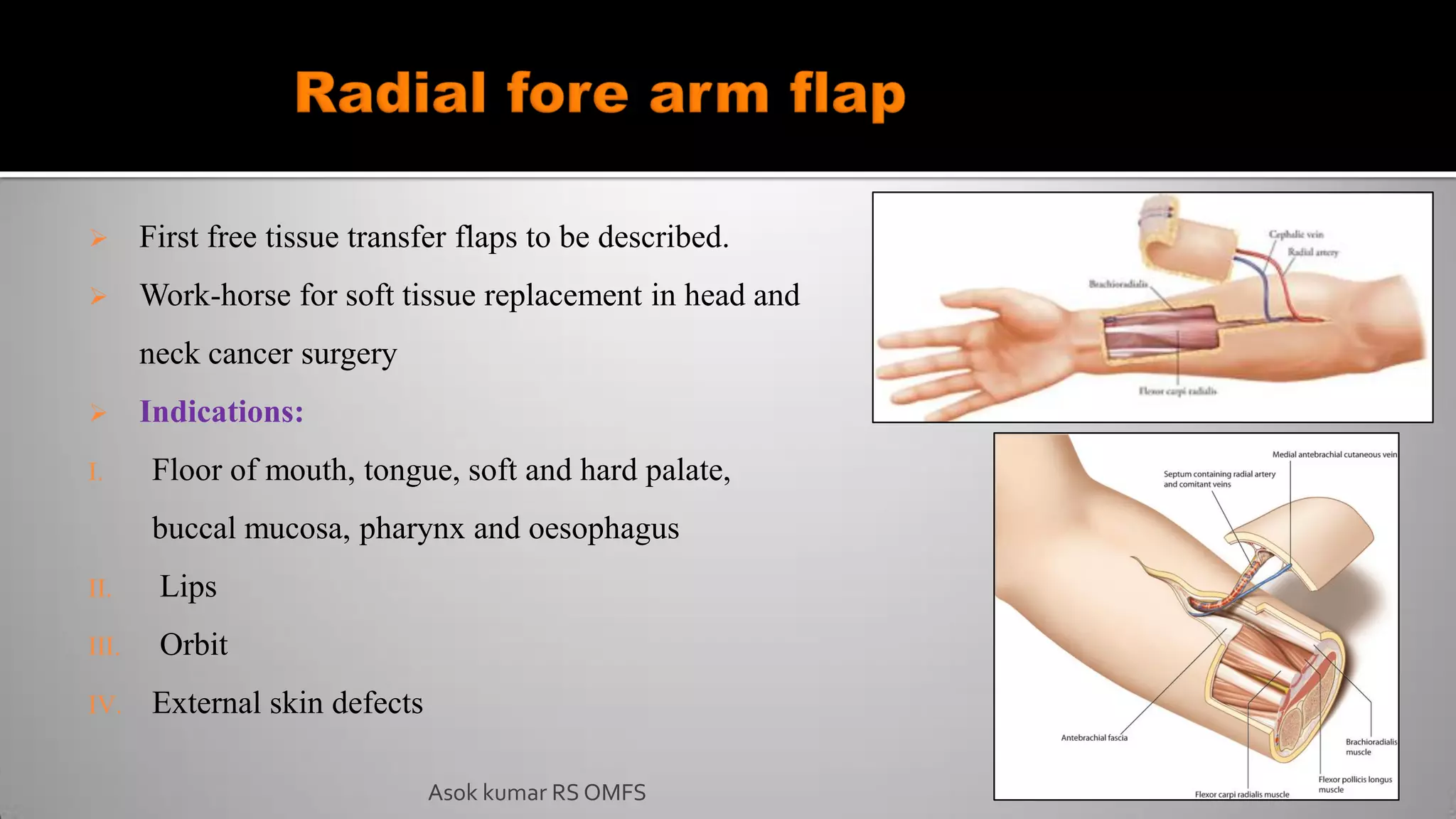
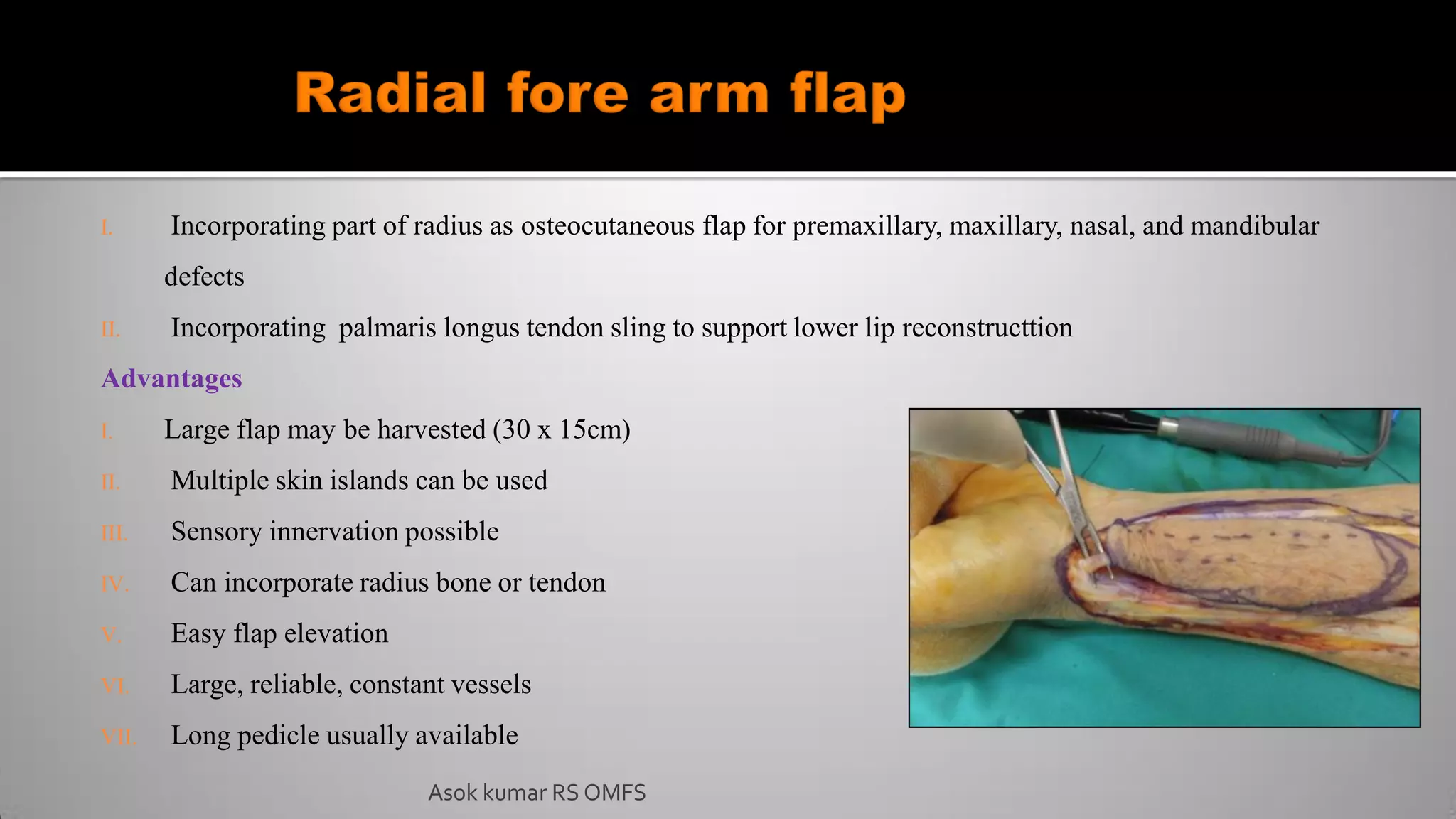
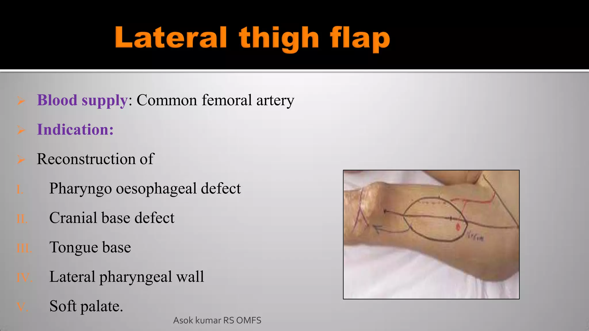
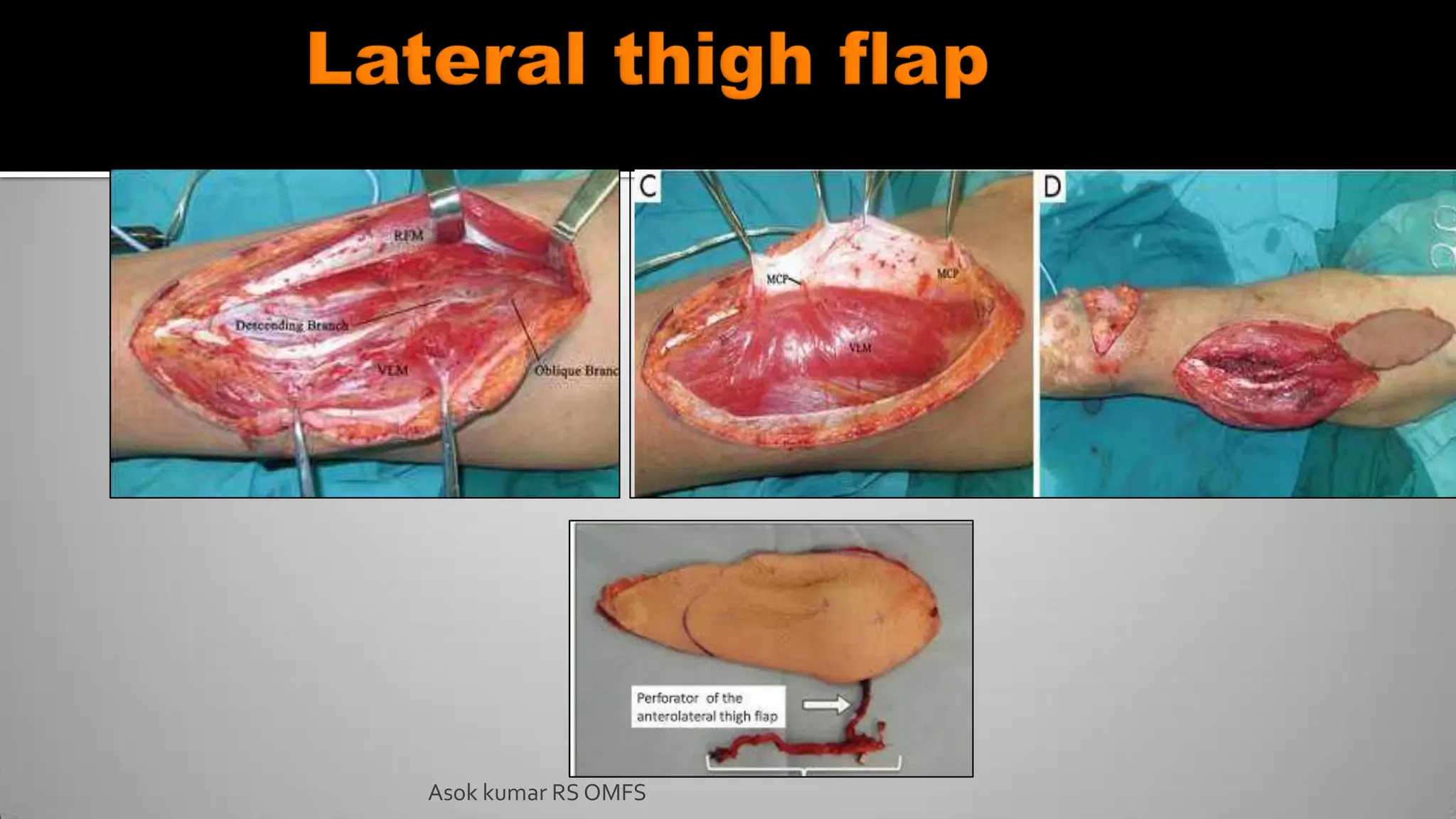
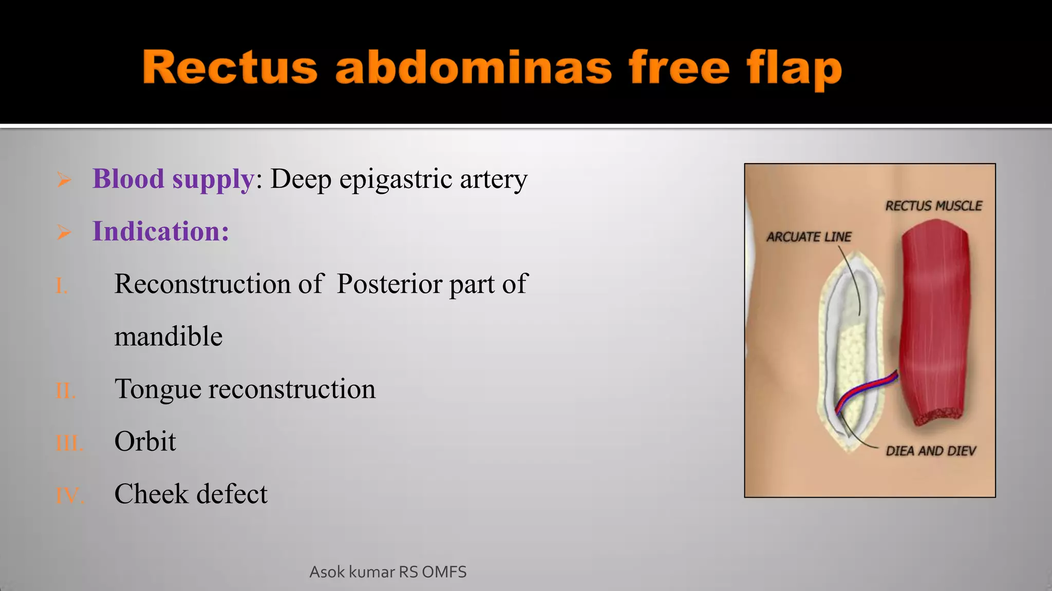
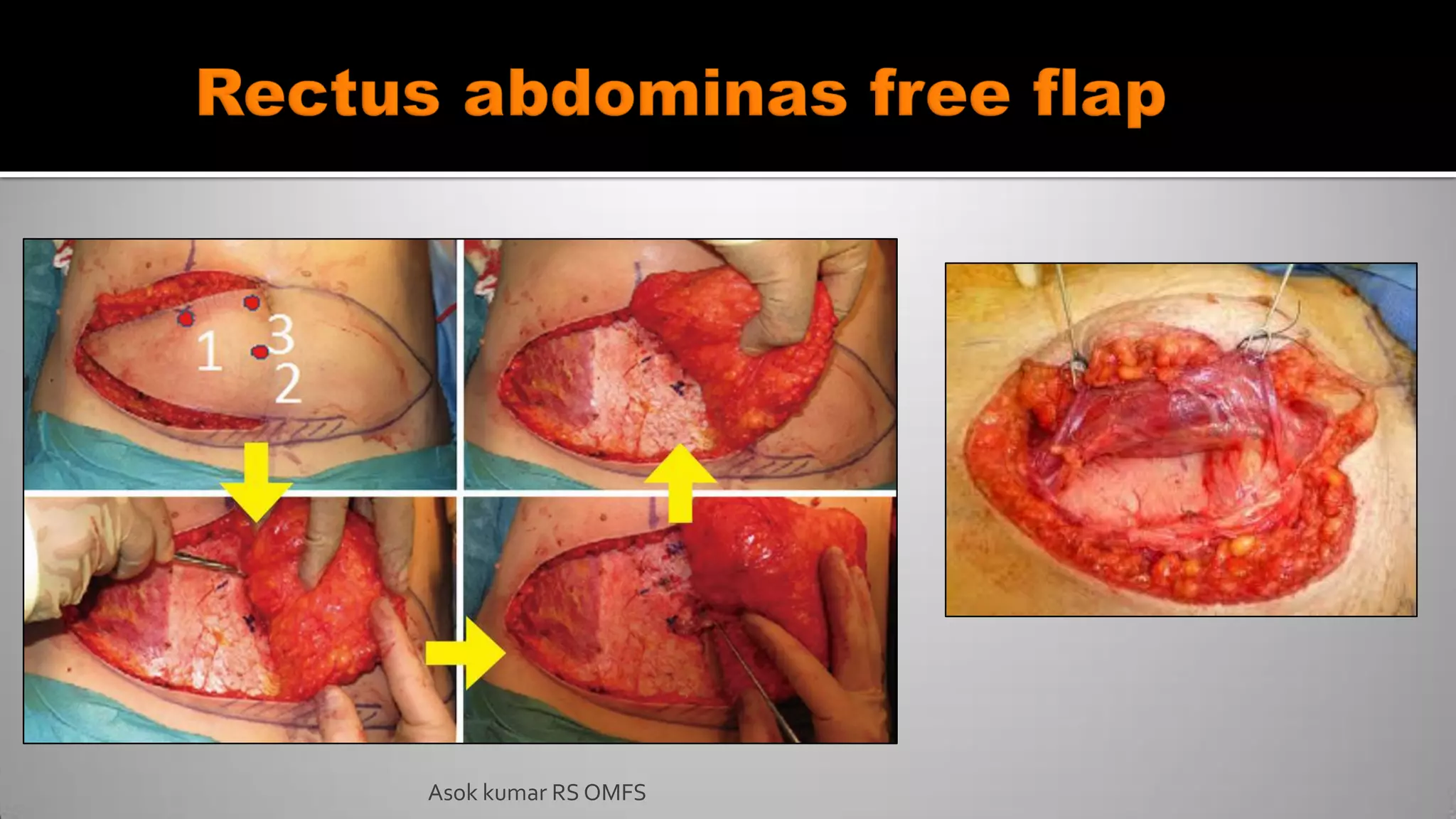
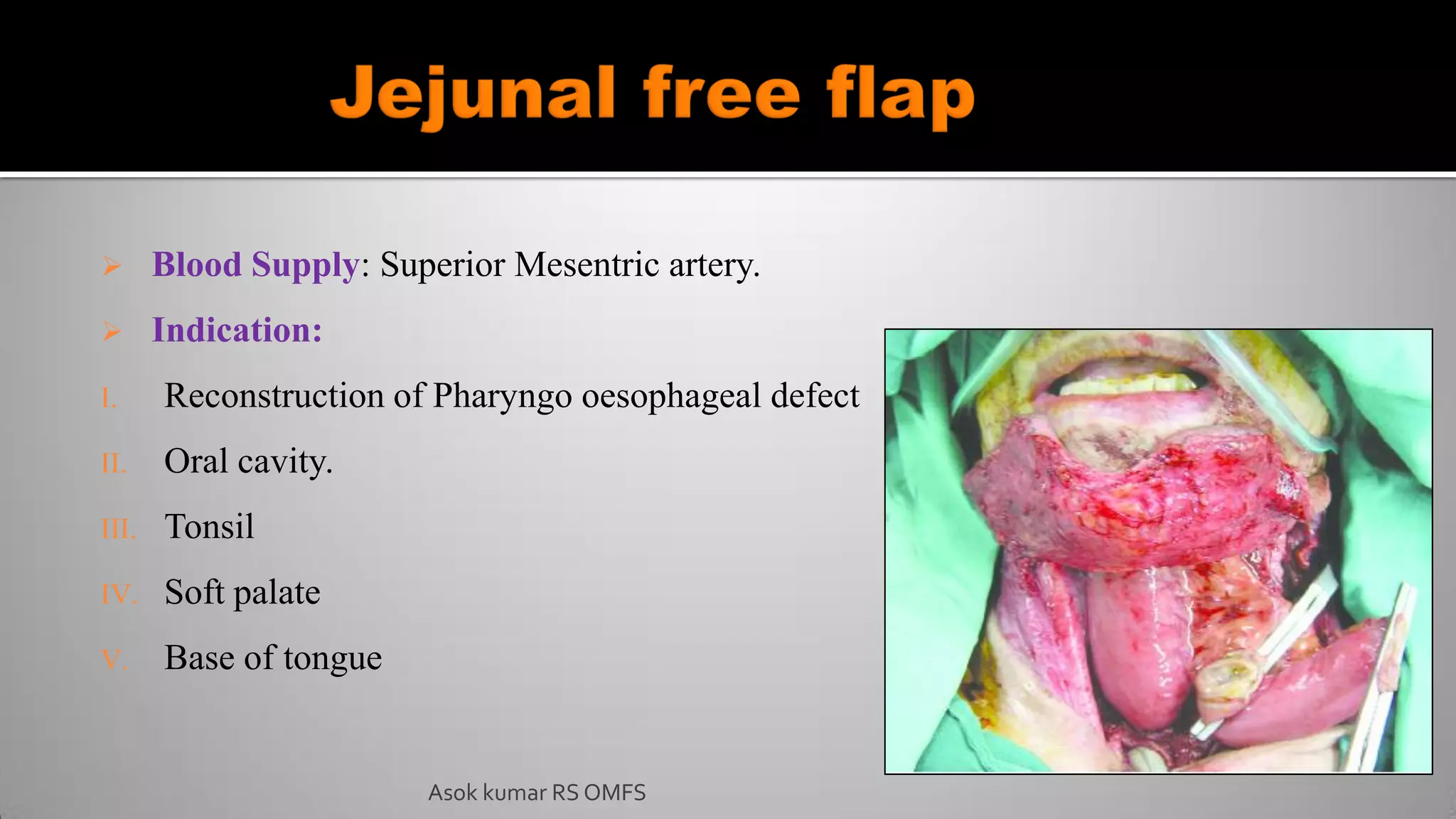
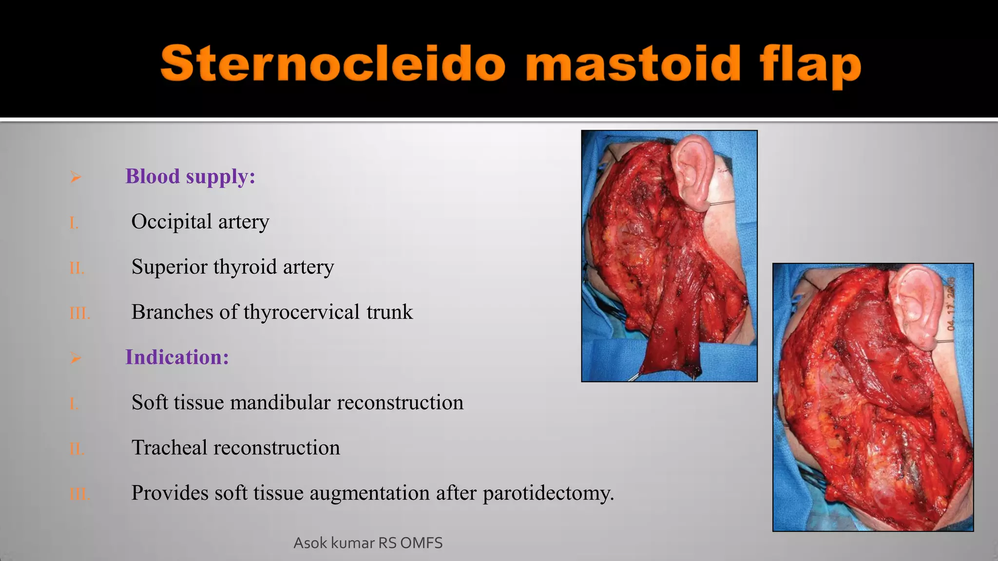
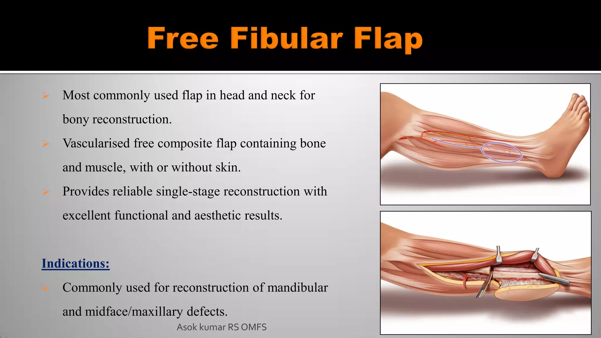
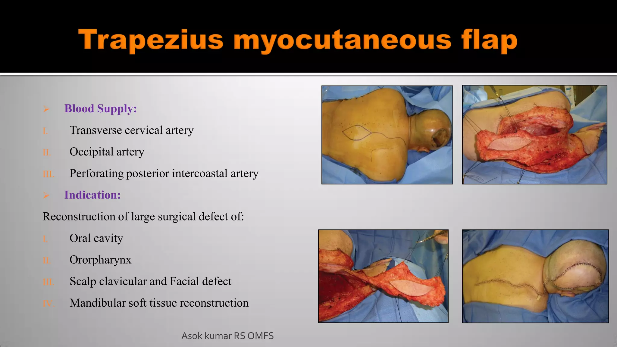
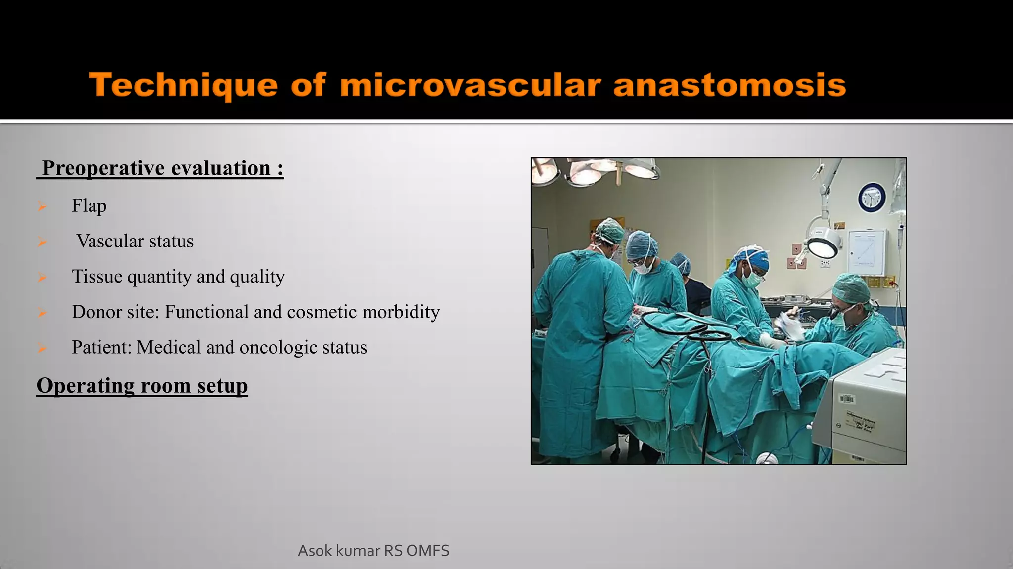
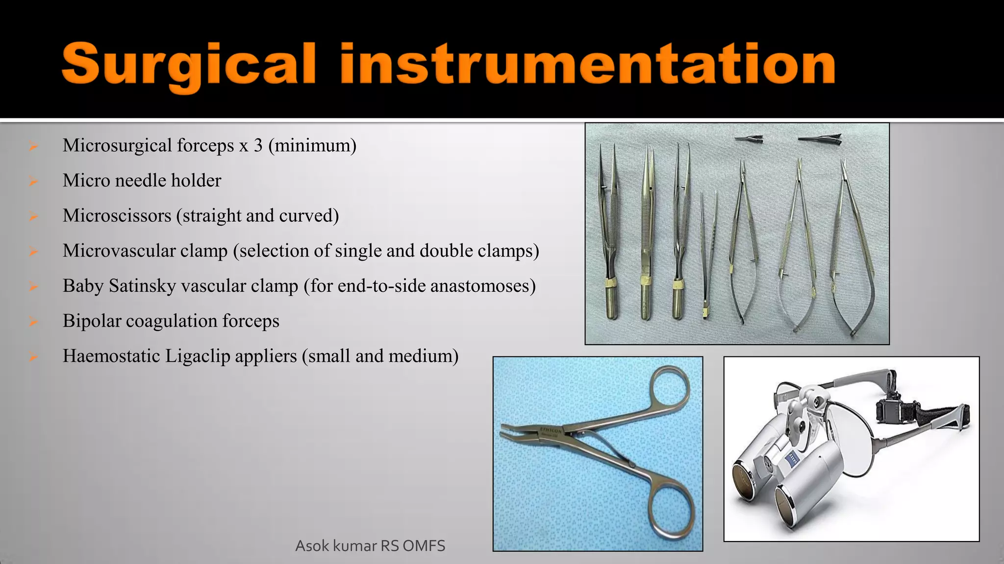
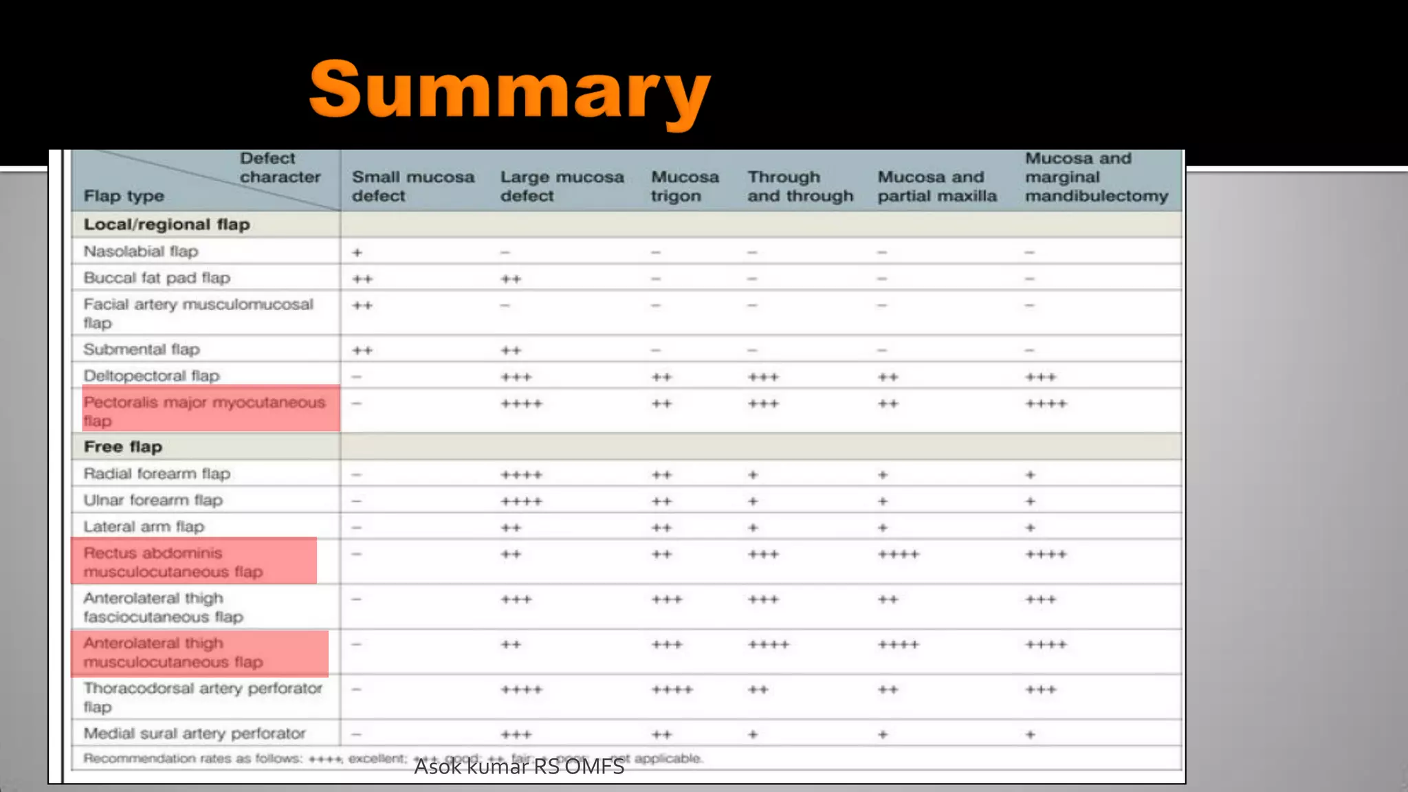
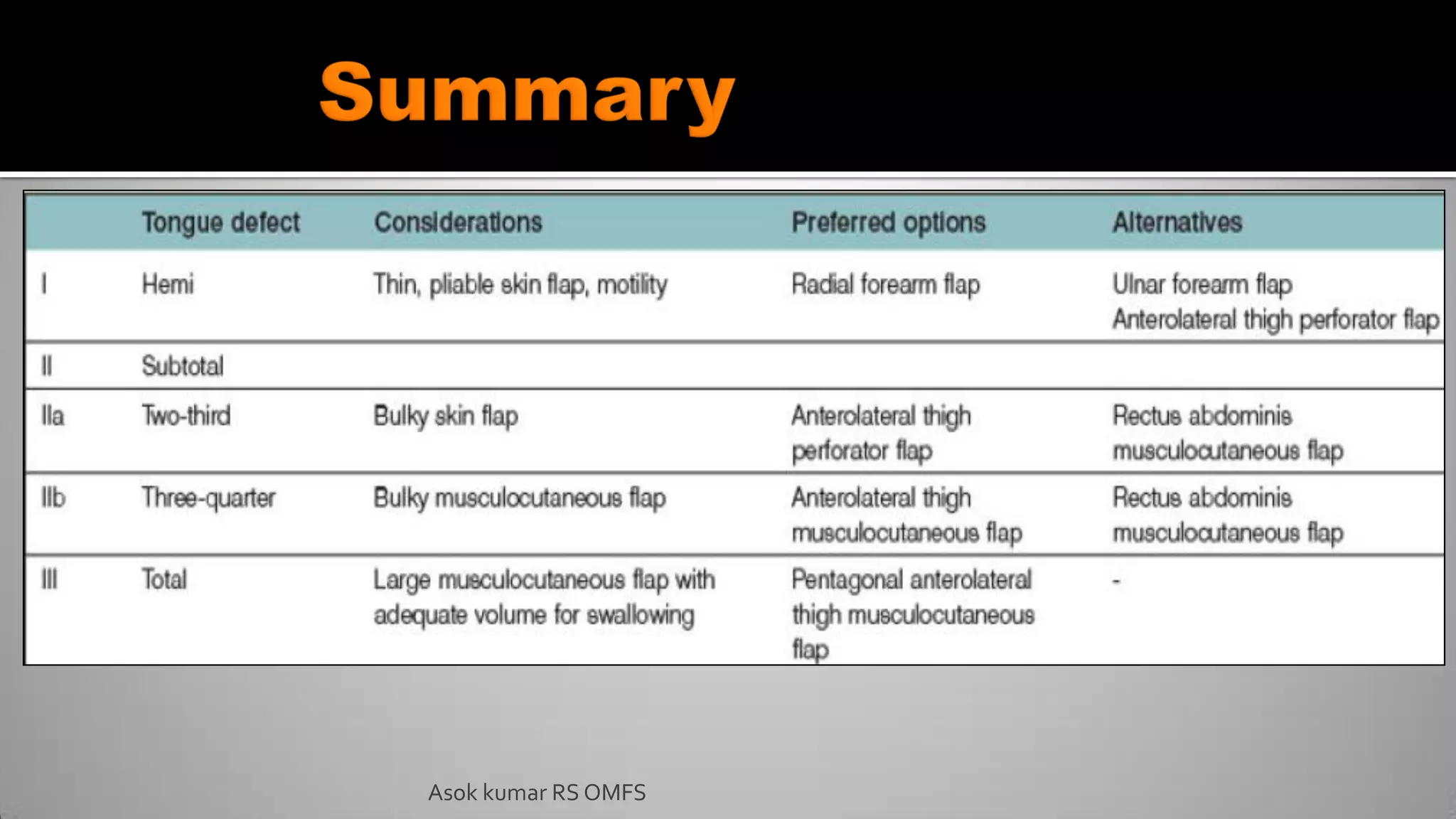
2. “When a part of one's person is lost, it should be replaced in kind, bone for bone, muscle for
muscle, hairless skin for hairless skin, an eye for an eye, a tooth for a tooth”. (Ralph
Millard)
Definition : A flap is a unit of tissue that is transferred from donor site to recipient site while
maintaining its own blood supply.
The Term Flap , originated from Dutch word “FLAPPE” which means “anything that hung
broad and loose, fastened only by one side”.
Flaps are usually used to repair structural defects following surgery for malignant conditions
of head and neck.
Asok kumar RS OMFS
3. 600 B.C : Susrutha performed nasal reconstruction using cheek flap
1440 A.D: Forehead rhinoplasty (India).
Pivotal flaps was preferred during early days. This involves rotation of the flap around its
vascular pedicle.
Advancement flap (French surgeons). This involves transfer of skin from adjacent area
without rotation.
Gaspar Tagliocozzi (1597), Italy: Experimented with the fabrication of noses from the
tissues of the upper arm.
Tansini (1896), described the concept of muscle and skin flap.
Asok kumar RS OMFS
4. Harold Gillies - Tube flaps in 1950s
Owens (1952): Stenocledomastoid flap.
Conley (1960): Regional flaps.
McGregor - Introduced the Forehead flap in 1963
Bakamjian - Introduced the Deltopectoral flap in 1965
Daniel & Taylor - Pioneered the Free flap in 1973
Ariyan - Pioneered the Pectoralis major myocutaneous flap in 1979
Asok kumar RS OMFS
5. The skin functions and properties include
a) Protection/ anatomic barrier
b) Thermoregulation
c) Protection against excessive fluid loss/evaporation
d) Storage areas (eg, lipids and water)
e) Sensation center and
f) Formation of an aesthetic zone, enhancing nonverbal communication/
expression.
Asok kumar RS OMFS
6. Nature of the wound determines the approach for closure.
Risk Factors: Location, Size,Adjacent structures, Etiology
(eg, trauma, malignancy, or cosmetic defect) and Medical
comorbidities
The goal of proper flap design is to closely restore the skin’s
functions and properties
Understanding of proper wound closure, wound healing,
relaxed skin tension lines, and the facial esthetic zones is
essential in this task
Asok kumar RS OMFS
7. Primary defect: Wound to be closed by local cutaneous flaps.
Secondary defect: Wound created when skin flap is transferred to repair primary defect
Wound closure tension: Amount of stress per unit along the suture line of a repaired
wound.
RSTL (Relaxed skin tension line)- Orientation of collagen fibres to skin and
manifested as a creases and wrinkles.
These lines are perpendicular to the lines of maximal extensibility. Knowledge of these
lines is essential for cosmetic and functional wound closure
Primary tissue movement : Transfer of skin to defect
Secondary tissue movement: Displacement of skin surrounding the defect.
Asok kumar RS OMFS
11. AREAS OF GREATER ESTHETIC CONCERN
Hair line (Forehead, Temple, Eyebrow)
Eyelid and orbital commissure
Nasal (Tip, Nares, and Ala)
Oral vermilion and commissure
Philtrum
Ear (lobe and helix)
GOOD TISSUE DONOR SITES
Neck and submental area , mental/
chin area
Cheek
Periauricular area
Forehead
Scalp
Asok kumar RS OMFS
12. Flaps may be classified according to their:
I. Blood supply
II. Location of donor site
III. Tissue content
IV. Configuration
V. Method of transfer
Asok kumar RS OMFS
13. MATHES AND NAHAI (1979)
Type I: One vascular pedicle
Type II: Dominant pedicle (s) + minor
pedicles
Type III: Two dominant pedicles
Type IV: Segmental vascular pedicles
Type V: Dominant pedicle + secondary
segmental pedicles.Based on vascular
pedicle types in muscles
Asok kumar RS OMFS
14. A. Axial pattern - Dominant vessels oriented in a
superficial axial position
B. Random pattern - Dermal and subdermal plexus
as its blood supply.
C. Pedicled flap: Supplied by arteries that supply
the skin paddle
D. Free tissue transfer: flap is harvested from a
distant site, and the vascular supply is
reestablished at the defect site
Asok kumar RS OMFS
15. A. Local Flaps: Use of tissue adjacent to defect
B. Regional flaps: Flaps located near to the defect but not in immediate proximity
to the defect
C. Distant flap: Flap harvested from different part of body
Asok kumar RS OMFS
16. A. Advancement flap
B. Rotational flap
C. Transpositional flap
D. Interpolated flap
E. Interpositioned flap
Asok kumar RS OMFS
17. A. Skin
B. Viscera
C. Muscle
D. Mucosa
Composite
A. Fasciocutaneous
B. Myocutaneous
C. Osseocutaneous
D. Tendocutaneous
E. Sensory/innervated flaps
F. Osseo-myocutaneous Asok kumar RS OMFS
19. Kinner & Jeter
Adequate color match
Adequate thickness – avoid protrusions or deficiencies
Preservation of clinically perceivable sensory innervation
Sufficient laxity – avoid retraction or deranged function
Resultant suture lines of either primary or secondary defects are restricted to
anatomic units and fall within natural skin lines
Asok kumar RS OMFS
20. Celsus of ancient Rome - First to perform advancement flap
Popularized by French surgeons in 1800 as sliding flaps
Used to cover skin defects close to an area of skin laxity
Blood supply – 1-2 ml / min / 100g of tissue is adequate.
Depend on random blood supply.
Flap length : 4:1 in Head and neck region
Commonly used in forehead, scalp, eyelid and upper lip
areas
Asok kumar RS OMFS
21. Travel in a single vector toward the
defect greater skin elasticity
Advancement flaps include:
A. Unipedicle or unilateral advancement
flaps,
B. Bilateral or bipedicle advancement
flaps,
C. V-Y advancement flaps, and
D. Island advancement flaps.
Asok kumar RS OMFS
22. Uses:
Reconstructing defects involving the forehead, lips and cheeks
Closure of oro antral fistula and alveolar clefts.
Advantage :Limit wound tension to a single vector.
Indications:
Single pedicle flap: defects of forehead ,helical rims,upper and lower lips and medial
cheek.
Bipedicle flap: large defects of the skull
V-Y advancement flap: lengthening the columnella in the repair of cleft lip nasal
deformities and used in releasing contracted scars .
Asok kumar RS OMFS
26. Derives its name from the pivot point at the base of
the flap as well as its arc of rotation
Transposition flap - Flap moves laterally into the
primary defect
Rotation flap – Flap is rotated into the defect..
Used in area with decreased skin elasticity.
Asok kumar RS OMFS
27. Semicircular flap
Rotates about a pivot point to fill the defect.
Place the arc closest to the defect to reach the most
distal point of the defect
Should be 5-8 times the width of the defect
Rotation of the flap decreases the effective length of
the flap.
For example, flap rotation of reduces the flap length
by 15%
Asok kumar RS OMFS
28. Asok kumar RS OMFS
Indications :
Rotational flap are useful in repairing
medial cheek defects.
Large rotational flaps are particularly
useful for reconstruction of posterior
cheek and upper neck defect.
29. Flap mobilised to primary defect over an incomplete bridge
of skin.
Eg: Rhombic (Linberg flaps). and bilobed flap
Flap design : Base of the flap should be continuous with the
primary (or revised) defect.
Length of the flap should not exceed 3 times the width
Asok kumar RS OMFS
30. Described by Esser (1918)
Popularized by Zimany
Reconstruct nasal and facial defects and even full thickness cheek defects.
Tension free closure of original and secondary defects.
90º is the optimal angle between the first and second flap
Maximum distortion occurs around the flap bases and the second donor lobe
closure sites
Disadvantages: Rotation pucker
Asok kumar RS OMFS
31. Repair a primary defect by means of both rotational and
linear movement
The flap traverse above or below adjacent tissue, but
connected to the base with a pedicle.
Indication:
A midforehead interpolation flap includes median and
paramedian flap effective in midface reconstruction.
It is used in medial canthal region, upper and lower
eyelids ,medial cheek, melolabial region and upper lip .
Asok kumar RS OMFS
32. Forehead flaps raised either laterally on temporal
region
Inferiorly on supraorbital region. large defects of
the nasal tip and nasal dorsum defects)
Axial blood supply by :
I. Supratrochlear artery
II. Supraorbital artery
The pedicle width should be 1 to 1.5 cm.
The pedicle can be divided after approximately 3
weeks. Asok kumar RS OMFS
33. Melolabial interpolated flaps are used to close
defects on the lower third of the lateral nose (ala
area).
Indications:
Superiorly based flap can be used to cover the
defects in the hard palate, alveolus, nasal lining,
upper lip and sometimes even the orbit.
The inferiorly based flap can be used for defects in
the alveolus, floor of mouth, lower lip and vermillion
and tonsillar fossa.
Average flap dimensions are 2.5cm in width and 6cm in length.
Asok kumar RS OMFS
34. Asok kumar RS OMFS
Blood supply:
I. Facial artery,
II. Angular artery,and
III. Nasal arteries.
ADVANTAGES:
Color and Texture of flap match to the defect site.
Allows for a near imperceptible reconstruction.
DISADVANTAGE:
Scar at the donor site.
Postoperative facial asymmetry in younger patients.
35. Axial flap
Encapsulated specialized fatty tissue
Used to fill small-to-medium sized soft
tissue and bony defects in the palate,
superior and inferior alveoli and buccal
mucosa.
Acts as gliding pads and cushions important
structures from forces generated by muscle
contraction.
Asok kumar RS OMFS
36. Asok kumar RS OMFS
The buccal fat pad has a body and four processes. The body is divided into
3 lobes – anterior, intermediate and posterior.
BLOOD SUPPLY:
1. Facial,
2. Transverse facial and
3. Internal maxillary arteries
37. Asok kumar RS OMFS
INDICATIONS
Reconstruction of small to medium (<5cm) includes oroantral
communication,surgical defects following tumour excision, excision of
leukoplakia and submucous fibrosis; and primary and secondary palatal clefts
Alternative or backup for failed buccal advancement flaps, palatal rotation
and transposition flaps, tongue and nasolabial flaps, and radial free forearm
flaps.
COMPLICATIONS:
Rare and may include partial necrosis and excessive scarring.
In case of larger the size of flap there is a risk of fibrosis and trismus.
38. Uses: Reconstruction of the lower lip
Blood supply: Labial artery.
Full thickness flap with skin / muscle / mucous
membrane.
The flap can be marked, rotated and sutured leading to
the formation of new commissure
Asok kumar RS OMFS
40. The procedure was first described initially by Eiselsberg for intraoral defects and soon after
by Lexer, who described its use for defects of the retromolar trigone and tonsillar areas.
Various types of tongue flaps
I. Dorsal tongue flap
II. Ventral tongue flap
III. Lateral tongue flap
IV. Sliding and island tongue flaps
Disadvantage:
Potential for color mismatch, especially in pigmented lips.
Asok kumar RS OMFS
41. Versatile flap used for the reconstruction of many oral, pharyngeal defects.
Reconstruction of congenital or traumatic defects of the lip; buccal
mucosa; palate; alveolus; tongue; floor of mouth
Reconstruct the vermillion border with excellent esthetic results
Reconstruction of buccal mucosa secondary to surgical management of
oral submucous fibrosis with similar results in postoperative mouth
opening compared with buccal fat pad grafts, nasolabial fold flaps
Asok kumar RS OMFS
42. Fan-shaped muscle covered by a strong fibrous aponuerotic
sheath.
Blood supply: Anterior and posterior deep temporal artery
Advantage :Ease of access to the muscle,
Quantity of muscle harvested, and
Ability to transfer the muscle to the oral cavity.
Disadvantage :Hollowing associated with the use of the
muscle.
Asok kumar RS OMFS
43. Indications:
Reconstruction of skull base and orbital defect
Reconstruction of defect of oral cavity;
Floor of mouth
Retromolar trigone
Nasopharynx
Soft palate
Asok kumar RS OMFS
44. Flaps located near to the defect but not in the immediate proximity.
INDICATION:
Large facial defect where local flaps doesn’t provide sufficient tissue to
restore .
Advantage : Availability of adequate amount of tissue.
Disadvantage:
1. Donor site morbidity
2. Poor color and texture match
3. Excessive bulkiness of flap
Asok kumar RS OMFS
45. Transfer of tissue from one site of body to the another one along with it
blood supply but detatched from the original location.
Blood supply is reconstituted using microsurgery to reconnect artery and
vein.
Asok kumar RS OMFS
46. Axial blood supply.
First described by Ariyan in 1979.
The flap may be employed either as a muscular or
musculocutaneous flap.
Blood supply: Pectoral branch of acromiothoracic artery
USES:
Reconstruction of soft tissue defects of the oropharynx, oral
cavity, hypopharynx, and skin of the neck;
To augment pharyngeal repairs following salvage laryngectomy
and
To cover carotid or jugular vein.
Asok kumar RS OMFS
47. Asok kumar RS OMFS
Advantage:
I. Reconstruction of defect can be done at a single stage
II. Can closed Primarily
III. Muscular component protects Carotid Artery in neck.
IV. Highly reliable due to consistent blood supply.
V. Can be used as a double skin paddle providing two layered
closure.
Disadvantage:
I. Too bulky
II. Distortion of donor site symmetry
III. Impairement of shoulder function due to loss of muscle.
IV. Less Satisfactory functional and esthetic results.
49. First free tissue transfer flaps to be described.
Work-horse for soft tissue replacement in head and
neck cancer surgery
Indications:
I. Floor of mouth, tongue, soft and hard palate,
buccal mucosa, pharynx and oesophagus
II. Lips
III. Orbit
IV. External skin defects
Asok kumar RS OMFS
50. Asok kumar RS OMFS
I. Incorporating part of radius as osteocutaneous flap for premaxillary, maxillary, nasal, and mandibular
defects
II. Incorporating palmaris longus tendon sling to support lower lip reconstructtion
Advantages
I. Large flap may be harvested (30 x 15cm)
II. Multiple skin islands can be used
III. Sensory innervation possible
IV. Can incorporate radius bone or tendon
V. Easy flap elevation
VI. Large, reliable, constant vessels
VII. Long pedicle usually available
51. Asok kumar RS OMFS
Disadvantage:
I. Potentially poor skin quality
II. Donor site morbidity
III. Poor cosmetic result
IV. Atherosclerosis and postoperative vascular
compromise.
52. Blood supply: Common femoral artery
Indication:
Reconstruction of
I. Pharyngo oesophageal defect
II. Cranial base defect
III. Tongue base
IV. Lateral pharyngeal wall
V. Soft palate.
Asok kumar RS OMFS
54. Blood supply: Deep epigastric artery
Indication:
I. Reconstruction of Posterior part of
mandible
II. Tongue reconstruction
III. Orbit
IV. Cheek defect
Asok kumar RS OMFS
56. Blood Supply: Superior Mesentric artery.
Indication:
I. Reconstruction of Pharyngo oesophageal defect
II. Oral cavity.
III. Tonsil
IV. Soft palate
V. Base of tongue
Asok kumar RS OMFS
57. Blood supply:
I. Occipital artery
II. Superior thyroid artery
III. Branches of thyrocervical trunk
Indication:
I. Soft tissue mandibular reconstruction
II. Tracheal reconstruction
III. Provides soft tissue augmentation after parotidectomy.
Asok kumar RS OMFS
58. Most commonly used flap in head and neck for
bony reconstruction.
Vascularised free composite flap containing bone
and muscle, with or without skin.
Provides reliable single-stage reconstruction with
excellent functional and aesthetic results.
Indications:
Commonly used for reconstruction of mandibular
and midface/maxillary defects.
Asok kumar RS OMFS
59. Advantages:
Well vascularised and Long vascular pedicle
Adequate length of donor bone (>25cm)
Adequate bone strength
Bony reconstruction can be shaped with multiple segmental
osteotomies
Stable bicortical osseointegrated dental implant fixation is
possible
Thin, pliable overlying skin
Very little soft tissue bulk (usually)
Simultaneous resection and harvesting of flap possible due to
distant location of the donor site from head and neck resection
Disadvantage:
Donor site morbidity:
Delayed wound healing and skin graft loss
especially following peroneal tendon
exposure;
muscle necrosis
Poor skin quality:
Asok kumar RS OMFS
60. Blood supply:
I. Internal mammary artery
Indication:
a. As an alternative to pectoralis major
myocutaneous flap for reconstruction of maxilla
and mandible. Asok kumar RS OMFS
61. Blood Supply:
I. Thoracodorsal artery
II. Peforators from posterior intercoastal and lumbar
vessels.
Indication:
I. Soft tissue reconstruction of larger defect
involving cheek ,maxilla, mandible
Asok kumar RS OMFS
62. Blood Supply:
I. Transverse cervical artery
II. Occipital artery
III. Perforating posterior intercoastal artery
Indication:
Reconstruction of large surgical defect of:
I. Oral cavity
II. Ororpharynx
III. Scalp clavicular and Facial defect
IV. Mandibular soft tissue reconstruction
Asok kumar RS OMFS
63. Preoperative evaluation :
Flap
Vascular status
Tissue quantity and quality
Donor site: Functional and cosmetic morbidity
Patient: Medical and oncologic status
Operating room setup
Asok kumar RS OMFS
64. Microsurgical forceps x 3 (minimum)
Micro needle holder
Microscissors (straight and curved)
Microvascular clamp (selection of single and double clamps)
Baby Satinsky vascular clamp (for end-to-side anastomoses)
Bipolar coagulation forceps
Haemostatic Ligaclip appliers (small and medium)
Asok kumar RS OMFS
65. Heparin-saline solution
5000 units in 50 ml saline
Lignocaine solution 1%
5ml/500mg ampoule in 50 ml saline
Lignocaine 10 % (undiluted)
Asok kumar RS OMFS
66. Asok kumar RS OMFS
Skin island, perforator,
fibula, and vascular pedicle
ready to be detached
Microvascular
clamp placed across
origin of superior
thyroid artery
Flap partially sutured and
bone secured prior to
commencing microvascular
anastomosis
Separating adventitia of
carotid sheath off internal
jugular vein with gentle
hydrodissection
68. Complete 1st anastomotic
suture line; commencing
2nd suture line
Pedicle anastomosed to facial
artery (FA) and internal jugular
vein (IJV)
Applying Baby satinsky
vascular clamp to
internal jugular vein
Asok kumar RS OMFS
69. Ensure an adequate blood pressure
Observe and feel pulsation of the arterial pedicle
Check for bleeding from the flap
Prick the skin flap with a needle if spontaneous bleeding is not observed
No bleeding suggests arterial in-flow obstruction
Dark blood suggests venous out-flow obstruction
Intra- and postoperative Doppler of the vascular pedicle (not routinely)
Continuous invasive monitoring of oxygenation of the flap e.g. Licox P02 microprobe
Asok kumar RS OMFS
70. Asok kumar RS OMFS
Flaps are most likely to fail in the 1st 48hrs
1. Colour
2. Temperature
3. Needle pricks to check bleeding (presence/absence an colour
4. Invasive monitoring of oxygen saturation (Licox)
5. Doppler monitoring of arterial inflow
73. Bakers Local flaps in facial reconstruction – Second edition
Stell and Marans textbook of head and neck surgery and oncology
Local and Regional Flaps in Head & Neck Reconstruction -A Practical Approach : Rui Fernandes.
Raising of microvascular flap. K.D.Wolff
Radial forearm free flap for reconstruction of the oral cavity: clinical experience in 55 cases :R.
González-García
Matthew H. Rigby and S. Mark Taylor Soft tissue reconstruction of the oral cavity:a review of current
options. Curr Opin Otolaryngol Head Neck Surg 2013, 21:311–317
Todd A. Schultz et al .Basic Flap Design Oral Maxillofacial Surg Clin N Am 26 (2014) 277–303
Krishna G. Patel et al Concepts in local flap design and classification Operative Techniques in
Otolaryngology (2011) 22, 13-23
Asok kumar RS OMFS