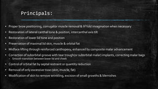
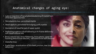
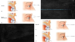
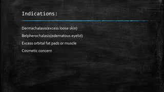
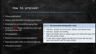
BLEpHaRoPLaStY
Dr.Akasha Amber
Plastic Surgery department,
BahawalVictoria Hospital, Bahawalpur
Introduction
▪ Rejuvination of upper and lower eyelids
▪ AIM:
– Youthful upper and lower eyelids
– Maintaining original shape
▪ Blepheraplasty; focus on entire periorbital structures
Principals:
▪ Proper brow positioning, corrugator muscle removal & lif fold invagination when necessary
▪ Restoration of lateral canthal tone & position, intercanthal axis tilt
▪ Restoration of lower lid tone and position
▪ Preservation of maximal lid skin, muscle & orbital fat
▪ Midface lifting through reinforced canthopexy, enhanced by composite malar advancement
▪ Correction of suborbital groove with tear trough(or suborbital malar) implants, correcting malar bags
– Smooth transition between lower lid and cheek
▪ Control of orbital fat by septal restraint or quantity reduction
▪ Removal of only excessive tisse (skin, muscle, fat)
▪ Modification of skin to remove wrinkling, excision of small growths & blemishes
Aging eyes
▪ Palpebral fissure: 28-30mm (H)
,8-10 MM (V)
▪ Lateral commissure; 2mm
superior to medial commissure
(canthal tilt)
▪ Upper lid margin arch peaks at
centre of pupil
– Lateral migration: (weakened
medial levator horn)
▪ Lid crease
– Occidental: 8-10 mm in Females,
7mm in Males
– Asian: absent or 4-6mm from
lash margin
▪ Lid fold( excess skin & muscle)
ANATOMY
▪ UPPER LID
– Skin
– Muscle
– Structural support
– Fat compartments
– Gland
▪ LOWER LID
– Skin
– Muscle
– Fat compartments
– Supportive framework
Upper lid
Anterior lamella
• Skin with subcutaneous tissue
• Muscle(orbicularis occuli)
Posterior lamella
• Tarsoligamentous sling
• (upper lid
retractors,capsulopalpebral
fascia,,tarsal plate,lateral &
medial canthal tendons)
• conjuntiva
Lower lid
Anterior lamella
• Skin with subcutaneous tissue
• Muscle(orbicularis occuli)
Posterior lamella
• Tarsal plate and capsulopalpebral
fascia with inferior tarsus
• conjuntiva
Anatomical changes of aging eye:
▪ Lateral migration of tarsal plate(weakening of medial horn
of levator apponeurosis
▪ Skin elasticity loss: periorbital rhytids
▪ Weak septum: periorbital fat bulging; puffy eyelids
▪ Herniation of nasal fat pad of upper eyelid
▪ Hollowing;superior sulcal hollowing or A-Frame deformity:
soft tissue volume loss
▪ Brow volume loss: brow flattening, brow ptosis; upper lid
hooding,lateral brow descent, narrowed brow lash distance
▪ Glabellar lines
▪ Eyelid bags, accentuation of lid cheek junction, tear trough
deformity
Indications:
Dermachalasis(excess loose skin)
Belpherochalasis(edematous eyelid)
Excess orbital fat pads or muscle
Cosmetic concern
How to proceed:
▪ Preop evaluation:
▪ history and detailed clinical examination
▪ Attention to surrounding aging face
▪ Rule out RED flags; conditions with high
postoperative risk
▪ Photographs
▪ Patient’s concern and expectations
▪ Operative plan with or without adjunts
▪ Informed consent
Preop evaluation:
▪ Visual acuity by snellen’s chart
▪ Visual field
▪ Occular movements (6 positions of gaze)
▪ Globe position: (vector +-), (Hertel exophthalmometry)
▪ Shirmer’s test (DRY EYE) for tear production
▪ Poor bell’s phenomenon
▪ Rule out occular pathologies
▪ Any history of contact lens intolerance
Brow and Upper Orbit:
▪ Brow:
– ptosis ,symmetry and shape
▪ Upper lid fold:
– Symmetry,ptosis
▪ Levator function:
– Excurtion
▪ Excess skin
▪ Excess preapponeurotic fat, ROOF
▪ Lacrimal gland ptosis
▪ Bony fullness or resorption
▪ FLOPPY EYELID SYNDROME
10-15mm(good)
6-9mm(fair)
<5mm
(poor)
Lower Orbit & Midface:
▪ Skin, muscle and orbital fat
– Excess/laxity
▪ Malar bags/ tear trough deformity
▪ Lateral canthal position:
– +ve or –ve tilt
▪ Lower lid tone/laxity
– Snap test
– Distraction test
▪ 1-2 mm minimal,
▪ 3-6mm moderate,
▪ >6mm significate laxity
▪ Scleral show
▪ Globe position
– Vector (+ve or –ve)
– Hertel exophthalmometry (16-18mm)
Systemic conditions:
▪ Any LASIK surgery during
previous 6 months
▪ DRY EYE SYNDROME
▪ PRIMARY ESSENTIAL
BLEPHAROSPASM
▪ GRAVES’ DISEASE
▪ EYELID INFLAMMATORY
DISORDERS
– Pemphigus, rosacea,
sarcoidosis,
Opertive Techniques:
▪ Upper lid blepharoplasty:
▪ Markings
▪ Lower lid blepharoplasty:
▪ Markings
▪ LA,GA
▪ MAC (monitored anesthesia care; LA with sedation and analgesia)
▪ Retrobulbar hemtoma or eyelid hematoma
▪ To avoid risk of post op ptosis, a supratarsal
fixationof pretarsal skin muscle to levator
apponeurosis in midpupillary line, 6-0 vicryl
,mattress suture
▪ Brow ptosis, excess brow fat pad corrected, lacrimal gland ptosis
corrected with fixation sutures
▪ ROOF addressed
▪ For Asian blepheraroplasty:
– low crease incision (4-6mm above lash line)
– Conservative resection of skin and fat
– To create a fold, multiple anchoring sutures through junction of upper tarsal
plate, levator insertion and dermis of upper lid skin
Lower lid blepharoplasty:
▪ Two approaches:
– Subciliary incision
– Tarsoconjuctival plus skin pinch approach
▪ Trans-septal
▪ retroseptal
▪ Through conjunctiva and capsulopalpebral fascia, through or behind
septum
▪ Minimal injury risks
Lower lid blepharoplasty:
▪ Two approaches:
– Subciliary incision
– Tarsoconjuctival plus skin pinch approach
▪ Subciliary Approach:
▪ Skin incision lateral to medial
▪ Stair-step incision through O.O muscle leaving 5mm pretarsal strip
▪ Skin muscle flap raised
▪ Dissection via septum to infraorbital rim
▪ Orbitomalar ligament released and tear trough deformity corrected.
▪ SOOF approached. Fat removed in conservative manner(all 3 compartments).
▪ Avoid injury to I.O.M (diplopia),avoid injury to arcuate expansion( further fat herniation)
▪ Arcus marginalis release, fat transposition, fat grafting
Canthoplasty and canthopexy
▪ Two layered canthopexy:
▪ A: suture fixating tarsal
tail into the drilled hole
▪ B: 2nd layer orbicularis
suture
▪ C: lateral sutures fix
lateral orbicularis to
deep temporal fascia
▪ D:if midface is elected,
inferior drill hole can be
made to fixate midface
tissues
▪ E: bury the knot into the
drill hole
Postop care
▪ Eye drops; moisturizing
▪ Ophthalmic lubricating
▪ Intermittent forced lid closure
▪ Head; elevated posture
▪ Ice cold packs for 24 hrs
▪ Ophthalmic ointment at night
▪ Sutures removal (5-7 days)
Complications:
▪ Bleeding
▪ Over correction
▪ Retrobulbar hemorrhage
▪ Chemosis
▪ Globe perforation
▪ Vision loss (periorbital filler injection/intra arterial embolization)
▪ Injury to SOM/IOM: diplopia
▪ Corneal irritation
▪ Inflammatory/infectious complications
▪ Hypersensitivity reactions
▪ Deep orbital cellulitis with intracavernous extension
▪ Subcutaneous epidermoid cysts
▪ epiphora
▪ Suture abscess,granuolma,
Complications:
▪ Dry eye
▪ Assymetry
▪ Lagophthalmos
▪ Lid malposition
▪ Ectropion/entropion
THANK YOU

Blepharoplasty

  • 1.
    BLEpHaRoPLaStY Dr.Akasha Amber Plastic Surgerydepartment, BahawalVictoria Hospital, Bahawalpur
  • 2.
    Introduction ▪ Rejuvination ofupper and lower eyelids ▪ AIM: – Youthful upper and lower eyelids – Maintaining original shape ▪ Blepheraplasty; focus on entire periorbital structures
  • 3.
    Principals: ▪ Proper browpositioning, corrugator muscle removal & lif fold invagination when necessary ▪ Restoration of lateral canthal tone & position, intercanthal axis tilt ▪ Restoration of lower lid tone and position ▪ Preservation of maximal lid skin, muscle & orbital fat ▪ Midface lifting through reinforced canthopexy, enhanced by composite malar advancement ▪ Correction of suborbital groove with tear trough(or suborbital malar) implants, correcting malar bags – Smooth transition between lower lid and cheek ▪ Control of orbital fat by septal restraint or quantity reduction ▪ Removal of only excessive tisse (skin, muscle, fat) ▪ Modification of skin to remove wrinkling, excision of small growths & blemishes
  • 4.
  • 5.
    ▪ Palpebral fissure:28-30mm (H) ,8-10 MM (V) ▪ Lateral commissure; 2mm superior to medial commissure (canthal tilt) ▪ Upper lid margin arch peaks at centre of pupil – Lateral migration: (weakened medial levator horn) ▪ Lid crease – Occidental: 8-10 mm in Females, 7mm in Males – Asian: absent or 4-6mm from lash margin ▪ Lid fold( excess skin & muscle)
  • 6.
    ANATOMY ▪ UPPER LID –Skin – Muscle – Structural support – Fat compartments – Gland ▪ LOWER LID – Skin – Muscle – Fat compartments – Supportive framework Upper lid Anterior lamella • Skin with subcutaneous tissue • Muscle(orbicularis occuli) Posterior lamella • Tarsoligamentous sling • (upper lid retractors,capsulopalpebral fascia,,tarsal plate,lateral & medial canthal tendons) • conjuntiva Lower lid Anterior lamella • Skin with subcutaneous tissue • Muscle(orbicularis occuli) Posterior lamella • Tarsal plate and capsulopalpebral fascia with inferior tarsus • conjuntiva
  • 16.
    Anatomical changes ofaging eye: ▪ Lateral migration of tarsal plate(weakening of medial horn of levator apponeurosis ▪ Skin elasticity loss: periorbital rhytids ▪ Weak septum: periorbital fat bulging; puffy eyelids ▪ Herniation of nasal fat pad of upper eyelid ▪ Hollowing;superior sulcal hollowing or A-Frame deformity: soft tissue volume loss ▪ Brow volume loss: brow flattening, brow ptosis; upper lid hooding,lateral brow descent, narrowed brow lash distance ▪ Glabellar lines ▪ Eyelid bags, accentuation of lid cheek junction, tear trough deformity
  • 18.
    Indications: Dermachalasis(excess loose skin) Belpherochalasis(edematouseyelid) Excess orbital fat pads or muscle Cosmetic concern
  • 19.
    How to proceed: ▪Preop evaluation: ▪ history and detailed clinical examination ▪ Attention to surrounding aging face ▪ Rule out RED flags; conditions with high postoperative risk ▪ Photographs ▪ Patient’s concern and expectations ▪ Operative plan with or without adjunts ▪ Informed consent
  • 20.
    Preop evaluation: ▪ Visualacuity by snellen’s chart ▪ Visual field ▪ Occular movements (6 positions of gaze) ▪ Globe position: (vector +-), (Hertel exophthalmometry) ▪ Shirmer’s test (DRY EYE) for tear production ▪ Poor bell’s phenomenon ▪ Rule out occular pathologies ▪ Any history of contact lens intolerance
  • 21.
    Brow and UpperOrbit: ▪ Brow: – ptosis ,symmetry and shape ▪ Upper lid fold: – Symmetry,ptosis ▪ Levator function: – Excurtion ▪ Excess skin ▪ Excess preapponeurotic fat, ROOF ▪ Lacrimal gland ptosis ▪ Bony fullness or resorption ▪ FLOPPY EYELID SYNDROME 10-15mm(good) 6-9mm(fair) <5mm (poor)
  • 22.
    Lower Orbit &Midface: ▪ Skin, muscle and orbital fat – Excess/laxity ▪ Malar bags/ tear trough deformity ▪ Lateral canthal position: – +ve or –ve tilt ▪ Lower lid tone/laxity – Snap test – Distraction test ▪ 1-2 mm minimal, ▪ 3-6mm moderate, ▪ >6mm significate laxity ▪ Scleral show ▪ Globe position – Vector (+ve or –ve) – Hertel exophthalmometry (16-18mm)
  • 23.
    Systemic conditions: ▪ AnyLASIK surgery during previous 6 months ▪ DRY EYE SYNDROME ▪ PRIMARY ESSENTIAL BLEPHAROSPASM ▪ GRAVES’ DISEASE ▪ EYELID INFLAMMATORY DISORDERS – Pemphigus, rosacea, sarcoidosis,
  • 24.
    Opertive Techniques: ▪ Upperlid blepharoplasty: ▪ Markings ▪ Lower lid blepharoplasty: ▪ Markings
  • 25.
    ▪ LA,GA ▪ MAC(monitored anesthesia care; LA with sedation and analgesia) ▪ Retrobulbar hemtoma or eyelid hematoma
  • 29.
    ▪ To avoidrisk of post op ptosis, a supratarsal fixationof pretarsal skin muscle to levator apponeurosis in midpupillary line, 6-0 vicryl ,mattress suture
  • 30.
    ▪ Brow ptosis,excess brow fat pad corrected, lacrimal gland ptosis corrected with fixation sutures ▪ ROOF addressed ▪ For Asian blepheraroplasty: – low crease incision (4-6mm above lash line) – Conservative resection of skin and fat – To create a fold, multiple anchoring sutures through junction of upper tarsal plate, levator insertion and dermis of upper lid skin
  • 31.
    Lower lid blepharoplasty: ▪Two approaches: – Subciliary incision – Tarsoconjuctival plus skin pinch approach ▪ Trans-septal ▪ retroseptal ▪ Through conjunctiva and capsulopalpebral fascia, through or behind septum ▪ Minimal injury risks
  • 36.
    Lower lid blepharoplasty: ▪Two approaches: – Subciliary incision – Tarsoconjuctival plus skin pinch approach ▪ Subciliary Approach: ▪ Skin incision lateral to medial ▪ Stair-step incision through O.O muscle leaving 5mm pretarsal strip ▪ Skin muscle flap raised ▪ Dissection via septum to infraorbital rim ▪ Orbitomalar ligament released and tear trough deformity corrected. ▪ SOOF approached. Fat removed in conservative manner(all 3 compartments). ▪ Avoid injury to I.O.M (diplopia),avoid injury to arcuate expansion( further fat herniation) ▪ Arcus marginalis release, fat transposition, fat grafting
  • 39.
  • 41.
    ▪ Two layeredcanthopexy: ▪ A: suture fixating tarsal tail into the drilled hole ▪ B: 2nd layer orbicularis suture ▪ C: lateral sutures fix lateral orbicularis to deep temporal fascia ▪ D:if midface is elected, inferior drill hole can be made to fixate midface tissues ▪ E: bury the knot into the drill hole
  • 44.
    Postop care ▪ Eyedrops; moisturizing ▪ Ophthalmic lubricating ▪ Intermittent forced lid closure ▪ Head; elevated posture ▪ Ice cold packs for 24 hrs ▪ Ophthalmic ointment at night ▪ Sutures removal (5-7 days)
  • 45.
    Complications: ▪ Bleeding ▪ Overcorrection ▪ Retrobulbar hemorrhage ▪ Chemosis ▪ Globe perforation ▪ Vision loss (periorbital filler injection/intra arterial embolization) ▪ Injury to SOM/IOM: diplopia ▪ Corneal irritation ▪ Inflammatory/infectious complications ▪ Hypersensitivity reactions ▪ Deep orbital cellulitis with intracavernous extension ▪ Subcutaneous epidermoid cysts ▪ epiphora ▪ Suture abscess,granuolma,
  • 46.
    Complications: ▪ Dry eye ▪Assymetry ▪ Lagophthalmos ▪ Lid malposition ▪ Ectropion/entropion
  • 47.

Editor's Notes

  • #3 Aging; changes in skin,muscle,fat,bone & ligamental structures,no single technique for all,common procedure but challenging, complications must be addressed
  • #4 1.Brow ptosis and lid ptosis corrected at this time ideally
  • #9 Superior arcade is 4 mm above lash line.. Lower arcade is 2 mm away from lash line… Muscleorigin n insertion pretarsal(superficial part;deep part as horner’s muscle) causes involuntary blinking, preseptal (superficial & deep part;jones’ muscle) voluntary blinking+lacrimal drainage, orbital: forceful eye closure; supplied by frontal, zygomatic & buccal br of facial n.
  • #10 Upper tarsal plate: 24mm(H), 8-10mm(V),anteriorly PTOO,LA, superiorly Muller’s m,conjunctiva posteriorly Lower TP: 24mm(H), 4mm(V), PTOO anter,capsulopalpebral fascia inferiorly, conjunctiva post.tarsal plates attach to orbit with lat n medial canthal tendons and retinacular structures
  • #11 Medial canthal tendon: ant reflection to nasolacrimal crest, post reflection to post. Lacrimal crest behind lacrimal sac. Lateral canthal tendon: superficial(lat orbital rim periosteum) n deep( whitnall’s tubercle) parts, tarsal straps are separate structures attached tarsus to orbit Superior transverse ligament of whitnall formed by facial thickening of Levator palpebrae superioris( medially to trochlea of SOM, laterally to lacrimal gland’s psedocapsule and lacrimal fossa Inferior suspensory/transverse ligament, lockwood’s ligament ,attached medially to med retinaculum, lat. to lat.retinaculum,fuses with inf tarsal border through capsulopalpebral fascia. Upper lid retractors: Levator muscle origin from lesser wing of sphenoid,apponeurosis inserts to ant surface of sup.tarsal late.apponerosis forms 5-7mm above tarsal plate..lateral horn(to lat orbital rim tubercle) separating lacrimal gland in two parts.medial horn inserts into deep part of medial canthal tendon.LPS supplied by occulomotor nerve Muller musle smooth,from deep surface of levator to superior border of tarsal plate,sympathetic innervation.
  • #12 Medial retinaculum: medial horn of LA, medial rectus check ligament, medial extensions of lockwood’s and whitnall’s ligaments, medial extension deep head of PTOO, orbital septum, medial canthal tendon Lateral retinaculum: lateral horn of LA, lateral rectus check ligament, larteral canthal tendon, lateral extensions of lockwood’s n whitnall’s ligament, orbital septum ,deep part of PTOO, tarsal starp
  • #13 Lower lid retractors: capsulopalpebral fascia;extension of inf rectus facscia, enclosing IOM, inseting into ant surface of tarsal plate, inf tarsal muscle(analogous to muller muscle) nserting into inf border of tarsal plate.
  • #14 Orbital septum fuses with LA several mm above tarsal plate, but fuses few mm above tarsal plate in lower lid… Nasal(pale white) and central/preapponeurotic(yellow) fat pads with interpad septum which is connected with trochlea of SOM, NASAL fat pad contains medial palpebral artery so careful, central is called preapponeurotic fat pad,responsible for lid fold fullness Lower lid fat pads:3; nasal( similar to upper lid nasal fat, central and lateral(temporal)..nasal n central separated by IOM, central n lateral by interpad septum n arcuate expansion(lockwood’s lig extension) Lacrimal gland;orbital and palpebral parts divided by lateral horn of levator connected by isthmus. GLAND PTOSIS:dehiscence of SOMMERING’s ligament(fibrous interlobular septa connecting gland to lac fossa of orbit. lateral to lacrimal gland, fat compartment” EISLER’S POCKET with EISLER’S fat, just above whitnall’s tubercle is landmark of this tubercle.
  • #15 Mendelson described prezygomatic space bounded by Orbitomalar ligament/orbicularis retaining ligament and the zygomaticocutaneous lig Orbitomalar lig n OO muscle origin from orbital rim define tear trough n lid-cheek junction Malar fat pads(subcutaneous) below it
  • #21 Supeior oblique: intortion, depression(adducted eye) and abduction Inf oblique: extortion, elevation(adducted eye), abduction Rule out diplopia, Shirmer’s test: tetracaine eye drops, shirmer strip, after 5 min, less than 10 mm wetting : no skin excision,lateral canthopexy n lubricants n temp tarsorraphy
  • #22 Lid fold asymmetry: lid ptosis, lid retraction, assyemtrical amount of soft tissue in upper lid,assymetrical brow position Ptosis: congenital(poor excurtion n levator function), acquired(ptosis; levator function better,high lid crease) apponeurotic dehiscence, myogenic (MG), neurogenic (H.S),mechanical( tr or trauma) 2-3mm, 3-5mm, >5mm:mild moderate n severe ptosis FES: large built males, upper lid eversion during forceful lid closure, post bleph chemosis,over-riding of lid and deparation from globe during closure,treat with tarsal resection n lat cathoplasty of upper lid
  • #23 -ve tilt: canthal disinsertion,laxity or prominent eye or it is hereditary trait -ve vector is relation of globe to the inf orbital rim n its overlying tissue in prominenet eye, globe projects anteriorly to it…poor globe support +ve vector is normal vector where ant most aspect of glove is behind the infraorbital rim n its overlying soft tissues Enophthalmos(deep set eyes): more internal placement of catho-pexy/plasty suture Exosphthalmos(prominent eye) high lateral canthal anchoring suture Prominent eye needs some infraorbital implants or lower lid spacers for suppoert
  • #27 Point A,on lid crease at midpupillary line (8-10mm in F ,7mm in M), following lid crease point B just medial to carumcle(prevent webbing), laterally point C 5-6mm from lash margin, Latera extension from point C in skin tension line( avoid extension to lateral orbital rim) Point D at level of lateral canthus(preserving 10-15mm skin between lower border of brow and lash margin) Crease drawn parallel to eyelid crease tapering medially
  • #28 Lenticular skin excision in young,more trapezoid lateral in elderly Skin excised Muscle strip removed(very thin) Septum opened Fat exposed If needed nasal fat pad removed with needle cautery Avoid inj to medial palpebral artery Avoid injuery to SOM and trochlea( diplopia and head tilting towards affected muscle side) Excess fat removal: peaked arch deformity of lid crease and A-frame deformity/more hollow and aged appearance.
  • #29 Interuupted nylon sutures lateral to lat.canthus, remaining continuous subcuticular stitches
  • #38 Single point below the lateral canthus,medially extension 2-3 mm below lash line Lateral extension of 6-10 in skin tension line,lateral extensions of upper n lower blepharoplasty incisons must be at least 10 mm away to avoid webbing
  • #46 Chemosis: ophthalmic steroids, decongestants,oral steroids, tarsorrhaphy Retrobulbar hematoma( immediate decompression,lateral canthotomy n cantholysis,surgical exploration,mannitol acetazolamide and oxygen)