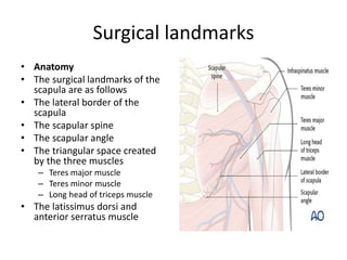
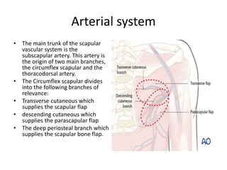
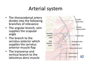
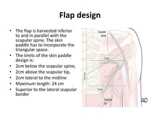
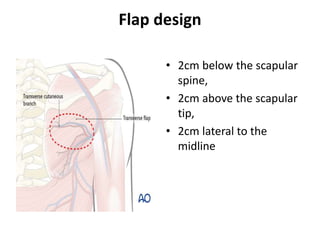
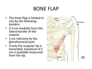
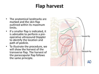
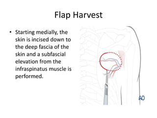
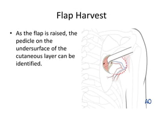
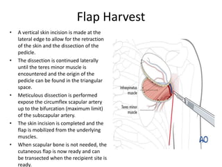
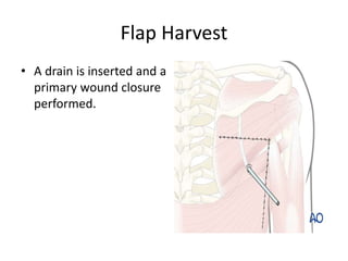
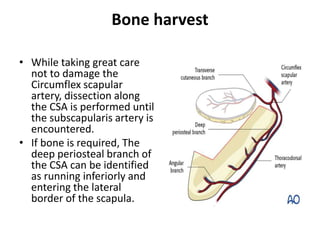
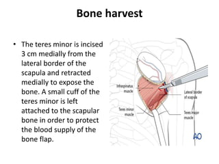
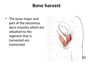
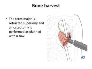
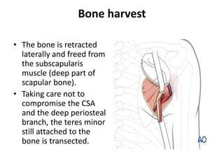
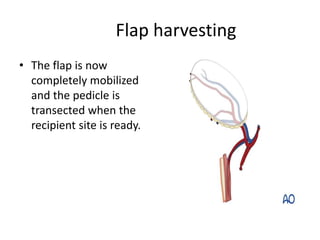
SCAPULAR FLAP
JAMEEL KIFAYATULLAH
Surgical landmarks
• Anatomy
• The surgical landmarks of the
scapula are as follows
• The lateral border of the
scapula
• The scapular spine
• The scapular angle
• The triangular space created
by the three muscles
– Teres major muscle
– Teres minor muscle
– Long head of triceps muscle
• The latissimus dorsi and
anterior serratus muscle
Arterial system
• The main trunk of the scapular
vascular system is the
subscapular artery. This artery is
the origin of two main branches,
the circumflex scapular and the
thoracodorsal artery.
• The Circumflex scapular divides
into the following branches of
relevance:
• Transverse cutaneous which
supplies the scapular flap
• descending cutaneous which
supplies the parascapular flap
• The deep periosteal branch which
supplies the scapular bone flap.
Arterial system
• The thoracodorsal artery
divides into the following
branches of relevance:
• The angular branch, vein
supplies the scapular
angle
• The branch to the
serratus anterior which
supplies the serratus
anterior muscle flap
• The transverse and
vertical branch to the
latissimus dorsi muscle
Flap design
• The flap is harvested inferior
to and in parallel with the
scapular spine. The skin
paddle has to incorporate the
triangular space.
• The limits of the skin paddle
design is:
• 2cm below the scapular spine,
• 2cm above the scapular tip,
• 2cm lateral to the midline
• Maximum length: 24 cm
• Superior to the lateral scapular
border
Flap design
• 2cm below the scapular
spine,
• 2cm above the scapular
tip,
• 2cm lateral to the
midline
BONE FLAP
• The bone flap is limited in
size by the following
borders:
• 2-3 cm medially from the
lateral border of the
scapula
• 2 cm inferiorly to the
glenohumeral joint
• If only the scapular tip is
harvested, maximum 4-5
cm is available measured
from the tip.
Flap harvest
• The anatomical landmarks are
marked and the skin flap
outlined within its maximum
limits.
• If a smaller flap is indicated, it
is advisable to perform a pre-
operative ultrasound Doppler
to identify the location and
path of pedicle.
• To illustrate the procedure, we
will show the harvest of the
transverse flap. The harvest of
the parascapular flap follows
the same principle.
Flap Harvest
• Starting medially, the
skin is incised down to
the deep fascia of the
skin and a subfascial
elevation from the
infraspinatus muscle is
performed.
Flap Harvest
• As the flap is raised, the
pedicle on the
undersurface of the
cutaneous layer can be
identified.
Flap Harvest
• A vertical skin incision is made at the
lateral edge to allow for the retraction
of the skin and the dissection of the
pedicle.
• The dissection is continued laterally
until the teres minor muscle is
encountered and the origin of the
pedicle can be found in the triangular
space.
• Meticulous dissection is performed
expose the circumflex scapular artery
up to the bifurcation (maximum limit)
of the subscapular artery.
• The skin incision is completed and the
flap is mobilized from the underlying
muscles.
• When scapular bone is not needed, the
cutaneous flap is now ready and can
be transected when the recipient site is
ready.
Flap Harvest
• A drain is inserted and a
primary wound closure
performed.
Bone harvest
• While taking great care
not to damage the
Circumflex scapular
artery, dissection along
the CSA is performed until
the subscapularis artery is
encountered.
• If bone is required, The
deep periosteal branch of
the CSA can be identified
as running inferiorly and
entering the lateral
border of the scapula.
Bone harvest
• The teres minor is incised
3 cm medially from the
lateral border of the
scapula and retracted
medially to expose the
bone. A small cuff of the
teres minor is left
attached to the scapular
bone in order to protect
the blood supply of the
bone flap.
Bone harvest
• The teres major and
part of the latissimus
dorsi muscles which are
attached to the
segment that is
harvested are
transected.
Bone harvest
• The teres major is
retracted superiorly and
an osteotomy is
performed as planned
with a saw.
Bone harvest
• The bone is retracted
laterally and freed from
the subscapularis
muscle (deep part of
scapular bone).
• Taking care not to
compromise the CSA
and the deep periosteal
branch, the teres minor
still attached to the
bone is transected.
Flap harvesting
• The flap is now
completely mobilized
and the pedicle is
transected when the
recipient site is ready.
ADVANTAGES
• Large volume supply of different soft tissue
components
• Rarely affected by vascular occlusive disease
Indications
• Very useful in situations where significant soft
tissue volume has to be replaced such as
segmental mandibular resections in
conjunction with glossectomy procedures or
extensive full thickness skin defects
• Composite scapula flap very useful for midface
reconstruction after extensive maxillectomies

Scapular flap

  • 1.
  • 2.
    Surgical landmarks • Anatomy •The surgical landmarks of the scapula are as follows • The lateral border of the scapula • The scapular spine • The scapular angle • The triangular space created by the three muscles – Teres major muscle – Teres minor muscle – Long head of triceps muscle • The latissimus dorsi and anterior serratus muscle
  • 3.
    Arterial system • Themain trunk of the scapular vascular system is the subscapular artery. This artery is the origin of two main branches, the circumflex scapular and the thoracodorsal artery. • The Circumflex scapular divides into the following branches of relevance: • Transverse cutaneous which supplies the scapular flap • descending cutaneous which supplies the parascapular flap • The deep periosteal branch which supplies the scapular bone flap.
  • 4.
    Arterial system • Thethoracodorsal artery divides into the following branches of relevance: • The angular branch, vein supplies the scapular angle • The branch to the serratus anterior which supplies the serratus anterior muscle flap • The transverse and vertical branch to the latissimus dorsi muscle
  • 5.
    Flap design • Theflap is harvested inferior to and in parallel with the scapular spine. The skin paddle has to incorporate the triangular space. • The limits of the skin paddle design is: • 2cm below the scapular spine, • 2cm above the scapular tip, • 2cm lateral to the midline • Maximum length: 24 cm • Superior to the lateral scapular border
  • 6.
    Flap design • 2cmbelow the scapular spine, • 2cm above the scapular tip, • 2cm lateral to the midline
  • 7.
    BONE FLAP • Thebone flap is limited in size by the following borders: • 2-3 cm medially from the lateral border of the scapula • 2 cm inferiorly to the glenohumeral joint • If only the scapular tip is harvested, maximum 4-5 cm is available measured from the tip.
  • 8.
    Flap harvest • Theanatomical landmarks are marked and the skin flap outlined within its maximum limits. • If a smaller flap is indicated, it is advisable to perform a pre- operative ultrasound Doppler to identify the location and path of pedicle. • To illustrate the procedure, we will show the harvest of the transverse flap. The harvest of the parascapular flap follows the same principle.
  • 9.
    Flap Harvest • Startingmedially, the skin is incised down to the deep fascia of the skin and a subfascial elevation from the infraspinatus muscle is performed.
  • 10.
    Flap Harvest • Asthe flap is raised, the pedicle on the undersurface of the cutaneous layer can be identified.
  • 11.
    Flap Harvest • Avertical skin incision is made at the lateral edge to allow for the retraction of the skin and the dissection of the pedicle. • The dissection is continued laterally until the teres minor muscle is encountered and the origin of the pedicle can be found in the triangular space. • Meticulous dissection is performed expose the circumflex scapular artery up to the bifurcation (maximum limit) of the subscapular artery. • The skin incision is completed and the flap is mobilized from the underlying muscles. • When scapular bone is not needed, the cutaneous flap is now ready and can be transected when the recipient site is ready.
  • 12.
    Flap Harvest • Adrain is inserted and a primary wound closure performed.
  • 13.
    Bone harvest • Whiletaking great care not to damage the Circumflex scapular artery, dissection along the CSA is performed until the subscapularis artery is encountered. • If bone is required, The deep periosteal branch of the CSA can be identified as running inferiorly and entering the lateral border of the scapula.
  • 14.
    Bone harvest • Theteres minor is incised 3 cm medially from the lateral border of the scapula and retracted medially to expose the bone. A small cuff of the teres minor is left attached to the scapular bone in order to protect the blood supply of the bone flap.
  • 15.
    Bone harvest • Theteres major and part of the latissimus dorsi muscles which are attached to the segment that is harvested are transected.
  • 16.
    Bone harvest • Theteres major is retracted superiorly and an osteotomy is performed as planned with a saw.
  • 17.
    Bone harvest • Thebone is retracted laterally and freed from the subscapularis muscle (deep part of scapular bone). • Taking care not to compromise the CSA and the deep periosteal branch, the teres minor still attached to the bone is transected.
  • 18.
    Flap harvesting • Theflap is now completely mobilized and the pedicle is transected when the recipient site is ready.
  • 19.
    ADVANTAGES • Large volumesupply of different soft tissue components • Rarely affected by vascular occlusive disease
  • 20.
    Indications • Very usefulin situations where significant soft tissue volume has to be replaced such as segmental mandibular resections in conjunction with glossectomy procedures or extensive full thickness skin defects • Composite scapula flap very useful for midface reconstruction after extensive maxillectomies