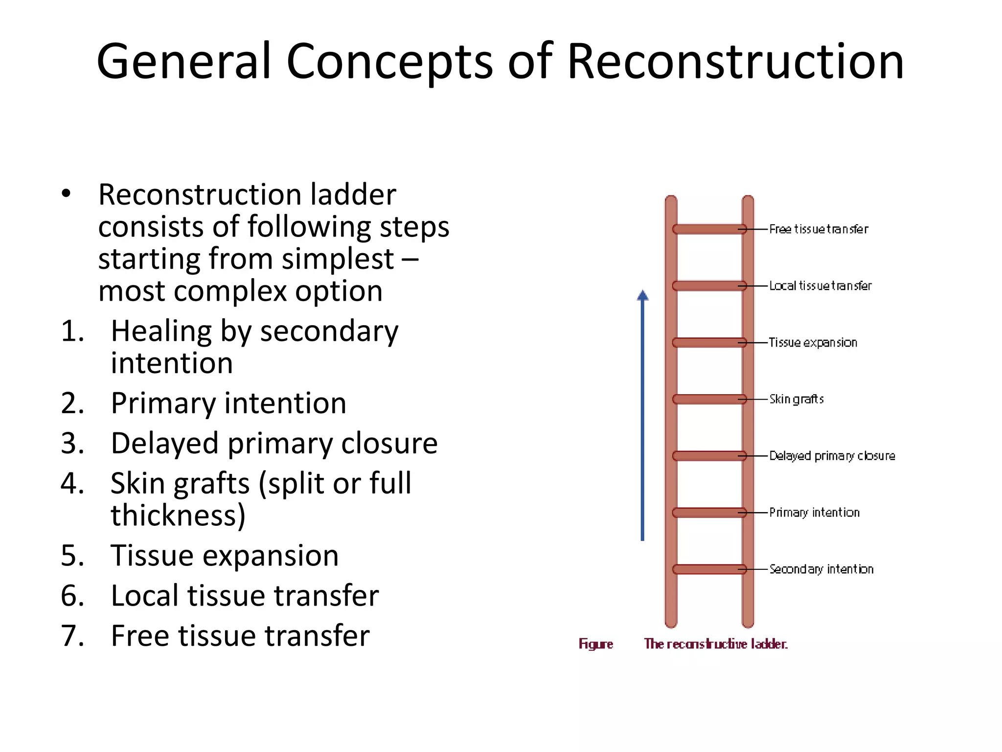
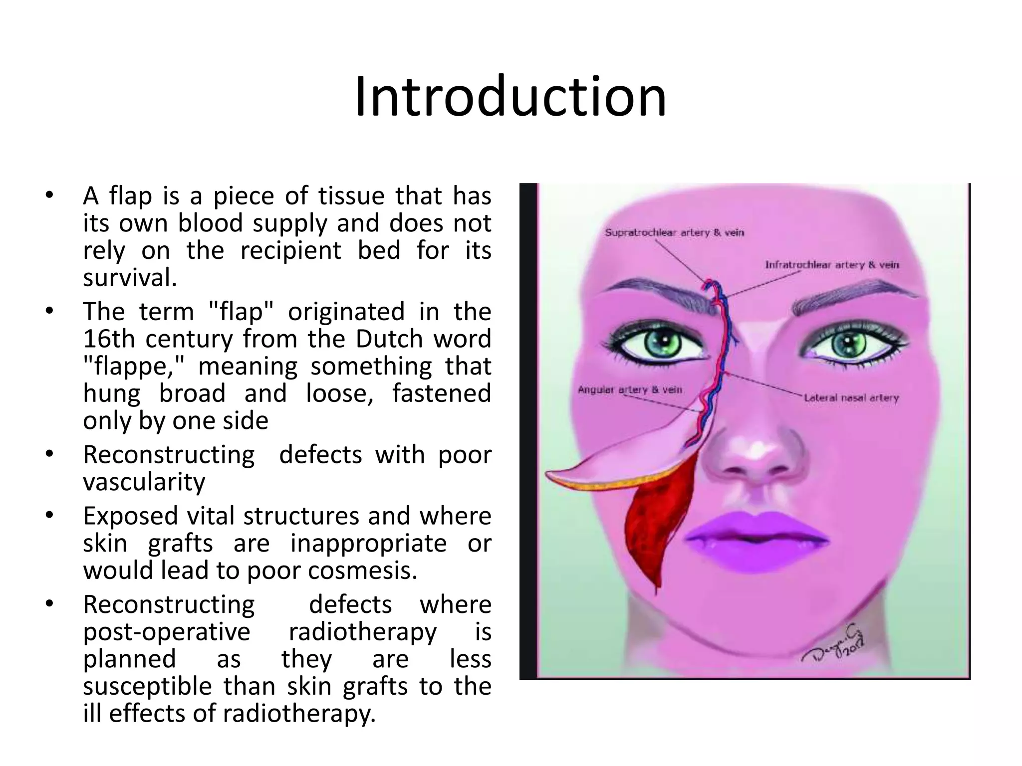
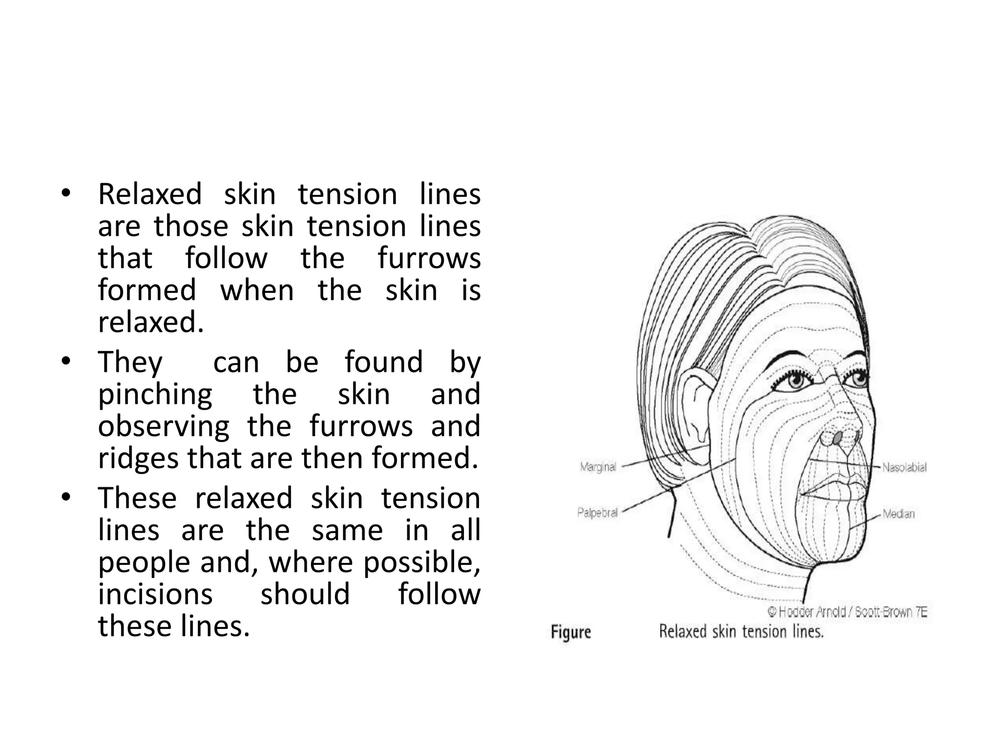
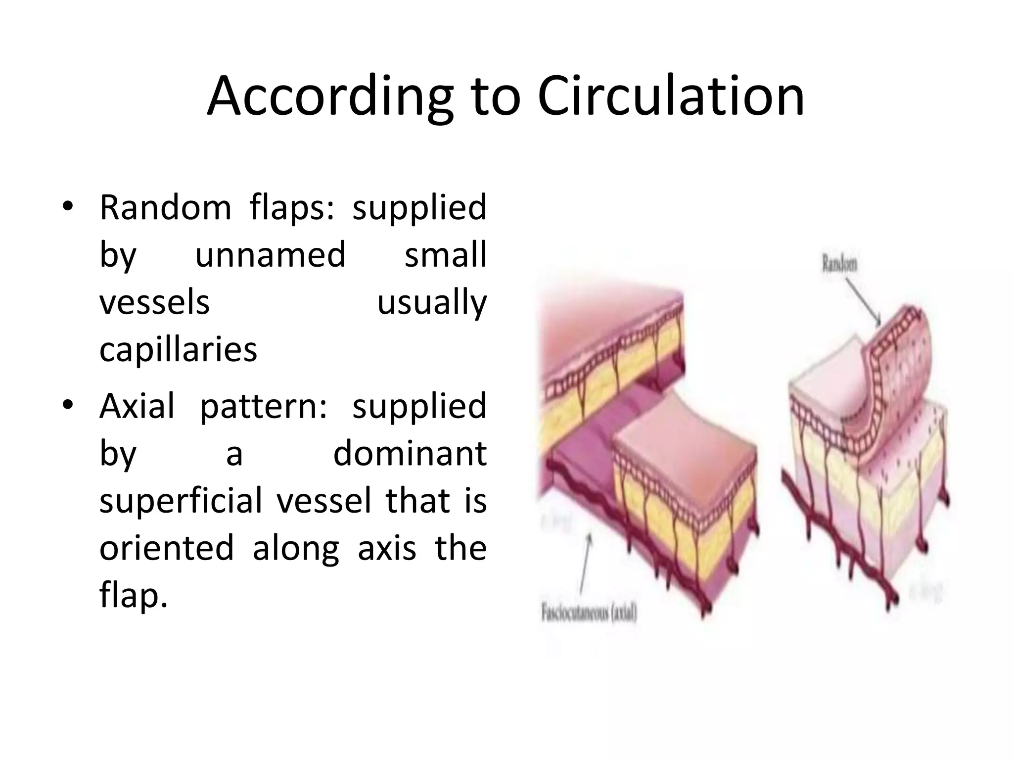
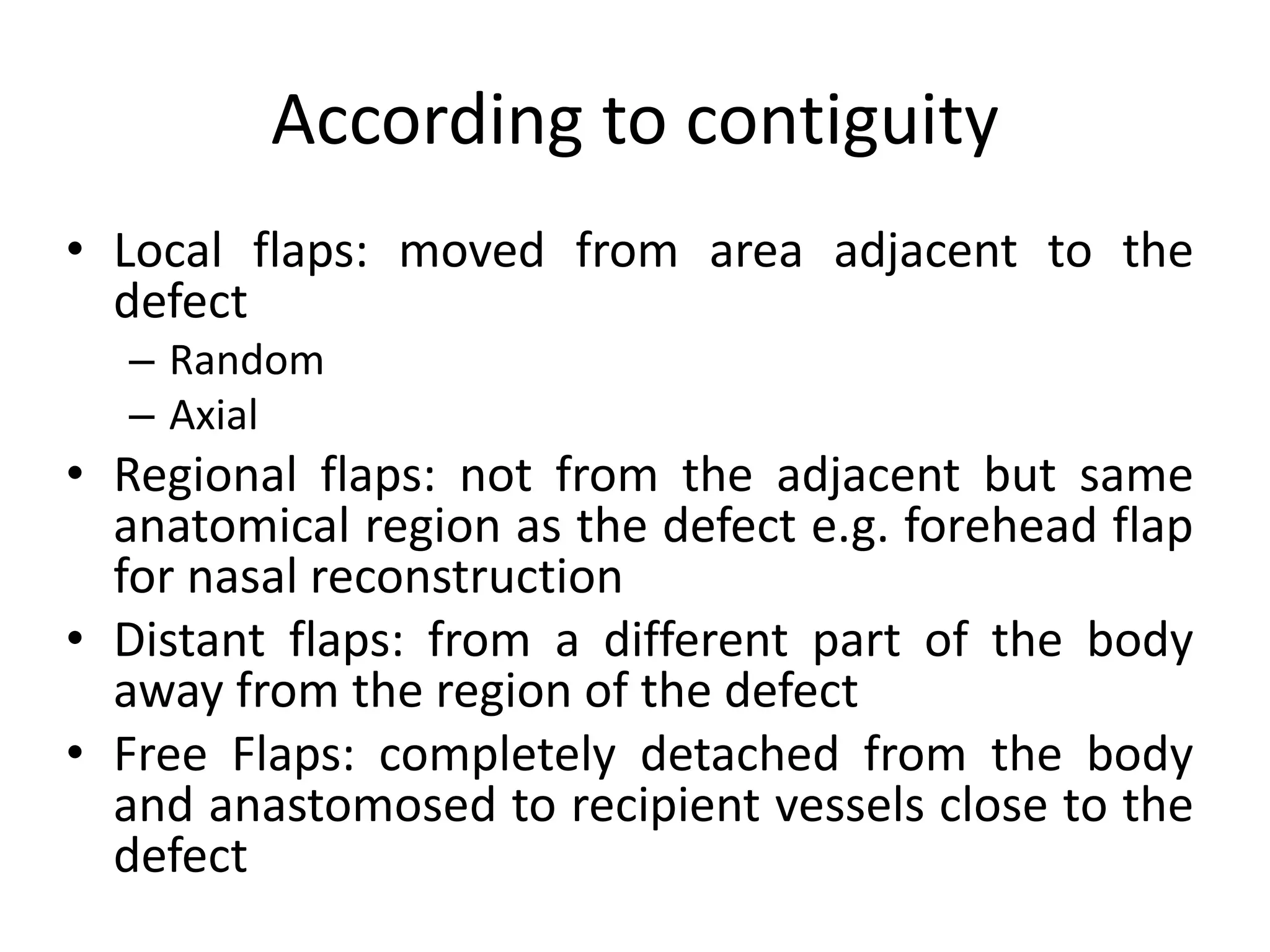
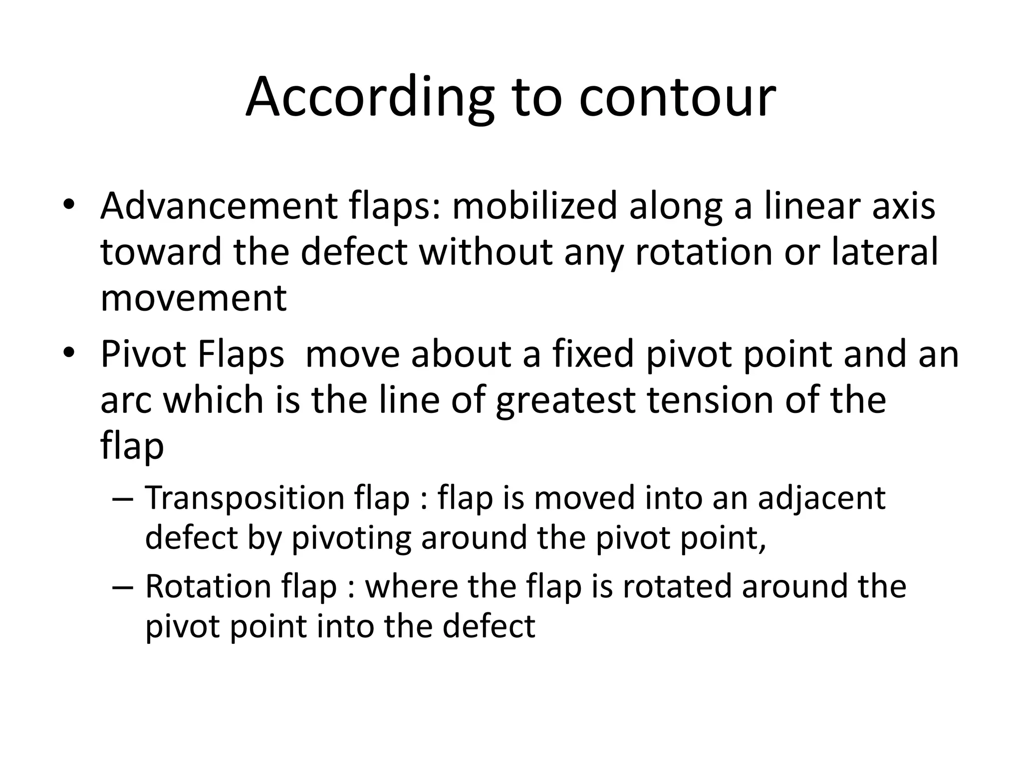
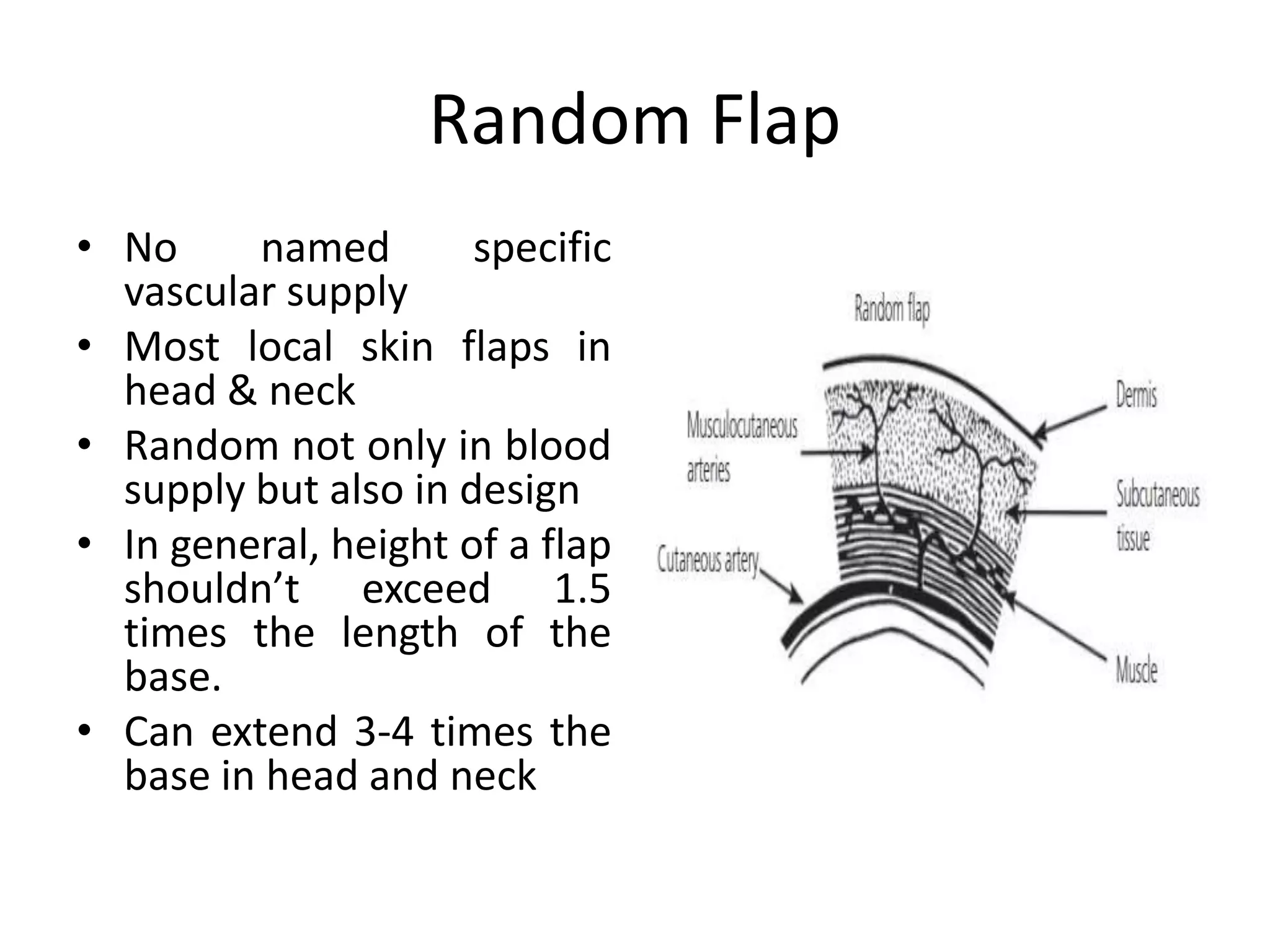
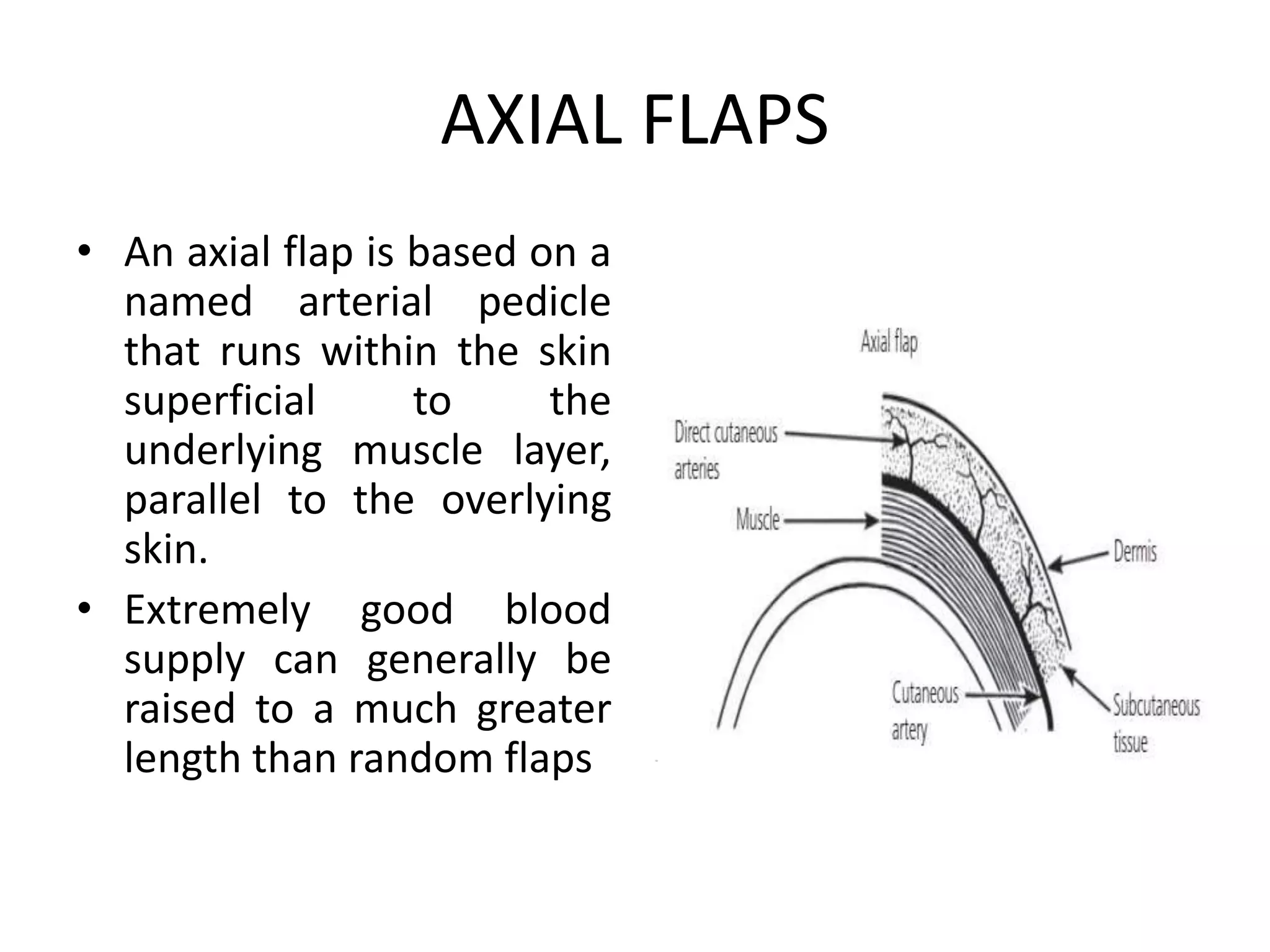
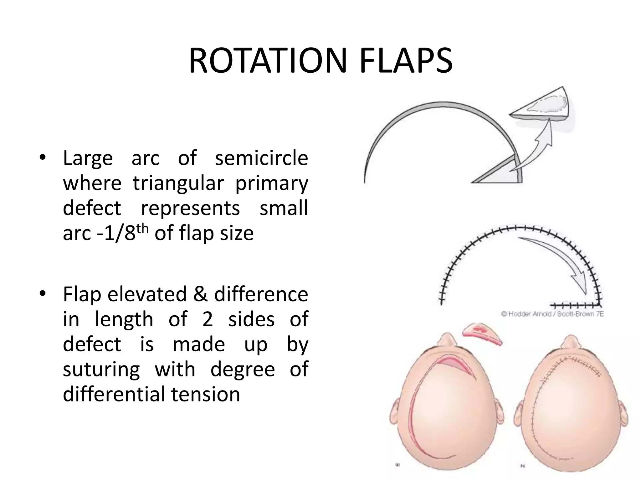
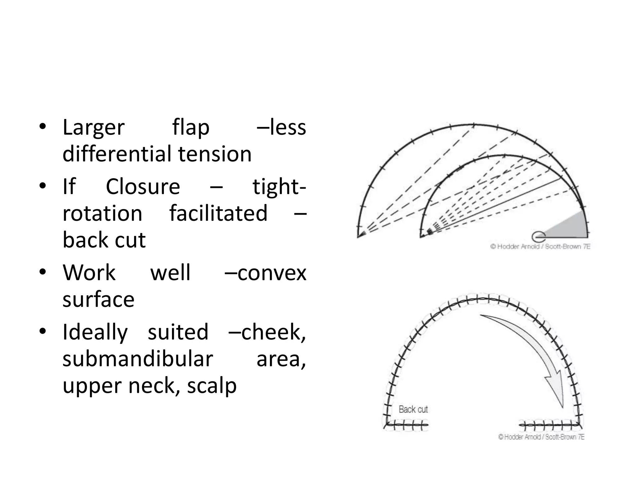
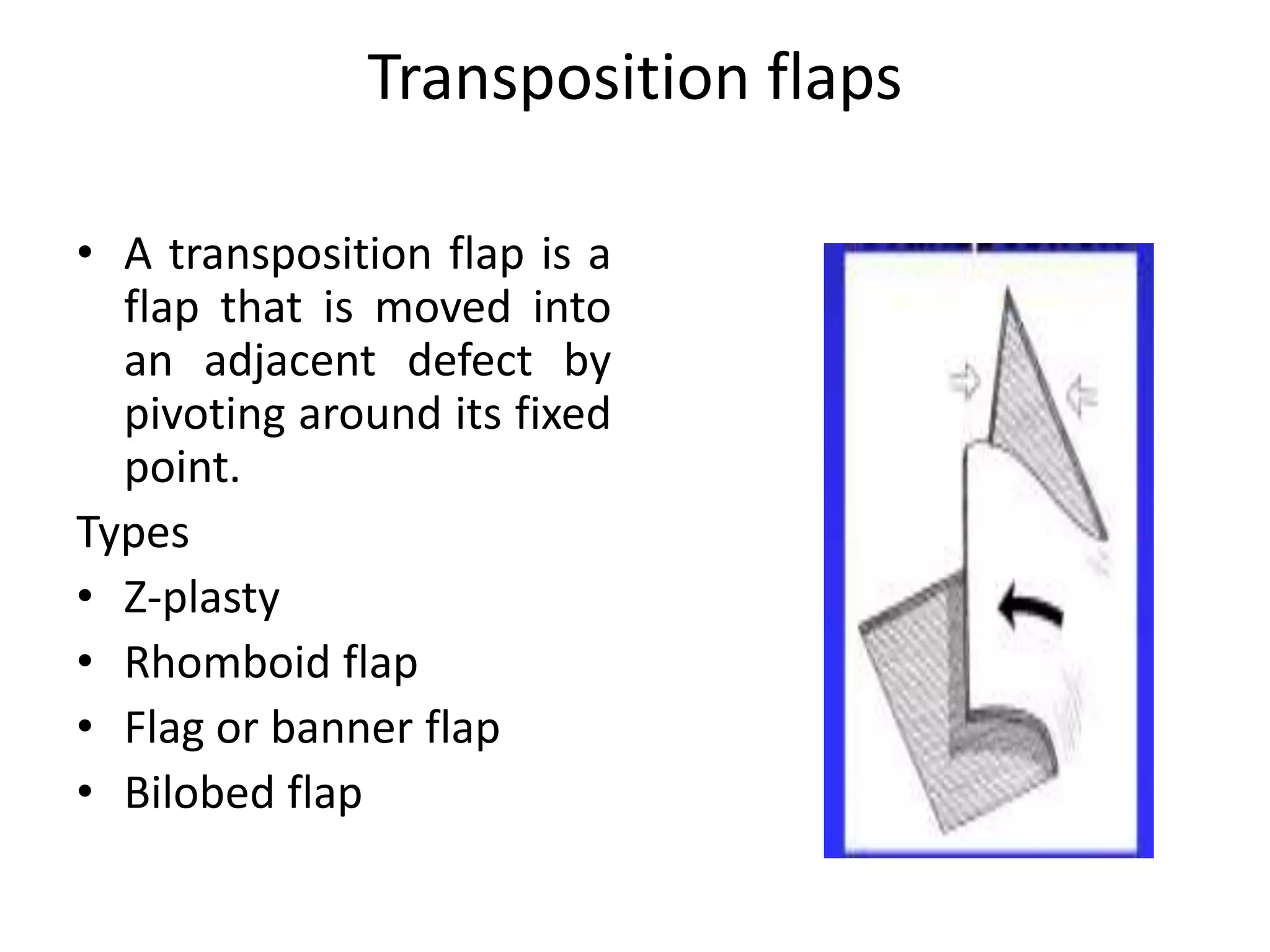
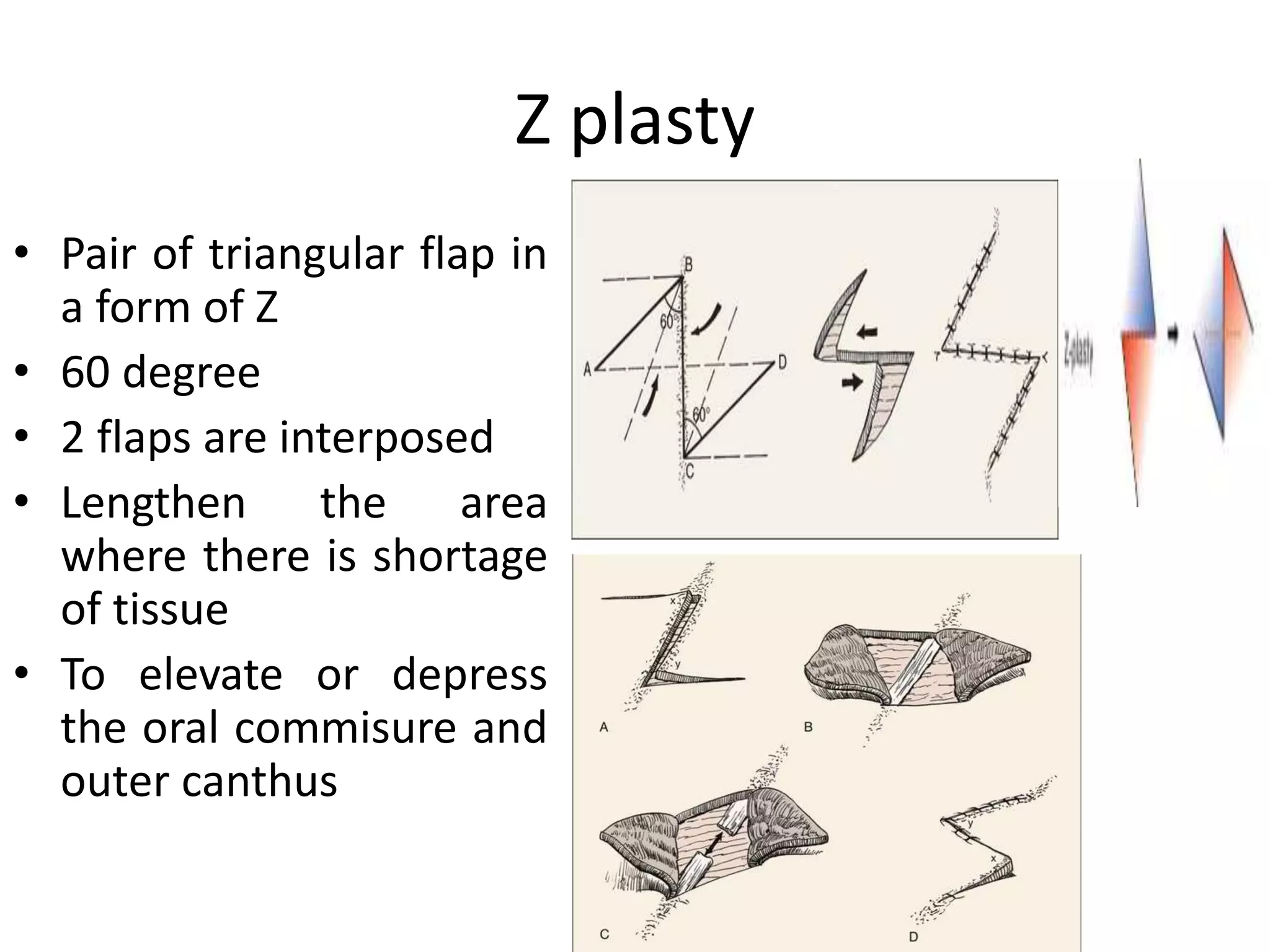
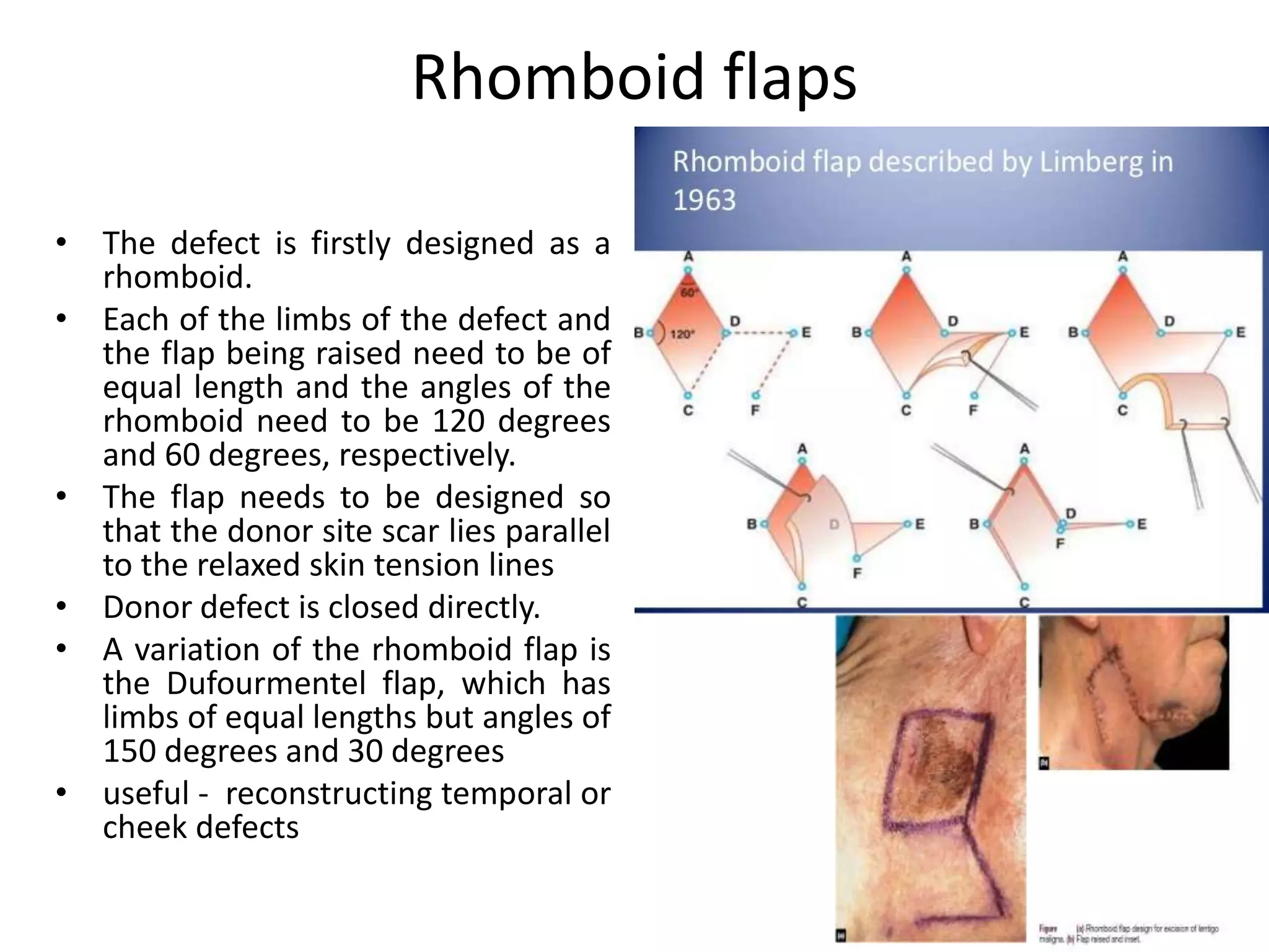
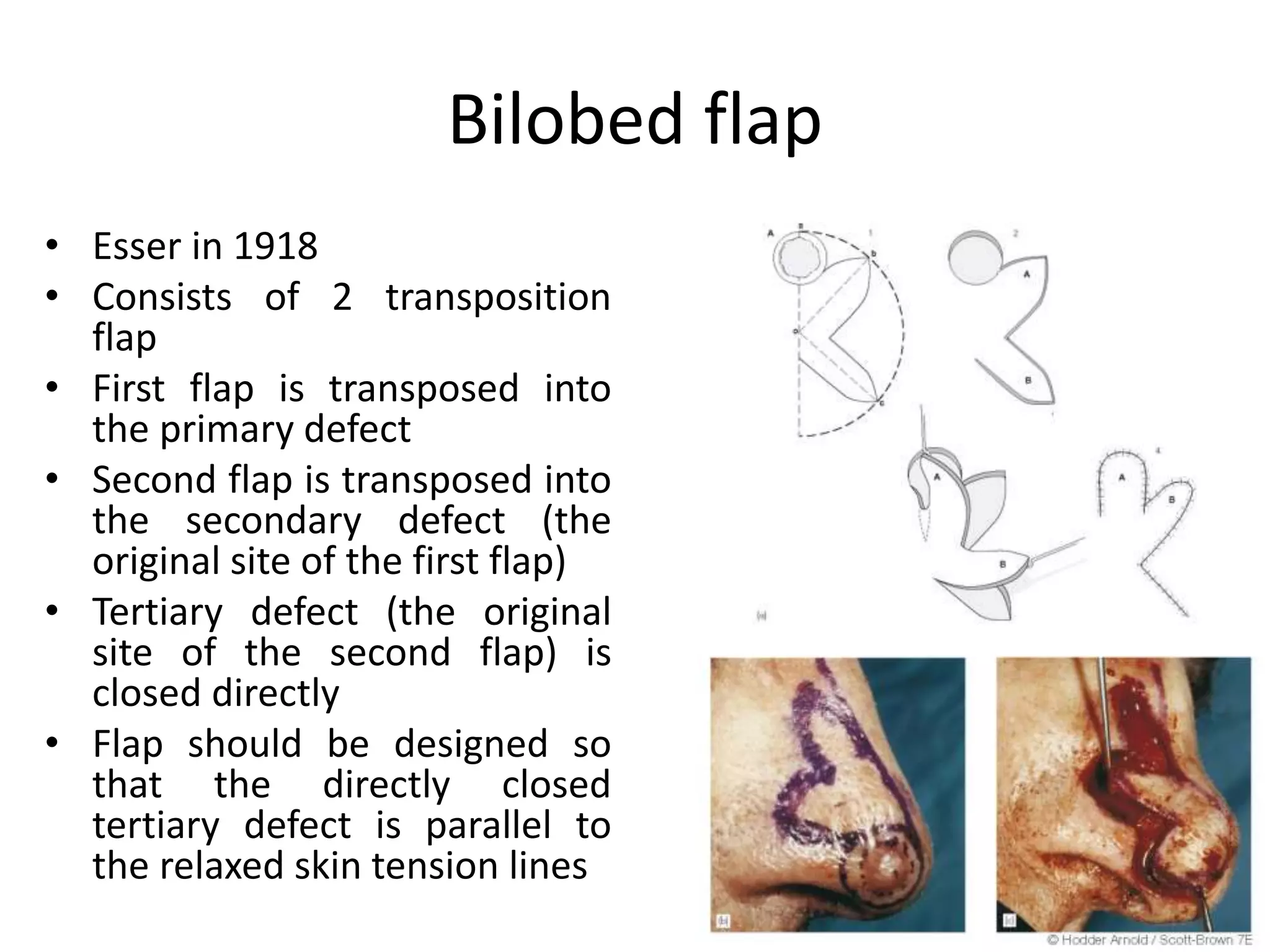
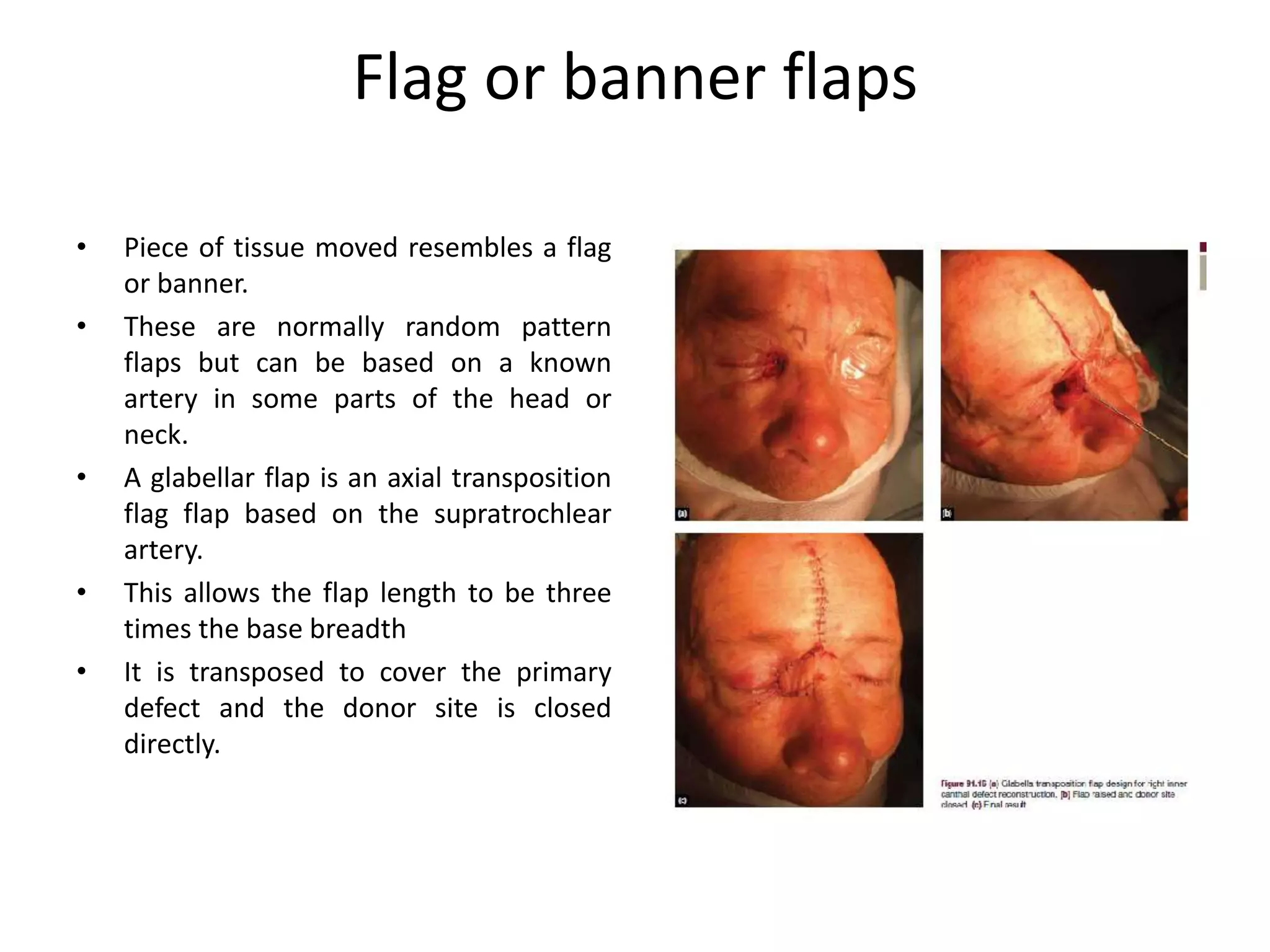
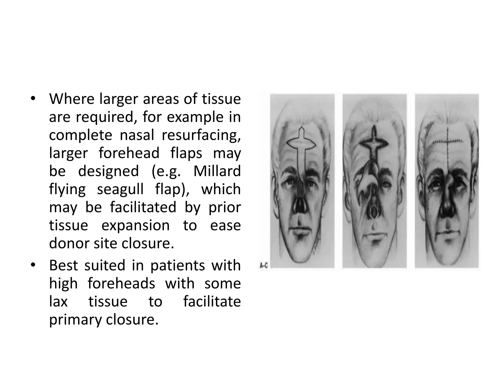
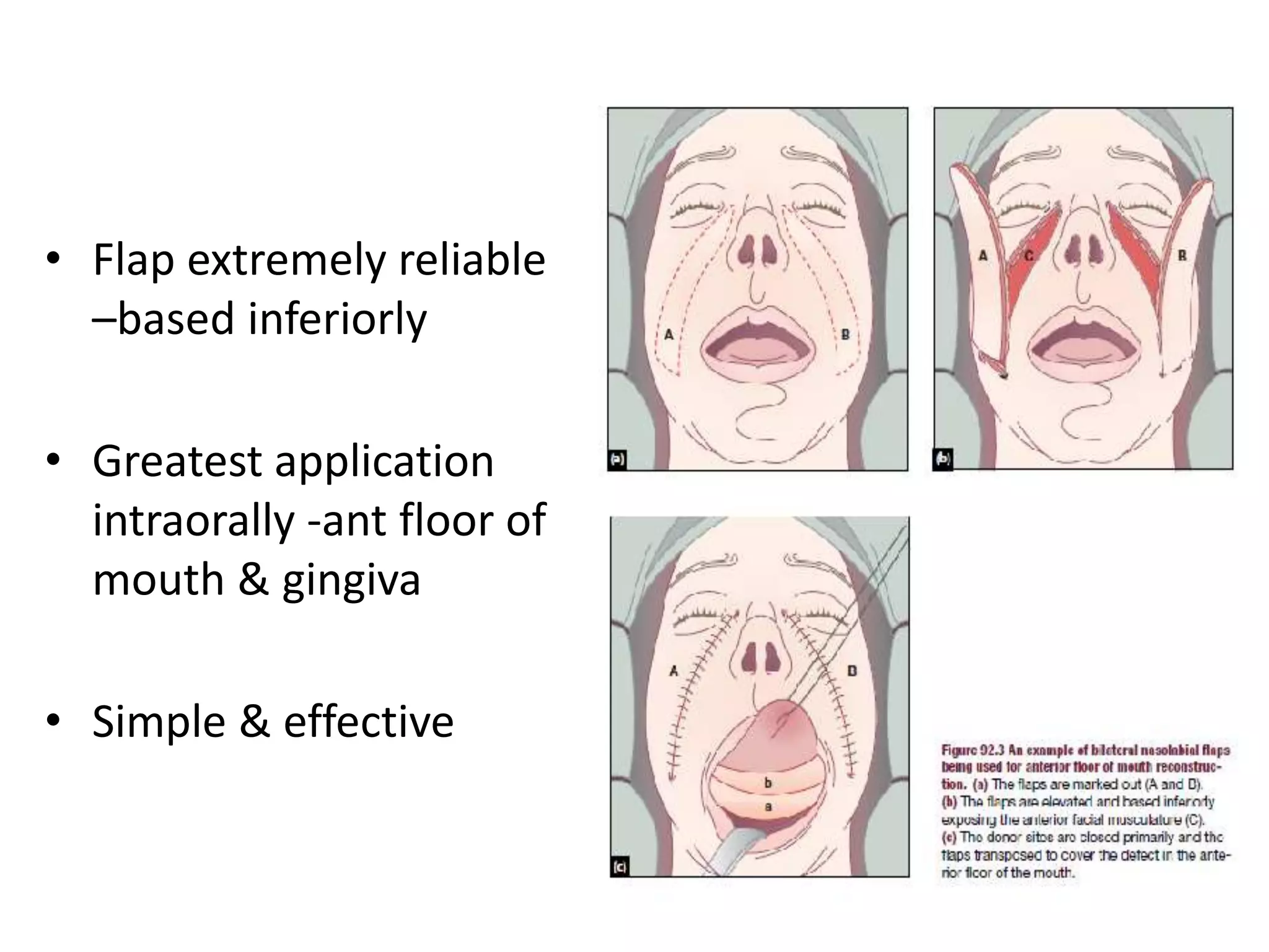
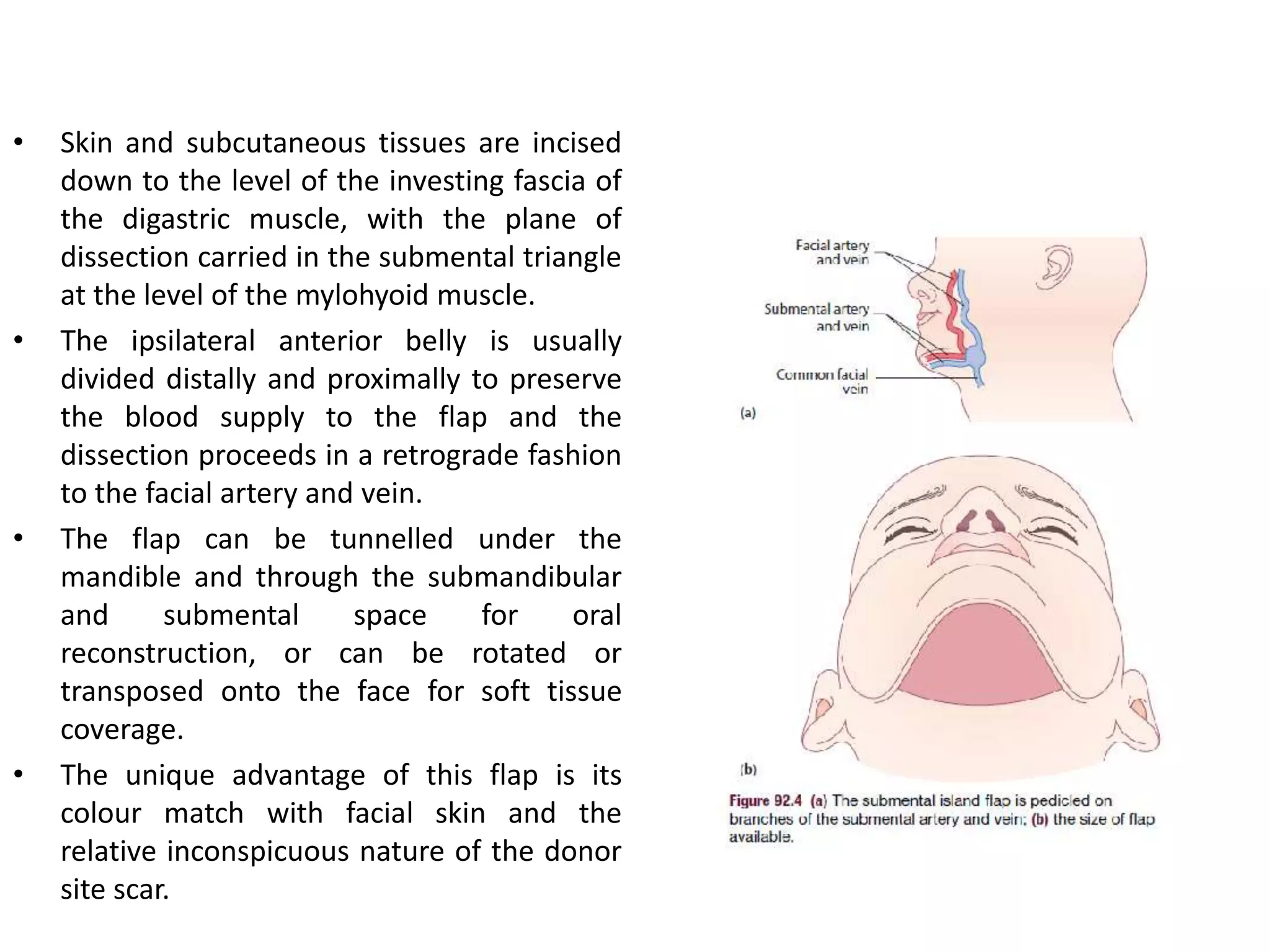
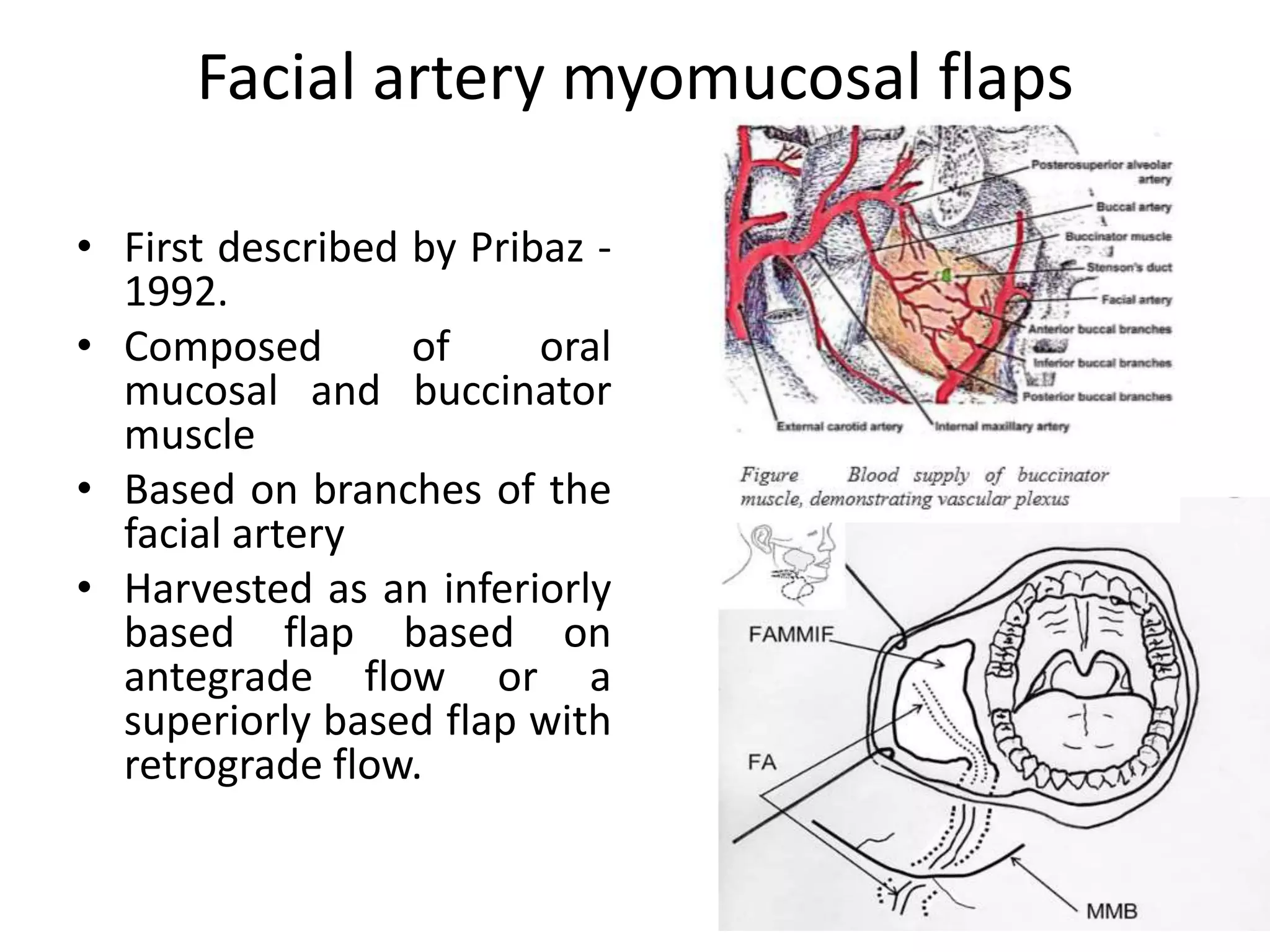
This document provides an overview of flaps used in head and neck surgery. It discusses the history of flap reconstruction from 600 BC to present day. It then covers general concepts in reconstruction including the reconstruction ladder. The rest of the document classifies and describes various types of flaps used in head and neck reconstruction including local, regional, distant, and free flaps. It discusses flap classification based on circulation, contiguity, composition, and contour. It provides details on specific flaps such as forehead, nasolabial, submental island, facial artery myomucosal, temporoparietal fascial, and deltopectoral flaps.
Head and neck cancer reconstruction is arguably the
most challenging area of reconstruction for the reconstructive
surgeon. A clear understanding of the principles of use of local flaps and a comprehensive understanding of the anatomy of these flaps provides the head and neck surgeon with a plethora of local and regional options for primary and secondary reconstruction.
Local & regional flaps /certified fixed orthodontic courses by Indian dental ...Indian dental academy
The Indian Dental Academy is the Leader in continuing dental education , training dentists in all aspects of dentistry and offering a wide range of dental certified courses in different formats.
Indian dental academy provides dental crown & Bridge,rotary endodontics,fixed orthodontics,
Dental implants courses.for details pls visit www.indiandentalacademy.com ,or call
0091-9248678078
Flap coverage in upper extremities in trauma VishalPatil483
SEMINAR PRESENTED BY DR VISHAL PATIL ,IN THE DEPT OF TRAUMA SURGERY AND CRITICAL CARE, AIIMS RISHIKESH
INCLUDES-INTRODUCTION-CLASSIFICATIONS OF FLAP-COMPLICATIONS RELATED TO FLAP COVERAGE- FLAP USED IN HAND AND UPPER EXTREMITY SOFT TISSUE RECONSTRUCTION WITH PICTURES OF IT
Title: Sense of Taste
Presenter: Dr. Faiza, Assistant Professor of Physiology
Qualifications:
MBBS (Best Graduate, AIMC Lahore)
FCPS Physiology
ICMT, CHPE, DHPE (STMU)
MPH (GC University, Faisalabad)
MBA (Virtual University of Pakistan)
Learning Objectives:
Describe the structure and function of taste buds.
Describe the relationship between the taste threshold and taste index of common substances.
Explain the chemical basis and signal transduction of taste perception for each type of primary taste sensation.
Recognize different abnormalities of taste perception and their causes.
Key Topics:
Significance of Taste Sensation:
Differentiation between pleasant and harmful food
Influence on behavior
Selection of food based on metabolic needs
Receptors of Taste:
Taste buds on the tongue
Influence of sense of smell, texture of food, and pain stimulation (e.g., by pepper)
Primary and Secondary Taste Sensations:
Primary taste sensations: Sweet, Sour, Salty, Bitter, Umami
Chemical basis and signal transduction mechanisms for each taste
Taste Threshold and Index:
Taste threshold values for Sweet (sucrose), Salty (NaCl), Sour (HCl), and Bitter (Quinine)
Taste index relationship: Inversely proportional to taste threshold
Taste Blindness:
Inability to taste certain substances, particularly thiourea compounds
Example: Phenylthiocarbamide
Structure and Function of Taste Buds:
Composition: Epithelial cells, Sustentacular/Supporting cells, Taste cells, Basal cells
Features: Taste pores, Taste hairs/microvilli, and Taste nerve fibers
Location of Taste Buds:
Found in papillae of the tongue (Fungiform, Circumvallate, Foliate)
Also present on the palate, tonsillar pillars, epiglottis, and proximal esophagus
Mechanism of Taste Stimulation:
Interaction of taste substances with receptors on microvilli
Signal transduction pathways for Umami, Sweet, Bitter, Sour, and Salty tastes
Taste Sensitivity and Adaptation:
Decrease in sensitivity with age
Rapid adaptation of taste sensation
Role of Saliva in Taste:
Dissolution of tastants to reach receptors
Washing away the stimulus
Taste Preferences and Aversions:
Mechanisms behind taste preference and aversion
Influence of receptors and neural pathways
Impact of Sensory Nerve Damage:
Degeneration of taste buds if the sensory nerve fiber is cut
Abnormalities of Taste Detection:
Conditions: Ageusia, Hypogeusia, Dysgeusia (parageusia)
Causes: Nerve damage, neurological disorders, infections, poor oral hygiene, adverse drug effects, deficiencies, aging, tobacco use, altered neurotransmitter levels
Neurotransmitters and Taste Threshold:
Effects of serotonin (5-HT) and norepinephrine (NE) on taste sensitivity
Supertasters:
25% of the population with heightened sensitivity to taste, especially bitterness
Increased number of fungiform papillae
Factory Supply Best Quality Pmk Oil CAS 28578–16–7 PMK Powder in Stockrebeccabio
Factory Supply Best Quality Pmk Oil CAS 28578–16–7 PMK Powder in Stock
Telegram: bmksupplier
signal: +85264872720
threema: TUD4A6YC
You can contact me on Telegram or Threema
Communicate promptly and reply
Free of customs clearance, Double Clearance 100% pass delivery to USA, Canada, Spain, Germany, Netherland, Poland, Italy, Sweden, UK, Czech Republic, Australia, Mexico, Russia, Ukraine, Kazakhstan.Door to door service
Hot Selling Organic intermediates
ARTIFICIAL INTELLIGENCE IN HEALTHCARE.pdfAnujkumaranit
Artificial intelligence (AI) refers to the simulation of human intelligence processes by machines, especially computer systems. It encompasses tasks such as learning, reasoning, problem-solving, perception, and language understanding. AI technologies are revolutionizing various fields, from healthcare to finance, by enabling machines to perform tasks that typically require human intelligence.
micro teaching on communication m.sc nursing.pdfAnurag Sharma
Microteaching is a unique model of practice teaching. It is a viable instrument for the. desired change in the teaching behavior or the behavior potential which, in specified types of real. classroom situations, tends to facilitate the achievement of specified types of objectives.
Anti ulcer drugs and their Advance pharmacology ||
Anti-ulcer drugs are medications used to prevent and treat ulcers in the stomach and upper part of the small intestine (duodenal ulcers). These ulcers are often caused by an imbalance between stomach acid and the mucosal lining, which protects the stomach lining.
||Scope: Overview of various classes of anti-ulcer drugs, their mechanisms of action, indications, side effects, and clinical considerations.
These simplified slides by Dr. Sidra Arshad present an overview of the non-respiratory functions of the respiratory tract.
Learning objectives:
1. Enlist the non-respiratory functions of the respiratory tract
2. Briefly explain how these functions are carried out
3. Discuss the significance of dead space
4. Differentiate between minute ventilation and alveolar ventilation
5. Describe the cough and sneeze reflexes
Study Resources:
1. Chapter 39, Guyton and Hall Textbook of Medical Physiology, 14th edition
2. Chapter 34, Ganong’s Review of Medical Physiology, 26th edition
3. Chapter 17, Human Physiology by Lauralee Sherwood, 9th edition
4. Non-respiratory functions of the lungs https://academic.oup.com/bjaed/article/13/3/98/278874
Tom Selleck Health: A Comprehensive Look at the Iconic Actor’s Wellness Journeygreendigital
Tom Selleck, an enduring figure in Hollywood. has captivated audiences for decades with his rugged charm, iconic moustache. and memorable roles in television and film. From his breakout role as Thomas Magnum in Magnum P.I. to his current portrayal of Frank Reagan in Blue Bloods. Selleck's career has spanned over 50 years. But beyond his professional achievements. fans have often been curious about Tom Selleck Health. especially as he has aged in the public eye.
Follow us on: Pinterest
Introduction
Many have been interested in Tom Selleck health. not only because of his enduring presence on screen but also because of the challenges. and lifestyle choices he has faced and made over the years. This article delves into the various aspects of Tom Selleck health. exploring his fitness regimen, diet, mental health. and the challenges he has encountered as he ages. We'll look at how he maintains his well-being. the health issues he has faced, and his approach to ageing .
Early Life and Career
Childhood and Athletic Beginnings
Tom Selleck was born on January 29, 1945, in Detroit, Michigan, and grew up in Sherman Oaks, California. From an early age, he was involved in sports, particularly basketball. which played a significant role in his physical development. His athletic pursuits continued into college. where he attended the University of Southern California (USC) on a basketball scholarship. This early involvement in sports laid a strong foundation for his physical health and disciplined lifestyle.
Transition to Acting
Selleck's transition from an athlete to an actor came with its physical demands. His first significant role in "Magnum P.I." required him to perform various stunts and maintain a fit appearance. This role, which he played from 1980 to 1988. necessitated a rigorous fitness routine to meet the show's demands. setting the stage for his long-term commitment to health and wellness.
Fitness Regimen
Workout Routine
Tom Selleck health and fitness regimen has evolved. adapting to his changing roles and age. During his "Magnum, P.I." days. Selleck's workouts were intense and focused on building and maintaining muscle mass. His routine included weightlifting, cardiovascular exercises. and specific training for the stunts he performed on the show.
Selleck adjusted his fitness routine as he aged to suit his body's needs. Today, his workouts focus on maintaining flexibility, strength, and cardiovascular health. He incorporates low-impact exercises such as swimming, walking, and light weightlifting. This balanced approach helps him stay fit without putting undue strain on his joints and muscles.
Importance of Flexibility and Mobility
In recent years, Selleck has emphasized the importance of flexibility and mobility in his fitness regimen. Understanding the natural decline in muscle mass and joint flexibility with age. he includes stretching and yoga in his routine. These practices help prevent injuries, improve posture, and maintain mobilit
Explore natural remedies for syphilis treatment in Singapore. Discover alternative therapies, herbal remedies, and lifestyle changes that may complement conventional treatments. Learn about holistic approaches to managing syphilis symptoms and supporting overall health.
Prix Galien International 2024 Forum ProgramLevi Shapiro
June 20, 2024, Prix Galien International and Jerusalem Ethics Forum in ROME. Detailed agenda including panels:
- ADVANCES IN CARDIOLOGY: A NEW PARADIGM IS COMING
- WOMEN’S HEALTH: FERTILITY PRESERVATION
- WHAT’S NEW IN THE TREATMENT OF INFECTIOUS,
ONCOLOGICAL AND INFLAMMATORY SKIN DISEASES?
- ARTIFICIAL INTELLIGENCE AND ETHICS
- GENE THERAPY
- BEYOND BORDERS: GLOBAL INITIATIVES FOR DEMOCRATIZING LIFE SCIENCE TECHNOLOGIES AND PROMOTING ACCESS TO HEALTHCARE
- ETHICAL CHALLENGES IN LIFE SCIENCES
- Prix Galien International Awards Ceremony
1. Flaps in Head and Neck Surgery 1
Dr Sandeep Shrestha
2nd year Resident
ENT
2. History
• 600 BC : Sushruta – cheek flap for nasal
reconstruction
• 1920 : Sir Harold Gillies used tubed flap
for head and neck reconstruction
• 1950 : defects were repaired using a
forehead flap or temporal flap
combined with split-thickness skin graft
• 1959 : Seidenberg described the first
revascularized flaps to the head and
neck
• 1963 : McGregor used forehead flap
based on the superficial temporal
artery for intraoral reconstruction.
• 1965 : Bakamjian first described the
deltopectoral flap.
3. • 1973: Daniel and Taylor
reported the first free flap
• 1976: Panje and Harashina
simultaneously described the
use of free flaps to
reconstruct defects of the oral
cavity.
• 1979: Ariyan described the
pedicled pectoralis major
myocutaneous flap.
• late 1980s and early 1990s,
the use of osteocutaneous
free flaps to reconstruct
mandibular defects was
advanced.
4. General Concepts of Reconstruction
• Reconstruction ladder
consists of following steps
starting from simplest –
most complex option
1. Healing by secondary
intention
2. Primary intention
3. Delayed primary closure
4. Skin grafts (split or full
thickness)
5. Tissue expansion
6. Local tissue transfer
7. Free tissue transfer
5. Introduction
• A flap is a piece of tissue that has
its own blood supply and does not
rely on the recipient bed for its
survival.
• The term "flap" originated in the
16th century from the Dutch word
"flappe," meaning something that
hung broad and loose, fastened
only by one side
• Reconstructing defects with poor
vascularity
• Exposed vital structures and where
skin grafts are inappropriate or
would lead to poor cosmesis.
• Reconstructing defects where
post-operative radiotherapy is
planned as they are less
susceptible than skin grafts to the
ill effects of radiotherapy.
6. Planning consideration
• Anatomy and physiology of the skin including
colour, texture, appearance and amount
• Local muscle anatomy, vascular supply, nerve
supply and lymphatic drainage
• Aesthetics of the area
• Possible sites for incision placement
• Area of local tissue availability in relation to
the area to be reconstructed
7. COSMETIC SUBUNITS OF
THE HEAD AND NECK
• The face is divided into multiple
cosmetic subunits.
• These cosmetic subunits should
be taken into account to
optimize cosmesis.
• Ideally, individual subunits
should be reconstructed
separately and scars should not
cross the subunits if possible.
• Graft or flap used to reconstruct
one cosmetic subunit and a
second graft or flap to
reconstruct an adjacent subunit.
• If more than 50% of a cosmetic
subunit is involved, it may be
desirable to excise the entire
subunit and reconstruct it to
obtain an optimal result.
8. • Relaxed skin tension lines
are those skin tension lines
that follow the furrows
formed when the skin is
relaxed.
• They can be found by
pinching the skin and
observing the furrows and
ridges that are then formed.
• These relaxed skin tension
lines are the same in all
people and, where possible,
incisions should follow
these lines.
9. Classification
1. According to Circulation
2. According to Contiguity
3. According to Composition
4. According to Contour
10. According to Circulation
• Random flaps: supplied
by unnamed small
vessels usually
capillaries
• Axial pattern: supplied
by a dominant
superficial vessel that is
oriented along axis the
flap.
11. According to contiguity
• Local flaps: moved from area adjacent to the
defect
– Random
– Axial
• Regional flaps: not from the adjacent but same
anatomical region as the defect e.g. forehead flap
for nasal reconstruction
• Distant flaps: from a different part of the body
away from the region of the defect
• Free Flaps: completely detached from the body
and anastomosed to recipient vessels close to the
defect
12. According to Composition
• Cutaneous flap: contain skin only
• Myocutaneous flap: contain skin + muscles
(pectoralis major/latissimus dorsi)
• Fasciocutaneous flap: contain skin + fascia
(radial forearm flap)
13. According to contour
• Advancement flaps: mobilized along a linear axis
toward the defect without any rotation or lateral
movement
• Pivot Flaps move about a fixed pivot point and an
arc which is the line of greatest tension of the
flap
– Transposition flap : flap is moved into an adjacent
defect by pivoting around the pivot point,
– Rotation flap : where the flap is rotated around the
pivot point into the defect
14. • Primary defect : A flap is placed
• Secondary defect : site from which the flap is
raised
15. Random Flap
• No named specific
vascular supply
• Most local skin flaps in
head & neck
• Random not only in blood
supply but also in design
• In general, height of a flap
shouldn’t exceed 1.5
times the length of the
base.
• Can extend 3-4 times the
base in head and neck
16. AXIAL FLAPS
• An axial flap is based on a
named arterial pedicle
that runs within the skin
superficial to the
underlying muscle layer,
parallel to the overlying
skin.
• Extremely good blood
supply can generally be
raised to a much greater
length than random flaps
17. ROTATION FLAPS
• Large arc of semicircle
where triangular primary
defect represents small
arc -1/8th of flap size
• Flap elevated & difference
in length of 2 sides of
defect is made up by
suturing with degree of
differential tension
18. • Larger flap –less
differential tension
• If Closure – tight-
rotation facilitated –
back cut
• Work well –convex
surface
• Ideally suited –cheek,
submandibular area,
upper neck, scalp
19. Transposition flaps
• A transposition flap is a
flap that is moved into
an adjacent defect by
pivoting around its fixed
point.
Types
• Z-plasty
• Rhomboid flap
• Flag or banner flap
• Bilobed flap
20. Z plasty
• Pair of triangular flap in
a form of Z
• 60 degree
• 2 flaps are interposed
• Lengthen the area
where there is shortage
of tissue
• To elevate or depress
the oral commisure and
outer canthus
21. Rhomboid flaps
• The defect is firstly designed as a
rhomboid.
• Each of the limbs of the defect and
the flap being raised need to be of
equal length and the angles of the
rhomboid need to be 120 degrees
and 60 degrees, respectively.
• The flap needs to be designed so
that the donor site scar lies parallel
to the relaxed skin tension lines
• Donor defect is closed directly.
• A variation of the rhomboid flap is
the Dufourmentel flap, which has
limbs of equal lengths but angles of
150 degrees and 30 degrees
• useful - reconstructing temporal or
cheek defects
22. Bilobed flap
• Esser in 1918
• Consists of 2 transposition
flap
• First flap is transposed into
the primary defect
• Second flap is transposed into
the secondary defect (the
original site of the first flap)
• Tertiary defect (the original
site of the second flap) is
closed directly
• Flap should be designed so
that the directly closed
tertiary defect is parallel to
the relaxed skin tension lines
23. Flag or banner flaps
• Piece of tissue moved resembles a flag
or banner.
• These are normally random pattern
flaps but can be based on a known
artery in some parts of the head or
neck.
• A glabellar flap is an axial transposition
flag flap based on the supratrochlear
artery.
• This allows the flap length to be three
times the base breadth
• It is transposed to cover the primary
defect and the donor site is closed
directly.
24. Advancement flaps
• Advancement flaps are moved forward into a defect by
stretching without any rotation or lateral movement.
• Depends on the elasticity of the tissue of the
flap(primary movement) and tissue adjacent to the
defect(secondary movement)
Techniques
• Burow’s triangle
• V-Y advancement
• Panthographic expansion
• Advancement Z-plasty
25. Burow’s triangles
• Single pedicled rectangular
flap –raised & advanced to
close defect
• Facilitated – excision of
Burow’s triangle –either
side of base of flap –
facilitates movement
• Length : breadth ratio 3 : 1
& 4 : 1
• Movement of tissue is
facilitated by laxity in
surrounding skin
26. V-Y advancement
• The flap itself is raised
as a triangular (or ‘V’-
shaped) flap.
• When is it advanced,
the secondary defect is
closed directly, leaving a
‘Y’-shaped scar.
27. Panthographic expansion
• Limbs of flap –designed
at 120 degree with back
cuts at bottom
• Looks - Inverted
tumbler
• Flap is advanced
• Donor site closed
primarily
• Useful –cheek & neck
28. Advancement Z - plasty
• Problems of skin
advancement- once
tissue advanced –unless
stopped –tends to
return
• Break up scar with Z-
plasty
• Useful – defects in
lower eye lids – excised
as triangle & tissue
advanced laterally
29. Bipedicled flaps
• Receives a blood supply from
both ends as it has two bases.
• less prone to necrosis than flaps
of similar dimensions which are
attached at only one end.
• Skin from the upper lid based
on both a medial and a lateral
attachment is swung down, like
a bucket handle, to provide
cover for a defect in the lower
lid.
• To increase the viability of this
flap, a strip of the orbicularis
oculi muscle can be included,
making it a musculocutaneous
flap.
31. Forehead flaps
• Axial flap
• Provides large areas of skin
& subcutaneous tissue
• Used –reconstruct defects
below level of eyes
• Based on ant br of
temporal artery -1st flaps –
intraoral reconstruction
32. • Rarely used nowadays as it
gives disfiguring donar site –
better alternatives
• Most common - Cutaneous
axial median forehead flap –
supratrochlear artery
• Raised & transposed to
reconstruct areas in upper
medial cheek region & lower
half of nose & alar rim
• Donor site –close primarily
• Cosmetic result excellent
33. • Where larger areas of tissue
are required, for example in
complete nasal resurfacing,
larger forehead flaps may
be designed (e.g. Millard
flying seagull flap), which
may be facilitated by prior
tissue expansion to ease
donor site closure.
• Best suited in patients with
high foreheads with some
lax tissue to facilitate
primary closure.
34.
35.
36. Nasolabial flap
• 1st documented –indian Sushruta -
600 BC
• True Myocutaneous flap
• Workhorse –reconstruction of
defects around face & anterior
oral cavity
• Based on distal br facial artery and
its venae commitantes.
• Flap designed with a inferior base
but can be based superiorly with a
more random vascular supply
40. Submental island flaps
• First described by Martin in 1983
• Axial pattern flap or a free flap for
reconstruction of the facial skin or
intraoral lining.
• Supplied by a branch or branches
of the facial artery.
• Venous drainage via the facial vein.
• Usually designed in the midline,
just below the margin of the
mandible, with the
superior/inferior dimension
determined by the ability to close
the submental skin.
• Dissection is usually initiated on
the contralateral side to the
planned vessel pedicle.
41. • Skin and subcutaneous tissues are incised
down to the level of the investing fascia of
the digastric muscle, with the plane of
dissection carried in the submental triangle
at the level of the mylohyoid muscle.
• The ipsilateral anterior belly is usually
divided distally and proximally to preserve
the blood supply to the flap and the
dissection proceeds in a retrograde fashion
to the facial artery and vein.
• The flap can be tunnelled under the
mandible and through the submandibular
and submental space for oral
reconstruction, or can be rotated or
transposed onto the face for soft tissue
coverage.
• The unique advantage of this flap is its
colour match with facial skin and the
relative inconspicuous nature of the donor
site scar.
42. Facial artery myomucosal flaps
• First described by Pribaz -
1992.
• Composed of oral
mucosal and buccinator
muscle
• Based on branches of the
facial artery
• Harvested as an inferiorly
based flap based on
antegrade flow or a
superiorly based flap with
retrograde flow.
43. • The basic harvest technique is to
Doppler out the facial artery through
the buccal mucosa and map the course
of the vessel.
• For the inferiorly based flap, dissection
begins anterosuperiorly to identify the
arterial supply to the upper lip with
division of the facial artery at this point
and then retrograde dissection, which
includes the mucosa, buccinator, facial
artery and the tissue and venous
plexus that lies between the artery and
the muscle.
• In the superiorly based flap, the
dissection begins inferiorly with
visualization and ligation of the facial
artery and then a retrograde dissection
of the tissues including the buccinator
muscle.
• A flap of 7–8 cm can be harvested with
a thickness of 8–10 mm.
44.
45. Temporoparietal fascial flaps
• First described by Golovine in
the 19th century for orbit
reconstruction.
• Versatile local rotation or
free fascial flap for
reconstruction of the head
and neck or extremities.
• Can be use for microtia
repair and auricular
reconstruction.
• Very thin and pliable flap
with minimal donor site
morbidity.
• The arterial supply -
superficial temporal artery
46. • The flap is harvested as an
elliptical or teardrop shape,
above the level of the
zygoma.
• The inferior limit of the
incision is usually the tragus
but inferior extensions can
be used for extended
rotations or if the surgeon
wishes to visualize the facial
nerve.
• The initial incision is started
just above the zygoma,
extending into the scalp.
47. • The plane of dissection is initiated
by defining the level of the
superficial temporal fascia just
below the subcutaneous fat layer
in the scalp.
• A good landmark is to look for the
hair follicles; if they are being
transected, the surgeon is
elevating the flap too superficially.
• Once fully mobilized from the
overlying skin, the flap is incised
around its periphery and elevated
in the plane just above the
temporal fascia.
• Dissection is carried down to
about 2 cm below the arch of the
zygoma to ensure an appropriate
arc of rotation.
48. Clinical applications
• Orbital reconstruction, including the
extenteration cavity
• Upper and lower eyelids and the eyebrow as a
fascio-cutaneous hair bearing flap
• Auricular reconstruction, including microtia and
traumatic or oncologic deformities
• Palate reconstruction
• Buccal mucosal reconstruction.
50. Deltopectoral flap
• Bakamjian & Littlewood -
1964
• Fasciocutaneous flap
• Axial pattern flap designed
on the ant chest wall
between line of clavicle &
level of ant axillary fold.
• Vascular supply – upper 3 or
4 perforating branches – int
mammary artery
51. Boundaries
• Superiorly- clavicle
• Laterally – acromium
• Inferiorly- line running ant
axillary fold to above the nipple
• Flap – extend any site in neck &
occasionally level – zygoma
• It retracts – side to side after –
elevated
• Elongate slightly over time –
particulary patients over 60 yrs
52. • Flexibility due to anomolous
pivot point
• Territory of perforator
vascular system – extend –as
far as groove seperating
deltoid from the pectoralis
major (deltopectoral groove)
• Extension beyond this –
failure – tip of flap
53. • Flap marked out
• Elevation – laterally
• Pectoral fascia left on flap leaving
muscle fibres below absolutely bare
• Any br thoracoacromial -
encountered- ligated
• Monopolar diathermy – judiciously
on flap & muscle
• Distant site-tube the bridge
segment in order to eliminate its
raw surface.
54.
55. Uses of deltopectoral flap
• To allow one stage reconstruction
of anterior neck skin
• To reconstruct defect by passing
as bridge over normal skin.
• Once take –occurred -3wks –
pedicle is divided & remaining
part of flap – returned to donor
site or discarded
• To reconstruct large defect –lower
face & upper neck
• Skin cover of an exposed carotid
artery
• Closure of a pharyngocutaneous
fistula
56. Contraindications
• Internal mammary artery that has previously
been used for coronary artery bypass surgery
• Prior trauma or surgery (mastectomy,
pacemaker, pectoralis major flap) to the
anterior chest wall.
57. Advantages
• Provides large area of skin cover
• Better colour and texture match compared to
free tissue transfer flaps from distant sites
• Less bulky than pectoralis major flap
• Technical simplicity
Disadvantages
• Limited arc of rotation
• Donor site defect requires skin graft
58.
59. Other distal axial flaps
• Cervical skin flaps
– Used –lower face & cheek
• Occipitomastoid based flaps
60. Supraclavicular artery island flaps
• First described in 1979 by
Lamberty
• Local fasciocutaneous flap
• Harvested from the skin
on the shoulder and
supraclavicular area.
• Based on a branch of the
tranverse cervical artery
61. FLAP HARVEST
• The supraclavicular artery is
identified using a hand-held
Doppler, usually originating in
a triangle bounded anteriorly
by the posterior border of the
sternocleidomastoid muscle,
posteriorly by the external
jugular vein and inferiorly by
the clavicle.
• The vessel is followed out over
the clavicle to identify the axis
and design of the flap.
• The flap is usually designed
with a width of 6–7 cm and a
length of 20–25 cm from the
rotation point.
62. • Dissection starts distally
and is carried
subfascially over the
deltoid until one
reaches the anterior
border of the trapezius.
• The proximal portion of
the flap is usually de-
epithelialized to allow
tunnelling into a facial
or intraoral defect.
63. CLINICAL APPLICATIONS
• The SAI flap has broad
application for external
skin defects extending
from the base of the neck
to the parotid.
• Use for reconstruction of
oral cavity, oropharynx,
hypopharynx
• Advantages -Ease of
elevation
, the transplant of an autologous skin flap to the lower extremity using the operating microscope.
They are not visible features of the skin, such as wrinkle lines.
The Langer's lines are important from a historical point of view. They represent the skin tension in rigor mortis.
The relaxed skin tension lines and Langer's lines do not correspond in many areas of the body
L:W:1:1
Face L:W:2:1
the dermal and subdermal plexus.
). Flaps located near the defect but are not in immediate proximity
Deltopectoral Cervical occipitomastoid
Rotation flaps: pivot around a point at the base of the flap.
Transposition flap: mobilized to an adjacent defect over an incomplete bridge of skin. Examples of transposition flaps include rhombic flaps and bilobed flaps.
Interposition flaps: mobilized to an adjacent defect over an incomplete bridge of skin whish is also elevated and mobilized. An example of an interposition flap is a Z-plasty.
Interpolated flaps: mobilized either over or beneath a complete bridge of intact skin via a pedicle.
Secondary defect can be closed directly or grafted or another flap is placed.
Cutaneous flap With in head and neck there is an extensive subdermal vascular plexus.E.g Bilobed flap, rhomboid,Z-palsty flap
Arterial flap
, which is determined not
only by their length and breadth ratio, but also by thevascular territory of the vessels that supply them. Because of
this, they can generally be raised to a much greater length
than random flaps and can therefore be used to move skin
over a greater distance
Large flaps that rotate into the primary defect.
The volume of tissue raised in the flap is high when compared to the defect being closed.
Normally, the flap is a semicircle and the primary defect is ‘triangulated’ or designed as a triangle, adjacent to the flap.
Geometrically, the flap circumference should be at least eight times the width of the defect to allow closure of the donor site;
However, in practice, it is best to design the flap as large as possible as the direct closure of the donor site relies on tissue redistribution and tissue elasticity.
These flaps are useful for dealing with defects of the scalp or cheek
Occasionally, the use of a Burow triangle or a back cut may be needed. A back cut releases a rotation flap that is too tight by decreasing the tension at the base of the flap, thus moving its pivot point towards the defect.
This decreases the tension of closure at the site of the primary defect.
A back cut that is taken too far, however, may significantly decrease the width of the flap, which will increase the chance of devascularizing it.
Limberg in 1966 Skin pinch
Rhomboids –can be designed as double or triple flap (swastika flap) – to close larger defects
Theoretically, the bilobed flap uses less tissue than any other method of wound closure and because of this minimizes tension at the primary defect.
Difficult flap to design
Long axis of larger flap is at right angle with the long axis of the smaller flap
Length is same where as width is half the larger one
Use In the nose or cheek or reconstruction of the auricular defect
Use in where there is insufficient soft tissue to close the
Excellent result – properly executed
Best way to reconstruct nose - with skin of nose
A number of small transposition flaps can be used around the face and generally the donor site is closed directly.
These help in allowing the flap to advance, as demonstrated by a Rintala flap being used to cover a defect of the nasal tip.
An example of one such flap is the nasolabial V–Y advancement flap shown, which is an axial flap based on either the superior labial artery or the angular artery.
An example of this is the Tripier flap for lower eyelid reconstruction The ideal use of a bilobed flap is where there is tissueavailable locally and where it is important to avoid producingtissue stretch. The bilobed flap is most commonly used to deal with defects at the tip of the nose. Although there is some spare tissue around this area, the nasal tip can become displaced if skin is imported to cover defects
here as a local flap.26
Original form as described by mcgregor
The flap includes epidermis, dermis, subcutaneous tissue, frontalis muscle and associated fascia. supratrochlear artery, which exits the orbit 1.7- 2.2cm lateral to the midline at the level of the superior orbital rim
Traumatic cause of nasal defect, excision of nonmelanotic cancer
The median forehead flap is harvested from the mid forehead and has a wide pedicle based in the center of the forehead, which originally captured both supratrochlear vessels as seen in the image below. The paramedian forehead flap is designed around a narrower pedicle based on the medial brow area over the superior/medial orbital rim. The skin paddle and pedicle are aligned vertically, with the supratrochlear notch in the paramedian position of the forehead as seen in the image above. The resultant donor scar is oriented vertically and aligns with the medial brow.
The midline forehead flap is a hybrid of median and paramedian flaps, with the skin paddle harvested from the precise center of the forehead. The associated pedicle runs obliquely and is based on a unilateral supratrochlear vessel and collaterals from the medial brow area
A is a nasal defect template (from suture package). B is the template transferred to the precise midline of the forehead. Pedicle division is performed safely after 3 weeks as seen in the image below. One must aggressively thin the cephalic border of the flap from above to best match surrounding skin thickness; however, exercise caution to avoid extending the level of undermining beyond 50% of the original skin paddle. The vascular supply to the flap is retrograde from the recipient bed, and excessive undermining can jeopardize viability. The original nasal defect wound is freshened, and the superior portion of the skin paddle trimmed to fit the remaining defect. The pedicle base is amputated, thinned, and then inset back into the medial brow region.
true myocutaneous flap pedicled on the facial artery, single-stage reconstruction of oral cavity defects or to staged reconstructions of facial defects
The redundant skin extending from the medial canthus of the eye to the inferior margin of the mandible (nasolabial sulcus and nasofacial groove) defines the donor site for the nasolabial flap. This area is relatively hairless except for the lower cheek in males, an important consideration in oral cavity reconstruction. The flap itself is comprised of skin, subcutaneous tissue and the underlying musculature. The
facial artery is deep to the platysma,
risorius, zygomaticus major and minor,
levator labii superioris, and levator labii
superioris alaeque nasi muscles. The
artery is superficial to the mandible,
buccinators muscle, and levator anguli oris muscle. The position of the artery deep to the majority of facial mimetic muscles suggests that the nasolabial flap may incorporate this muscle layer and be developed as a true musculocutaneous flap.
The design usually places the most medial limit of the flap in the nasolabial fold with the superior limit approximating the medial canthus of the eye. The medial to lateral dimension of the flap is determined by the defect to be reconstructed and the ability to primarily close the donor site. Flap elevation is usually initiated distally with a retrograde dissection above the plane of the facial musculature.
In the intraoral application a tunnel is placed traversing the facial muscles and buccinators, allowing the inferiorly based flap to enter the oral cavity
This flap is extremely reliable when based inferiorly, with a relatively inconspicuous donor site, particularly when bilateral flaps are used. Its greatest application intraorally is for the anterior floor of mouth and gingiva, where its use is both simple and effective. The donor site for this flap is best tolerated in elderly patients with rhytids that mask the donor site. The flap is then tunnelled through the buccal space to repair the intraoral defect,
A fusiform flap is marked ensuring that the medial border of the flap is on the nasofacial groove (Figures 1, 4, 5). A pencil Doppler probe may be useful to locate and delineate the course of the artery. Average flap dimensions are 2.5cm in width and 6cm in length. The width may be closer to 5cm when the facial skin is very redundant. The superior border of the flap is inferior to the medial canthus along the nasofacial junction. Placement of the inferior border depends on the nature of the defect. For floor of mouth reconstruction, the inferior border of the flap should be at the superior border of the mandible; reconstruction of palatal defects, however, requires the inferior border to be approximately at the level of the oral commissure.
Tumour excised, flap is outlined famale :nasolabial fold,male laterally.flap is elevated skin,subdermal flap and underlying facial muscle .primary suturing.3 weeks later pedicle is ready for division and insetting of flap.
that pass either over or through the submandibular gland, traversing medially on the mylohyoid muscle and then deep to the anterior belly of the digastric muscle to provide a perforator- based arterial supply and venous drainage to the submental skin. The flap comprises skin, subcutaneous fat, platysma muscle, submental fat, and lymph nodes; the anterior belly of digastric and mylohyoid muscles may also be included. Axial fasciocutaneous flap that includes skin, subcutaneous tissue, platysma, and fat and is pedicled on the submental artery and veins
There can be occasional problems with venous congestion, particularly in the reverse flow design,
because of valves in the facial vein
Used for facial, oral cavity, oro- and hypopharyngeal soft tissue defects.
The anatomy of this flap is based on the buccinator muscle and its relationship to the facial artery. The buccinator is covered medially by the submucosa and mucosa and laterally by the external lamina of the muscles of facial expression, the masseter, the buccal fat pad, and the facial artery and vein. The flap is about 5mm thick, and comprises buccal mucosa, submucosa and buccinator muscle, with the feeding vessels and vascular plexus.
. The facial artery, a branch of the external carotid artery, enters the face by curving around the lower border of the mandible at the anterior edge of the masseter muscle. It then follows a tortuous course, passing superiorly and anteriorly to a position just lateral to the commissure of the mouth. At this point it lies deep to the risorius, zygomaticus major muscle and the superficial lamina of the orbicularis oris muscle. It lies superficial to the buccinator muscle and the lateral edge of the deep lamina of the orbicularis oris muscle.
At this point in its course it gives off multiple perforating vessels to the cheek and the superior labial artery. It continues superiorly to the angular artery, which reaches the medial canthus. It has communicating branches with the buccal and infraorbital branches.
The FAMM flap is ideally suited for reconstruction of small mucosal defects in the oral cavity and in particular the mucosa of the lip.
The flap can also be rotated across the alveolus to close small defects of the floor of mouth or tongue, as well as the palate. via
the pterygoid plexus and internal maxillary (7*5cm)
Anterolateral floor of the mouth
There is some variation in venous anatomy, with a small percentage of patients having venous drainage through the post-auricular vein or occipital veins. The temporoparietal fascia (TPF) lies just under the subcutaneous tissue of the lateral scalp. The fascia has an inner and an outer layer, with the artery and vein entering between the inner and outer layers and then coursing vertically in the outer layer of the fascia.
The outer layer of the TPF extends as the superficial muscular aponeurotic system (SMAS) below the zygoma. A thin muscular layer (the superficial auricular muscle) separates two parts of the outer layer of the fascia below the temporal line. The inner layer of the TPF contains a dense vascular network, which originates from the outer layer. Two nerves have an anatomic relation to the flap: the auriculotemporal nerve, a branch of V3, lies within the superficial layer of the TPF and theoretically could provide for a sensate flap; and the frontal branch of the facial nerve traverses over the zygoma in the same plane as the frontal branch of the superficial temporal artery and can be injured if the dissection is carried too far forward in the plane of this vessel. For flap harvest the patient is usually positioned in the supine position, with the drape line along the vertex of the scalp leaving the post-auricular area exposed.
The important landmarks :arch of the zygoma, the pinna and the usual landmarks of the facial nerve.The artery usually lies just in front of the pinna and is easily palpated or detected with the Doppler in the this location. The artery ascends vertically to the apex with a frontal branch coming off 1–3 cm above the zygomatic arch. The incision is placed just posterior to the position of the vertical branch and can be either a straight or curvilinear incision into the scalp or ‘Y’ shaped for larger teardrop-shaped flaps.
The surgeon harvesting this flap for the first time must take great care not to incise too deeply as the pedicle can easily be divided during the incision
Internal mammary artery emerges from the medial end of the intercostal space.
Not from end to end
Extension – flap beyond this – not axial flap
Monopolar diathermy is used judiciously on the flap and the muscle, as this could damage the flap and any diathermy marks on the muscle may compromise the subsequent take of any skin graft. When raising the flap, retraction is upwards by an assistant using skin hooks; it must not be doubled back on itself as this could lead to buttonholing
After 3 weeks pedicle is divided, the bridge segment of the flap is being untubed and returned to its original site on the chest,excising enough of the graft to accommodate it.mastoid area,ear,parotid area,cheek,lower lip, chin and the neck within the arc.
The deltopectoral flap has an anomalous pivot point. There is considerable laxity of the skin on the anterior axillary fold when the arm is abducted. This means that the lower border of the flap is considerably longer than the upper part. The pivot point on the flap is thus at the medial end of the upper limb and not the lower limb. This needs to be taken into account when planning the flap. The donor site is usually covered with a split skin graft.
Fasciocutaneous pedicle limits the reach of the flap
The spinal accessory nerve is identified and dissection continues in a subfascial plane.
The dissection then turns to elevation of the infraclavicular portion of the flap in the subfascial plane; this dissection in the subfascial plane is maintained until over the clavicle.
Once the acromion is reached the dissection is continued with careful dissection, with branch ligation with either bipolar cautery or surgical clips.
Skeletonization of pedicle is not usually necessary, unless wider arcs of rotation are required. The distal extension of the transverse cervical artery may restrict rotation and can be ligated if necessary.
simple and quick procedure, usually done in less than an hour, and is a relatively easy to learn procedure. The flap is highly reliable and since the procedure is shorter than free flap reconstruction and does not require expertise in microsurgery, the supraclavicular flap is beneficial for high risks patients who can’t sustain long operative time The supraclavicular flap is an excellent choice for reconstruction of skin defects in the lower face and neck because of a good texture and color match with the skin of the neck and lower face. It also has great pliability which makes it suitable for reconstruction of areas with high mobility, such as the neck and oral cavity. Also, since the skin in the supraclavicular area is generally hairless, it is an appropriate for mucosal defects. The skin has a thin dermis and little fat, which accounts for the flap's thinness which makes it appropriate for reconstruction of contour sensitive areas such as the anterior neck and for pharyngeal defects in which a bulky flap may impair swallowing.
It can easily reach distant sites, such as the oropharynx, due to its long pedicle and broad arc of rotation. It can also be used for large defects, since the flap can reach a considerable size, with maximum dimensions of approximately 10 x 20 cm.