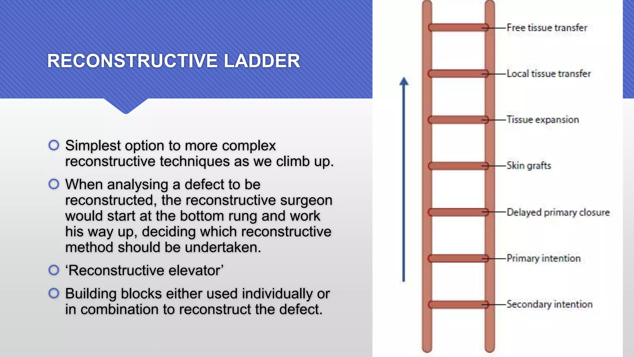
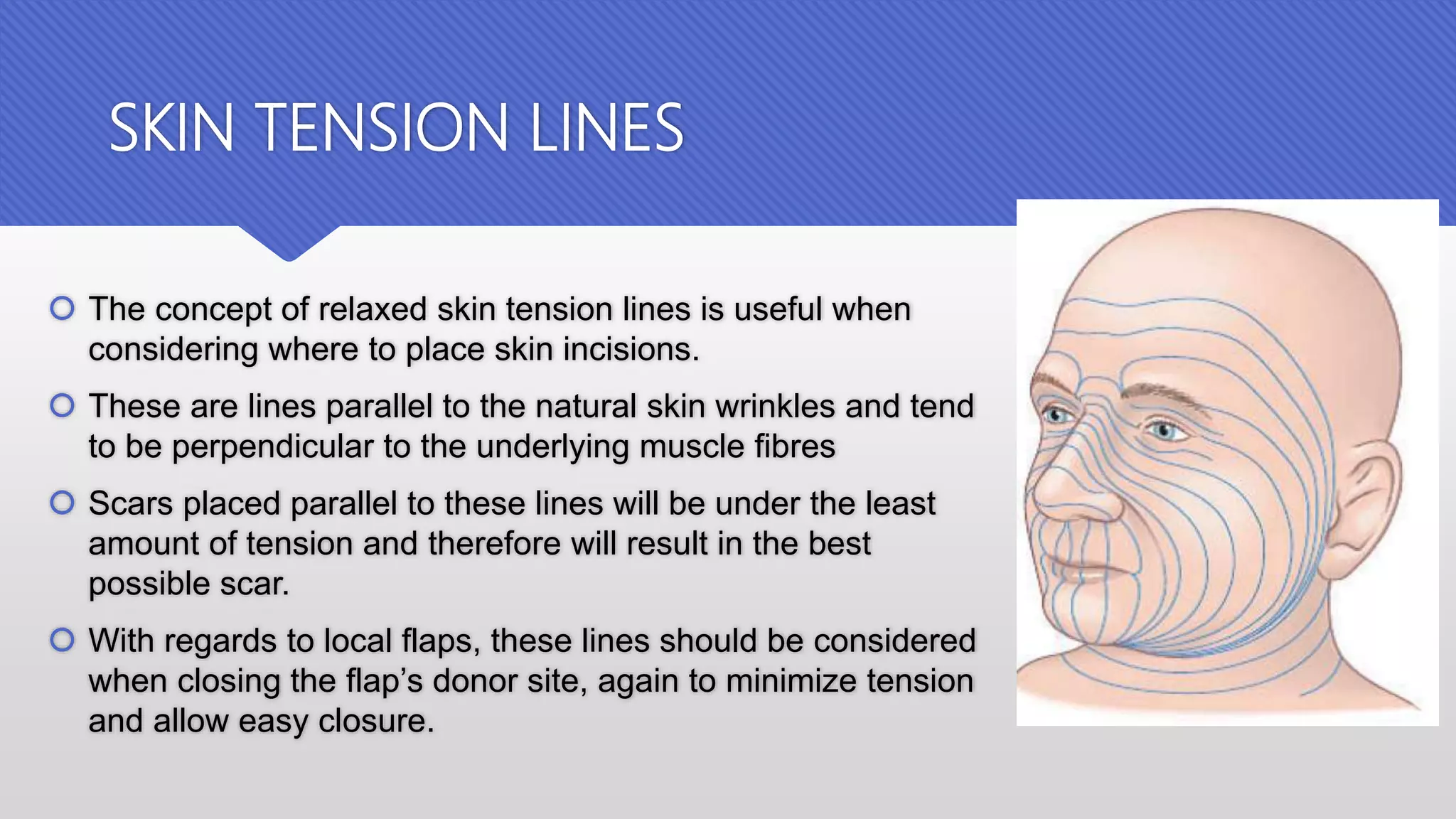
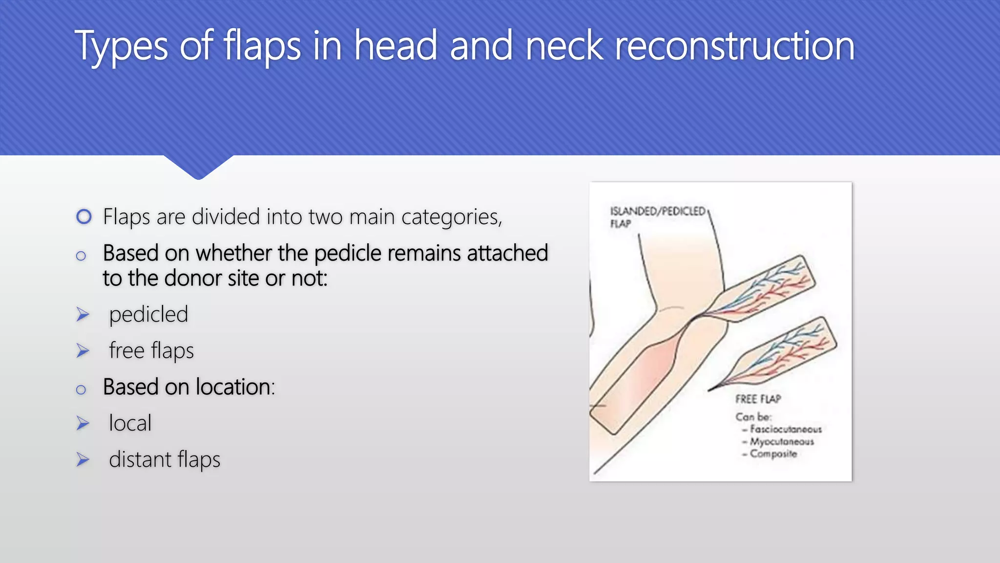
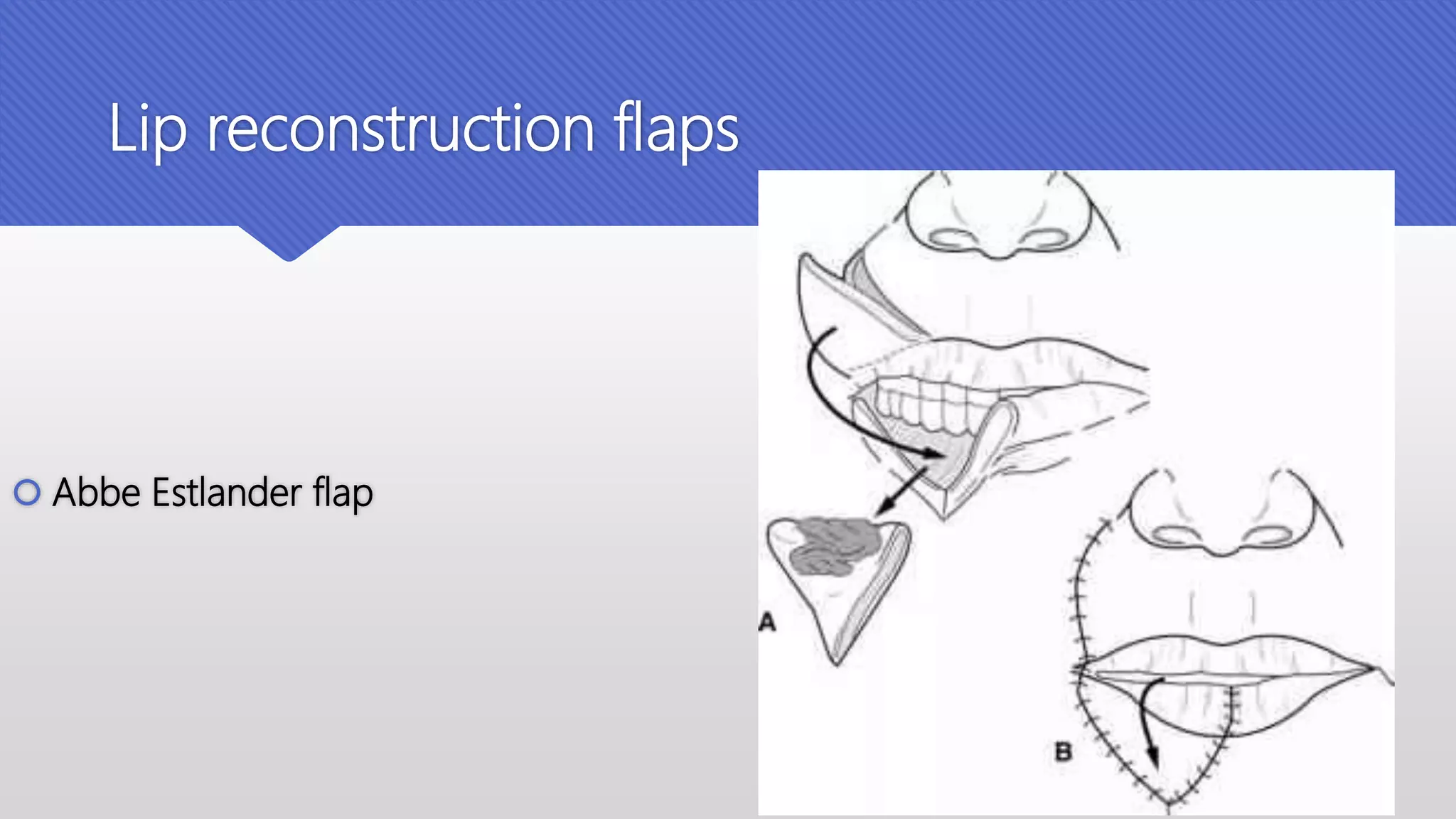
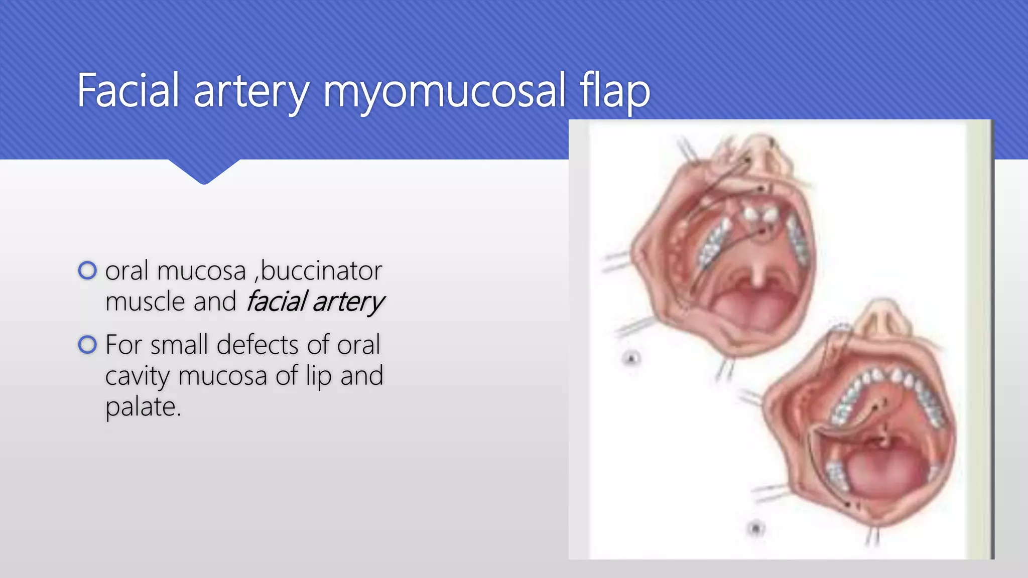
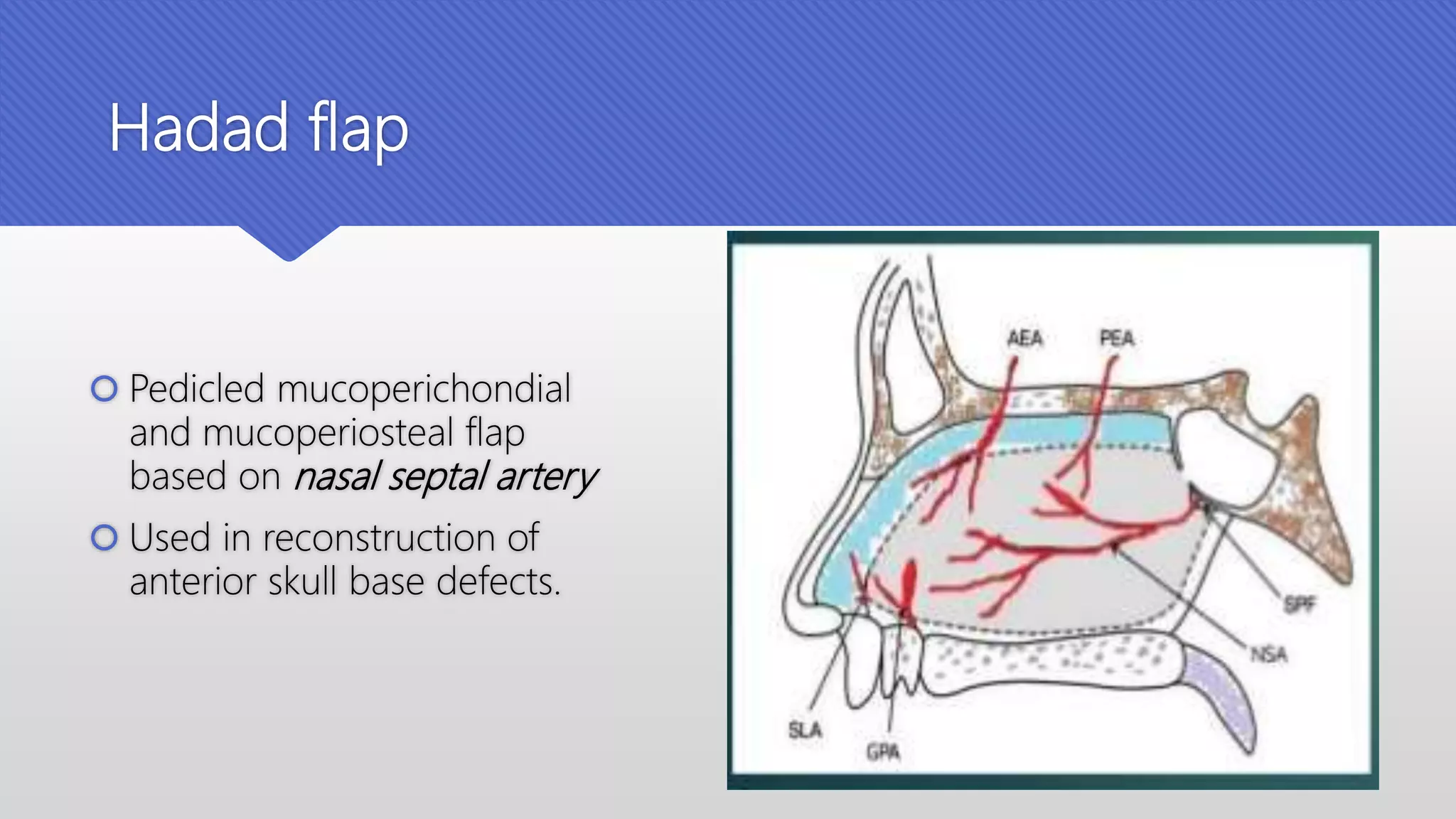
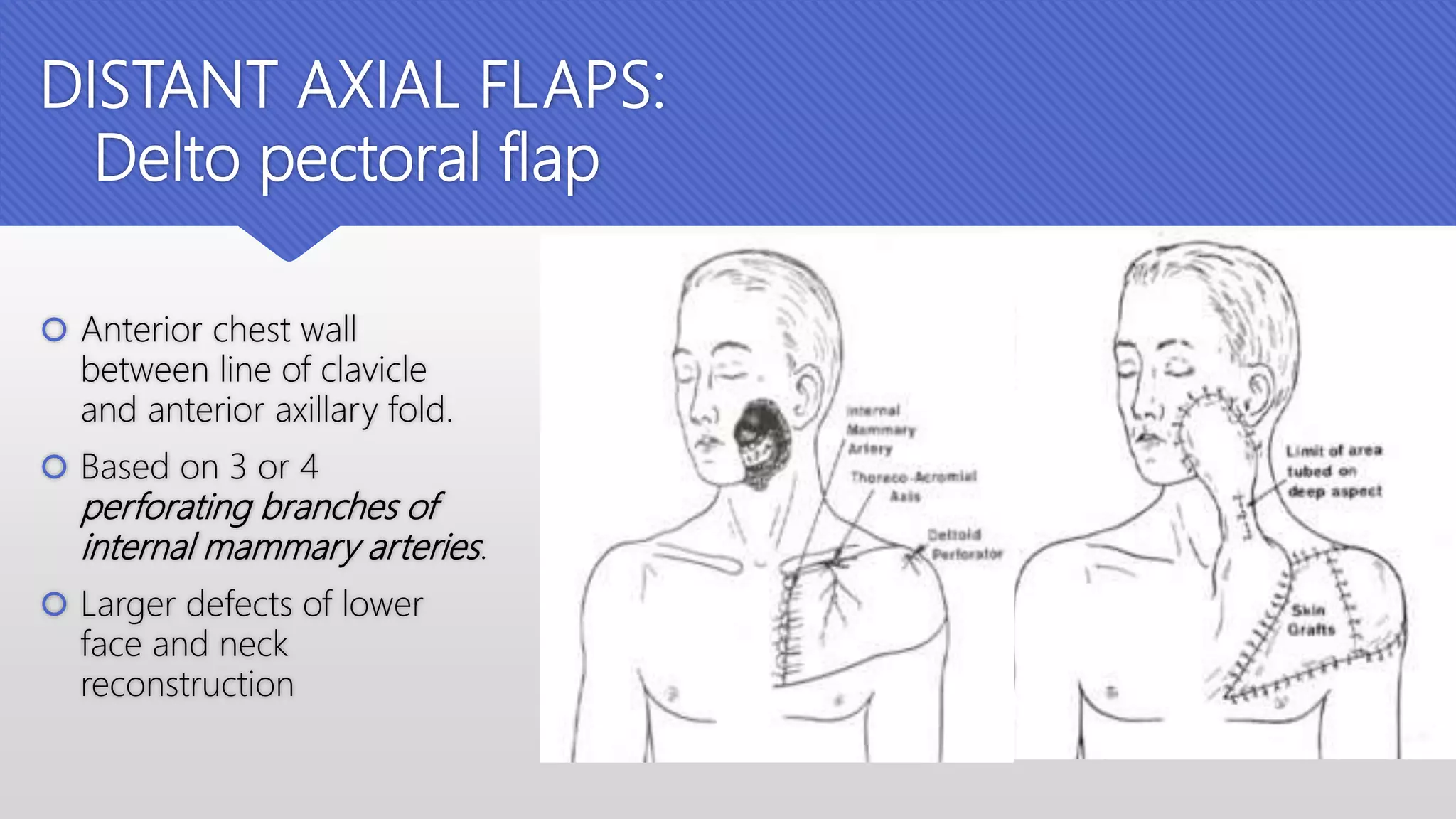
GRAFTS AND FLAPS IN
HEAD AND NECK
RECONSTRUCTION
Dr. R. Grace Vandana
Postgraduate
Dept Of ENT, NMCH
HISTORY
 Origins of head and neck reconstructive surgery
began in Egypt and ancient India dating back over
5000 years.
 Sushrutha – father of Indian surgery laid foundations
for a variety of pedicled and rotation flaps.
 The first human skin graft was performed by Astley
cooper in 1817.
 Josef Dieffenbach (berlin) – father of plastic surgery.
 Harlod gilles – pioneered techniques for facial
reconstructive surgery.
INTRODUCTION
 Defects following head and neck surgery can often be closed using the
technique of direct suture.
 Used when the defect is small and where local conditions mean that
enough tissue is available.
 For larger defects or in situations where direct suture is neither applicable
nor available, surgical defects in the head and neck can be reconstructed
with grafts, local flaps, regional flaps or free-tissue transfer.
RECONSTRUCTIVE LADDER
 Simplest option to more complex
reconstructive techniques as we climb up.
 When analysing a defect to be
reconstructed, the reconstructive surgeon
would start at the bottom rung and work
his way up, deciding which reconstructive
method should be undertaken.
 ‘Reconstructive elevator’
 Building blocks either used individually or
in combination to reconstruct the defect.
GILLIES’ PRINCIPLES OF
RECONSTRUCTIVE SURGERY
 The commandments of Sir Harold Gillies that relate to reconstruction in
general:
 Losses must be replaced in kind.
 Treat the primary defect first.
 Thou shalt provide thyself with a lifeboat.
 Thou shalt not throw away a living thing.
 Replace things into their normal position by recreation of the defect.
SKIN TENSION LINES
 The concept of relaxed skin tension lines is useful when
considering where to place skin incisions.
 These are lines parallel to the natural skin wrinkles and tend
to be perpendicular to the underlying muscle fibres
 Scars placed parallel to these lines will be under the least
amount of tension and therefore will result in the best
possible scar.
 With regards to local flaps, these lines should be considered
when closing the flap’s donor site, again to minimize tension
and allow easy closure.
WEDGE EXCISION
 Lesions located at the free edge of
tissues such as the eyelid, lips and helical
rims can be excised as a wedge and
repaired.
 The separate components of these
composite tissues must be repaired
individually to optimize function and
appearance.
SUBUNITS
 Ideally, individual subunits should be
reconstructed separately and scars
should not cross the subunits if
possible.
 It is therefore preferable to use a graft
or a flap to reconstruct one cosmetic
subunit and a second graft or flap to
reconstruct an adjacent subunit.
 If more than 50% of a cosmetic subunit
is involved, it may be desirable to
excise the entire subunit and
reconstruct it to obtain an optimal
result.
 A graft is a piece of tissue that has no blood supply of its own and its
survival depends on it gaining a blood supply from the recipient bed.
 A flap is a piece of tissue that has its own blood supply and does not rely
on the recipient bed for its survival.
GRAFTS
 A graft is a piece of tissue that is transferred from one site to another, devascularizing it
in the process.
 The area from which the graft is taken is known as the donor site and the area to which
the graft is applied is known as the recipient site.
 CLASSIFICATION:
 according to their composition – skin, bone, cartilage, fat, mucosa, or composite grafts
which consist of two or more different tissue types (e.g. a septal mucosal graft consisting
of septum and mucosa).
 according to their source autograft – same individual
Iso Graft- genetically identical individuals
Allograft – same species
Xenograft – different species
 Skin grafts are of two types:
split-thickness skin grafts
(STSGs) or full-thickness skin
grafts (FTSGs) dependent on
the dermal content of the graft.
 FTSGs contain epidermis and
all of the dermis whereas
STSGs contain epidermis and a
variable amount of dermis (thin,
intermediate or thick)
GRAFT HEALING
Factors affecting graft healing
 Metabolic demand – FTSG > STSG
 Local factors
 Type of recipient bed –
 readily accepting – healthy granulation tissue, muscle and fascia, bone covered with
periosteum
 Less suitable – fat, exposed bone and tendon.
 Shearing forces
 Most common cause of graft failure is formation of hematoma or seroma between the graft
and the recipient bed.
 Prevented by meticulous hemostasis and meshing or fenestrating the graft to allow escape.
 Other factors include infection
 Systemic conditions
 Radio and chemotherapy
 Nutritional status
Donor sites
STSG
FTSG
COMPOSITE GRAFTS
 Contain two or more different tissue
types.
 Examples
 Septal mucosal graft which contains
septum and mucosa.
 Chondromucosal graft from the ear
which contains cartilage and skin.
 Useful in reconstructing composite
defects, either for complete
reconstruction, as in the reconstruction of
an alar rim or small defect of an ear, or as
partial reconstruction of a more complex
composite defect.
FLAPS
 The term ‘flap’ is alleged to originate from the dutch word flappe, meaning something
broad and loose that hangs and attached only at one side.
 A flap, unlike a skin graft, has its own blood supply.
 Flaps are useful in reconstructing defects with poor vascularity, exposed vital
structures and where skin grafts are inappropriate or would lead to poor cosmesis.
 Flaps are also useful in reconstructing defects where post-operative radiotherapy is
planned as they are less susceptible than skin grafts to the ill effects of radiotherapy.
Types of flaps in head and neck reconstruction
 Flaps are divided into two main categories,
o Based on whether the pedicle remains attached
to the donor site or not:
 pedicled
 free flaps
o Based on location:
 local
 distant flaps
Pedicle
Refers to the vascular bundle
Types
 Major or dominant vascular pedicle: larger vascular pedicle
location and size usually constant
if divided – leads to avascular necrosis
 Minor vascular pedicle: small
location and size not constant
if divided- perfusion maintained by dominant pedicle
 Secondary segmental pedicle: series of pedicles, which supply a well defined segments
Classification based on vascular anatomy
 5 types
 Type I: single vascular pedicle e.g. lateral and medial gastrocnemius; rectus
femoris
 Type II: dominant + minor vascular pedicles e.g. platysma; SCM
 Type III: 2 dominant vessels e.g. temporalis
 Type IV: segmental vasculature e.g. sartorius; tibialis anterior
 Type V: 1 dominant + 1 segmental vasculature e.g. pectoralis major; latissimus
dorsi
 Based on muscle used:
 37 muscle flaps and 20 myocutaneous flaps
 Head and neck: platysma, SCM, temporalis
 Trunk: anterior: rectus abdominis, pectoralis major
 posterior: latissimus dorsi, trapezius
Classification of local flaps
Based on method of
movement
Advancement Pivotal
Transposition Rotational
Advancement flap
Transpositional flap
Rotational flap
Advancement flaps:
 Relies on skin elasticity
for the closure of
primary defect
 Ex:naso labial V-Y flap
Glabellar flap
Pivot flaps:
 A pivot flap moves about a fixed pivot point
and can either be a
 Transposition flap where the flap moves
laterally across the pivot point or
 Rotation flap where the flap is rotated
around the pivot point
Rhomboid flap
Used in lateral nasal and chee
defects
Bi lobed flap
Lip reconstruction flaps
 Abbe Estlander flap
Axial flaps
 Based on named arteriovenous pedicle
 Examples : forehead flaps – Based on
anterior branches of temporal artery
 Cutaneous axial median forehead flap –
based on supratrochlear artery
 Used for reconstruction of upper medial
cheek and external nose defects.
Facial artery myomucosal flap
 oral mucosa ,buccinator
muscle and facial artery
 For small defects of oral
cavity mucosa of lip and
palate.
Sub mental island flap
 Facial artery branches
 Used for facial skin or intraoral
lining
Temporo parietal fascial flap
 Based on superficial temporal
artery.
 Used for orbital reconstruction
Eyelids
Auricle
Palate
Buccal mucosa
Hadad flap
 Pedicled mucoperichondial
and mucoperiosteal flap
based on nasal septal artery
 Used in reconstruction of
anterior skull base defects.
DISTANT AXIAL FLAPS:
Delto pectoral flap
 Anterior chest wall
between line of clavicle
and anterior axillary fold.
 Based on 3 or 4
perforating branches of
internal mammary arteries.
 Larger defects of lower
face and neck
reconstruction
Pectoralis major myocutaneous flap
 Based on Pectoral branch of
acromiothoracic artery
branch of axillary artery
Latissimus dorsi flap
 Based on thoracodorsal vessels branches
of subscapular artery
 Advantages
 Large amount of tissue can be transferred
 Pedicled or free tissue transfer
 Cosmetic advantage
 Versatile
 Disadvantages
 Bulky
 Occasional donor site dehiscence
 Reduction in upper limb power
FREE FLAPS
 Autologous tissue transfer from a distant donor site to reconstruct a defect after pedicle has
been completely detached
 Blood supply is reinstalled by means of microsurgical anastomoses between donor and recipient
vessels.
Forearm flaps
 Based on radial artery
 Used for reconstruction of oral tongue
Buccal mucosa
Palate reconstruction
Upper and lower lip
Nasal reconstruction
Rectus abdominis flap
 Based on deep inferior epigastric artery (DIEA)
 Used in large volume reconstructions of oral cavity and oropharynx
Anterolateral thigh flap
 Based on descending branch of
lateral circumflex femoral artery
 Used for tube conduit for total
pharyngeal reconstruction
Free osseocutaneous fibular flap
 Based on peroneal artery
 Used for oromandibular reconstruction
 Others:
 Free jejunal flap- total pharyngeal
reconstruction
 Gastro omental flap - total pharyngeal
reconstruction
Flap survival
 Flap viability is initially dependent on its robust blood supply via the
pedicle so that metabolic demands of the mobilized tissue are met.
 Multiple anatomical and physiological factors that hamper adequate
perfusion, before the collateral capillary network develops, can be
detrimental for the flap survival.
 Flap survival depends on flow though the pedicle of the flap
Compromised flap
 The most common cause for the flap failure being failure to recognize a
compromised circulation.
 Vascular occlusion (thrombosis) of one of the vessels was the primary
(4.5% arterial, 6.8% venous) reason for flap loss, with venous thrombosis
being more common than arterial occlusion. The majority of flap failures
occurred within the first 36 hours
Factors affecting flap survival
Extrinsic :
 External compression of the circulation to the flap is either due to a tight dressing, tension in the
skin wound closure
 The prompt release of thigh bandages, dressings and removal of wound sutures and hematoma
drainage may salvage the situation
Intrinsic :
 Co morbidities like age, systemic diseases
 Vasospasm in the pedicle
Take home message
 Make use of the reconstructive ladder.
 Understand the process of wound and graft healing.
 Replace like with like.
 Reconstruct individual cosmetic units where possible.
THANK YOU

Grafts and flaps in head and neck

  • 1.
    GRAFTS AND FLAPSIN HEAD AND NECK RECONSTRUCTION Dr. R. Grace Vandana Postgraduate Dept Of ENT, NMCH
  • 2.
    HISTORY  Origins ofhead and neck reconstructive surgery began in Egypt and ancient India dating back over 5000 years.  Sushrutha – father of Indian surgery laid foundations for a variety of pedicled and rotation flaps.  The first human skin graft was performed by Astley cooper in 1817.  Josef Dieffenbach (berlin) – father of plastic surgery.  Harlod gilles – pioneered techniques for facial reconstructive surgery.
  • 3.
    INTRODUCTION  Defects followinghead and neck surgery can often be closed using the technique of direct suture.  Used when the defect is small and where local conditions mean that enough tissue is available.  For larger defects or in situations where direct suture is neither applicable nor available, surgical defects in the head and neck can be reconstructed with grafts, local flaps, regional flaps or free-tissue transfer.
  • 4.
    RECONSTRUCTIVE LADDER  Simplestoption to more complex reconstructive techniques as we climb up.  When analysing a defect to be reconstructed, the reconstructive surgeon would start at the bottom rung and work his way up, deciding which reconstructive method should be undertaken.  ‘Reconstructive elevator’  Building blocks either used individually or in combination to reconstruct the defect.
  • 5.
    GILLIES’ PRINCIPLES OF RECONSTRUCTIVESURGERY  The commandments of Sir Harold Gillies that relate to reconstruction in general:  Losses must be replaced in kind.  Treat the primary defect first.  Thou shalt provide thyself with a lifeboat.  Thou shalt not throw away a living thing.  Replace things into their normal position by recreation of the defect.
  • 6.
    SKIN TENSION LINES The concept of relaxed skin tension lines is useful when considering where to place skin incisions.  These are lines parallel to the natural skin wrinkles and tend to be perpendicular to the underlying muscle fibres  Scars placed parallel to these lines will be under the least amount of tension and therefore will result in the best possible scar.  With regards to local flaps, these lines should be considered when closing the flap’s donor site, again to minimize tension and allow easy closure.
  • 7.
    WEDGE EXCISION  Lesionslocated at the free edge of tissues such as the eyelid, lips and helical rims can be excised as a wedge and repaired.  The separate components of these composite tissues must be repaired individually to optimize function and appearance.
  • 8.
    SUBUNITS  Ideally, individualsubunits should be reconstructed separately and scars should not cross the subunits if possible.  It is therefore preferable to use a graft or a flap to reconstruct one cosmetic subunit and a second graft or flap to reconstruct an adjacent subunit.  If more than 50% of a cosmetic subunit is involved, it may be desirable to excise the entire subunit and reconstruct it to obtain an optimal result.
  • 9.
     A graftis a piece of tissue that has no blood supply of its own and its survival depends on it gaining a blood supply from the recipient bed.  A flap is a piece of tissue that has its own blood supply and does not rely on the recipient bed for its survival.
  • 10.
    GRAFTS  A graftis a piece of tissue that is transferred from one site to another, devascularizing it in the process.  The area from which the graft is taken is known as the donor site and the area to which the graft is applied is known as the recipient site.  CLASSIFICATION:  according to their composition – skin, bone, cartilage, fat, mucosa, or composite grafts which consist of two or more different tissue types (e.g. a septal mucosal graft consisting of septum and mucosa).  according to their source autograft – same individual Iso Graft- genetically identical individuals Allograft – same species Xenograft – different species
  • 11.
     Skin graftsare of two types: split-thickness skin grafts (STSGs) or full-thickness skin grafts (FTSGs) dependent on the dermal content of the graft.  FTSGs contain epidermis and all of the dermis whereas STSGs contain epidermis and a variable amount of dermis (thin, intermediate or thick)
  • 12.
  • 13.
    Factors affecting grafthealing  Metabolic demand – FTSG > STSG  Local factors  Type of recipient bed –  readily accepting – healthy granulation tissue, muscle and fascia, bone covered with periosteum  Less suitable – fat, exposed bone and tendon.  Shearing forces  Most common cause of graft failure is formation of hematoma or seroma between the graft and the recipient bed.  Prevented by meticulous hemostasis and meshing or fenestrating the graft to allow escape.
  • 14.
     Other factorsinclude infection  Systemic conditions  Radio and chemotherapy  Nutritional status
  • 16.
  • 17.
    COMPOSITE GRAFTS  Containtwo or more different tissue types.  Examples  Septal mucosal graft which contains septum and mucosa.  Chondromucosal graft from the ear which contains cartilage and skin.  Useful in reconstructing composite defects, either for complete reconstruction, as in the reconstruction of an alar rim or small defect of an ear, or as partial reconstruction of a more complex composite defect.
  • 19.
    FLAPS  The term‘flap’ is alleged to originate from the dutch word flappe, meaning something broad and loose that hangs and attached only at one side.  A flap, unlike a skin graft, has its own blood supply.  Flaps are useful in reconstructing defects with poor vascularity, exposed vital structures and where skin grafts are inappropriate or would lead to poor cosmesis.  Flaps are also useful in reconstructing defects where post-operative radiotherapy is planned as they are less susceptible than skin grafts to the ill effects of radiotherapy.
  • 20.
    Types of flapsin head and neck reconstruction  Flaps are divided into two main categories, o Based on whether the pedicle remains attached to the donor site or not:  pedicled  free flaps o Based on location:  local  distant flaps
  • 21.
    Pedicle Refers to thevascular bundle Types  Major or dominant vascular pedicle: larger vascular pedicle location and size usually constant if divided – leads to avascular necrosis  Minor vascular pedicle: small location and size not constant if divided- perfusion maintained by dominant pedicle  Secondary segmental pedicle: series of pedicles, which supply a well defined segments
  • 22.
    Classification based onvascular anatomy  5 types  Type I: single vascular pedicle e.g. lateral and medial gastrocnemius; rectus femoris  Type II: dominant + minor vascular pedicles e.g. platysma; SCM  Type III: 2 dominant vessels e.g. temporalis  Type IV: segmental vasculature e.g. sartorius; tibialis anterior  Type V: 1 dominant + 1 segmental vasculature e.g. pectoralis major; latissimus dorsi
  • 23.
     Based onmuscle used:  37 muscle flaps and 20 myocutaneous flaps  Head and neck: platysma, SCM, temporalis  Trunk: anterior: rectus abdominis, pectoralis major  posterior: latissimus dorsi, trapezius
  • 24.
  • 27.
    Based on methodof movement Advancement Pivotal Transposition Rotational
  • 28.
  • 29.
    Advancement flaps:  Relieson skin elasticity for the closure of primary defect  Ex:naso labial V-Y flap
  • 30.
  • 31.
    Pivot flaps:  Apivot flap moves about a fixed pivot point and can either be a  Transposition flap where the flap moves laterally across the pivot point or  Rotation flap where the flap is rotated around the pivot point Rhomboid flap Used in lateral nasal and chee defects
  • 32.
  • 33.
    Lip reconstruction flaps Abbe Estlander flap
  • 34.
    Axial flaps  Basedon named arteriovenous pedicle  Examples : forehead flaps – Based on anterior branches of temporal artery  Cutaneous axial median forehead flap – based on supratrochlear artery  Used for reconstruction of upper medial cheek and external nose defects.
  • 35.
    Facial artery myomucosalflap  oral mucosa ,buccinator muscle and facial artery  For small defects of oral cavity mucosa of lip and palate.
  • 37.
    Sub mental islandflap  Facial artery branches  Used for facial skin or intraoral lining
  • 38.
    Temporo parietal fascialflap  Based on superficial temporal artery.  Used for orbital reconstruction Eyelids Auricle Palate Buccal mucosa
  • 39.
    Hadad flap  Pedicledmucoperichondial and mucoperiosteal flap based on nasal septal artery  Used in reconstruction of anterior skull base defects.
  • 40.
    DISTANT AXIAL FLAPS: Deltopectoral flap  Anterior chest wall between line of clavicle and anterior axillary fold.  Based on 3 or 4 perforating branches of internal mammary arteries.  Larger defects of lower face and neck reconstruction
  • 42.
    Pectoralis major myocutaneousflap  Based on Pectoral branch of acromiothoracic artery branch of axillary artery
  • 43.
    Latissimus dorsi flap Based on thoracodorsal vessels branches of subscapular artery  Advantages  Large amount of tissue can be transferred  Pedicled or free tissue transfer  Cosmetic advantage  Versatile  Disadvantages  Bulky  Occasional donor site dehiscence  Reduction in upper limb power
  • 44.
    FREE FLAPS  Autologoustissue transfer from a distant donor site to reconstruct a defect after pedicle has been completely detached  Blood supply is reinstalled by means of microsurgical anastomoses between donor and recipient vessels.
  • 45.
    Forearm flaps  Basedon radial artery  Used for reconstruction of oral tongue Buccal mucosa Palate reconstruction Upper and lower lip Nasal reconstruction
  • 46.
    Rectus abdominis flap Based on deep inferior epigastric artery (DIEA)  Used in large volume reconstructions of oral cavity and oropharynx
  • 47.
    Anterolateral thigh flap Based on descending branch of lateral circumflex femoral artery  Used for tube conduit for total pharyngeal reconstruction
  • 48.
    Free osseocutaneous fibularflap  Based on peroneal artery  Used for oromandibular reconstruction
  • 49.
     Others:  Freejejunal flap- total pharyngeal reconstruction  Gastro omental flap - total pharyngeal reconstruction
  • 50.
    Flap survival  Flapviability is initially dependent on its robust blood supply via the pedicle so that metabolic demands of the mobilized tissue are met.  Multiple anatomical and physiological factors that hamper adequate perfusion, before the collateral capillary network develops, can be detrimental for the flap survival.
  • 51.
     Flap survivaldepends on flow though the pedicle of the flap
  • 52.
    Compromised flap  Themost common cause for the flap failure being failure to recognize a compromised circulation.  Vascular occlusion (thrombosis) of one of the vessels was the primary (4.5% arterial, 6.8% venous) reason for flap loss, with venous thrombosis being more common than arterial occlusion. The majority of flap failures occurred within the first 36 hours
  • 53.
    Factors affecting flapsurvival Extrinsic :  External compression of the circulation to the flap is either due to a tight dressing, tension in the skin wound closure  The prompt release of thigh bandages, dressings and removal of wound sutures and hematoma drainage may salvage the situation Intrinsic :  Co morbidities like age, systemic diseases  Vasospasm in the pedicle
  • 54.
    Take home message Make use of the reconstructive ladder.  Understand the process of wound and graft healing.  Replace like with like.  Reconstruct individual cosmetic units where possible.
  • 55.