Downloaded 122 times


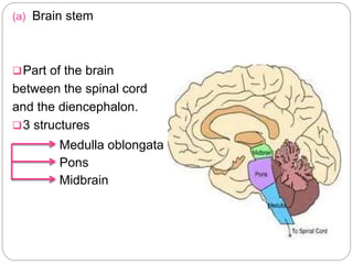

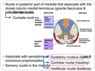
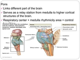
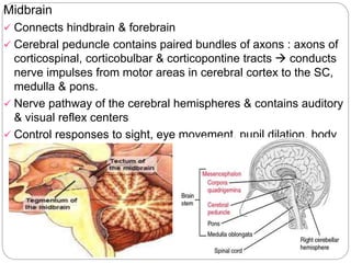
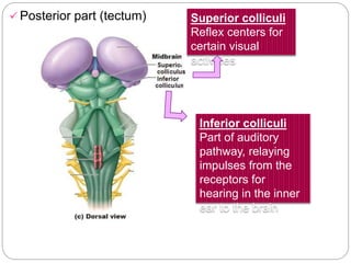


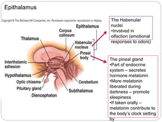
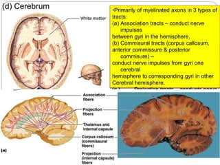


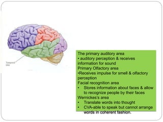

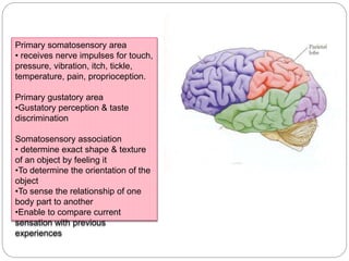
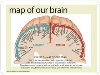

The document summarizes the major parts and structures of the human brain. It discusses the four major parts: (1) brain stem, (2) cerebellum, (3) diencephalon, and (4) cerebrum. Within each part, it describes the key substructures and their functions. For example, it notes that the brain stem contains the medulla, pons, and midbrain and regulates vital functions. The cerebellum coordinates movement and balance. The diencephalon includes the thalamus, hypothalamus, and epithalamus and relays sensory information. The cerebrum is the largest part and contains areas associated with motor control, language, and the five