15.ppt
•Download as PPT, PDF•
0 likes•216 views

gross anatomy of the brain and cranial nerves of the human.

Recommended
More Related Content
What's hot
What's hot (20)
Similar to 15.ppt
Similar to 15.ppt (20)
More from Medical Knowledge
More from Medical Knowledge (20)
Recently uploaded
Recently uploaded (20)
15.ppt
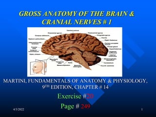
- 1. 4/3/2022 1 GROSS ANATOMY OF THE BRAIN & CRANIAL NERVES # 1 MARTINI, FUNDAMENTALS OF ANATOMY & PHYSIOLOGY, 9TH EDITION, CHAPTER # 14 Exercise #20 Page # 249
- 2. 4/3/2022 2 NOTE: THIS IS A STUDY GUIDE, NOT AN ALL INCLUSIVE REVIEW. THERE MIGHT BE THINGS NOT COVERED BY THIS STUDY GUIDE THAT MIGHT BE ASKED IN YOUR PRACTICUMS / QUIZZES. STUDENTS ARE RESPONSIBLE FOR READING THEIR TEXBOOK (S) AND FOR ALL THE MATERIAL COVERED DURING THE LABORATORY PERIOD, AS PER THE COURSE SYLLABUS
- 3. 4/3/2022 3 OBJECTIVES – Identifying the major regions of the brain and state their function. – Identifying the meninges. – Locating and naming selected cranial nerves.
- 4. CENTRAL NERVOUS SYSTEM (CNS)- BRAIN & SPINAL CORD PERIPFERAL NERVOUS SYSTEM (PNS)- NERVES & ASSOCITED GANGLIA CEREBRAL HEMIPHERES- 2 HALF OF THE CEREBEUM, RIGHT & LEFT DIVIDED BY LONGITUDINAL FISSURE GYRI (PLURAL)- THICK AREAS GYRUS- (SINGULAR)- FOLDS PRECENTRAL GYRUS POSTCENTRAL GYRUS FISSURES: DEEP GROOVES SULCI- SHALLOW GROOVES SEPARATING GYRI SULCY- PLURAL SULCUS- SINGULAR LONGITUDINAL FISSURE- IT SEPARATES THE RIGHT & LEFT HEMISPHERES CENTRAL SULCUS- IT DIVIDES THE CEREBRAL HEMISPHERS IN ANTERIOR & POSTERIOR
- 5. 4/3/2022 5 LATERAL SULCUS- IT SEPARATES FRONTAL AND PARIETAL LOBES FROM TEMPORAL LOBE PARIETO OCCIPITAL SULCUS- IT SEPARATES THE PARIETAL LOBE FROM OCCIPITAL LOBE FRONTAL LOBE- FROM CENTRAL SULCUS ANTERIORLY PREFRONTAL CORTEX- MOST ANTERIOR PART OF THE FRONTAL LOBE F- CENTER OF THE INTELECT FOR RAZONALIZATION TEMPORAL LOBE- UNDER THE TEMPORAL BONES UNDER THE LATERAL SULCUS PARIETAL LOBE- UNDER THE PARIETAL BONES & BEHIND THE FRONTAL LOBE OCCIPITAL LOBE- POSTERIOR LOBE OF THE CEREBRUM UNDER OCCIPITAL BONE
- 6. 4/3/2022 6 PRE CENTRAL GYRUS- GYRUS BEFORE THE CENTRAL SULCUS (ORANGE IN MODEL) IT CONTEINS THE PRIMARY MOTOR AREA- F- TO CONTROL VOLUNTARY MUSCLE MOVEMENT POSTCENTRAL GYRUS- GYRUS AFTER THE CENTRAL SULCUS (LAVANDER IN MODEL) IT CONTEIS THE PRIMARY SOMATOSENSORY AREA F- TO RECEIVE SENSORY INFO FROM GENERAL SENSORY RECEPTORS EX- TOUCH, PAIN, TEMP SOMATOSENSORY ASSOCIATION AREA- BEHIND THE POST CENTRAL GYRUS (LAVANDER) F- TO INTERPRET SENSORY INFORMATION BROCA’S AREA- CONTEIS THE SPEECH CENTER F- TO CONTROL MUSCLES OF SPEECH
- 7. 4/3/2022 ALFONSO A. PINO. MD. 7
- 8. 4/3/2022 ALFONSO A. PINO. MD. 8
- 9. 4/3/2022 ALFONSO A. PINO. MD. 9
- 10. 4/3/2022 ALFONSO A. PINO. MD. 10
- 11. 4/3/2022 ALFONSO A. PINO. MD. 11
- 12. 4/3/2022 12 TURN THE MODEL OVER OLFACTORY BULBS- THE BULBS TO THE FRONT F- CARRY SENSORY INFO ABOUT SMELL OLFACTORY TRACS – WHERE THE BULB ARE ATTACHED TO F- TO CARRY SENSORY INFO ABOUT SMELL OPTIC CHIASMA- WHERE THE OPTIC NERVES CROSS (IN THE MIDDLE) OPTIC TRACTS- FROM OPTIC NERVES BACK TO THE BRAIN F- TO CARRY VISUAL INFORMATION PITUITARY GLAND (HYPOFISIS)- F- TO PRODUCE MANY HORMONES MAMiILARY BODIES- (ORANGE & WHITE IN MODEL) F-TO CARRY SMELL IMPULSE
- 13. 4/3/2022 ALFONSO A. PINO. MD. 13
- 14. 4/3/2022 14 OPEN THE BRAIN IN HALF CEREBRAL PEDUNCLES- POSTERIOR PART OF THE PONS F- TO CARRY MOTOR INFORMATION PONS- CONNECTS THE CEREBRUM WITH THE CEREBELUM F-RELAYS SENSORY INFO TO CEREBELLUM & THALAMUS AND CONTAIN BREATHING CENTER SUBCONSCIOUS SOMATIC & VISCERAL MOTOR CENTERS MEDULA OBLONGATA- F- RELAYS SENSORY INFO TO THALAMUS & BRAIN STEM TO CONTROL VITAL SIGNS (BLOOD PRESSURE, HEART RATE, RESPIRATORY RATE)
- 15. 4/3/2022 ALFONSO A. PINO. MD. 15
- 16. 4/3/2022 16 Corpora cuadrigemina- 2 superior colliculies- F- to process visual sensation for visual reflex To control reflex mov of eyes, head & neck 2 inferior colliculies- F- to process auditory sensation for auditory reflex To control reflex mov of head, neck, & trunk (loud voice) Corpus callosum- F-to carry info between cerebral hemispheres Fornix- tracts of white matter that connects limbic system
- 17. Septum pellucidum- it separates the lateral Ventricles (1 & 2) Thalamus- from anterior commisure to pineal gland F- relays & processing centers for sensory information Intermediate mass-(brown model); F- to connect both sides of the thalamus Hypothalamus- from superior to the optic chiasm to posterior margin of mammillary bodies F- to control emotions- feeding, hunger & thirst centers Autonomic f-heart rate, blood pressure, Respiration & digestive funct To secrete hormones (adh, oxytocine) To control circadian rhythms
- 18. 4/3/2022 18 Epithalamus (pineal body- or gland)- F- to produce melatonin h Setting circadian rhythm Timming human sexual maturation Protects brain against free radicals Location- lies posterior portion of roof of 3rd ventricle Cerebellum- Vermis- (narrow band of cortex along the middline) F- to separate right & left cerebellar hemispheres arbor vitae- white mater of cerebellum Function of cerebellum- adjust postural movement balance and equilibrium Programming fine tuning movement to make movements smoo
- 19. 4/3/2022 ALFONSO A. PINO. MD. 19
- 20. 4/3/2022 ALFONSO A. PINO. MD. 20
- 21. 4/3/2022 ALFONSO A. PINO. MD. 21
- 22. 4/3/2022 ALFONSO A. PINO. MD. 22
- 23. 4/3/2022 ALFONSO A. PINO. MD. 23 THE CEREBELLUM
- 24. 4/3/2022 24 THE CEREBELLUM (ARBOR VITAE)
- 25. 4/3/2022 25 MENINGES Meninx= membrane: for protection Cranial meninges (From exrternal to internal) Dura mater- endosteal layer Dural sinus Dura mater- meningeal layer Subdural space Arachnoid mater Subarcnoid space contains csf Pia mater- for nourishment cerebral cortex
- 26. 4/3/2022 26 CRANIAL MENINGES (CONTINUATION) Dura mater Falx cerebri- formed by dura mater along longitudinal fissure F- to separate the cerebral hemispheres Superior sagital sinus- venous sinus within the falx cerebri Tentorium cerebelli- on top of the cerebellum F- to separate the cerebral hemispheres from those of the cerebellum
- 27. 4/3/2022 ALFONSO A. PINO. MD. 27
- 28. 4/3/2022 ALFONSO A. PINO. MD. 28
- 29. 4/3/2022 29 VENTRICLES Function of the ventricles- to contain CSF Lateral ventricles (1 & 2) 3rd ventricle Interventricular foramen- to connect lateral ventricle with 3rd ventricle Cerebral aqueduct- to connect 3rd ventricle with fourth ventricle Fourth ventricle Choroid plexus- function- it contains the ependimal cells for production of CSF
- 30. 4/3/2022 ALFONSO A. PINO. MD. 30
- 31. 4/3/2022 ALFONSO A. PINO. MD. 31
- 32. CRANIAL NERVES I- olfactory n- (not seen in charts, just in model) F- special sensory, to carry smell impulse II- optic n- special sensory, to carry visual info III- oculomotor-motor-to control movement of the muscles of the eyes IV-troclear-motor, eye movement (superior oblique muscles of the eyes V-trigeminal- (sensory & motor)- to face chewing muscles VI-abducens-motor- eye movement. Loc : pons & medulla F- lateral rectus muscle of the eyes VII- facial-(sensory & motor)- F- muscles of facial expression Carries motor info for face
- 33. 4/3/2022 ALFONSO A. PINO. MD. 33
- 34. 4/3/2022 34 VIII- vestibulococlear n- special sensory Vestibular branch- balance & equilibrium Cochlear branch- hearing IX- glosopharyngeal n- sensory & motor- Head & neck X- vagus n- sensory & motor- Organs in thorax & abdomen XI-accessory n- motor- Neck, upper back, trapezius & sternocleidomastoid muscles XII- hypoglossal n- motor- tongue movement
- 35. 4/3/2022 35 OLFACTORY EPITHELIUM OLFACTORY FIBERS OLFACTORY BULB OLFACTORY TRACT (chapter #17)
- 36. 4/3/2022 ALFONSO A. PINO. MD. 36
- 37. 4/3/2022 ALFONSO A. PINO. MD. 37
- 38. AUTONOMIC NERVOUS SYSTEM Parasympathetic or craniosacral division- To maintain body under resting conditions Preganglionic neuron: located in CNS, found in brain and sacral part of spinal cord (s2-s4) Pregaglionic fibers: from CNS to ganglia Intramural or terminal ganglion- contain ganglionic neurons postganglionic fibers: from ganglia to organ Sympathetic or thoraco lumbar division- To put the body in alert state Preganglionic neuron: in CNS (spinal cord t1-l2) White ramus communicans: contain preganglionic unmyelinatesd fibers Splanchnic nerves – preganglionic fibers Sympathetic chain ganglia: contain ganglionic neurons collateral ganglion: contain ganglionic neurons Gray ramus communicans: contain postganglionic unmylinated fibers
- 39. 4/3/2022 39 REMEMBER! GO TO THE TUTORING ROO AND PRACTICE WITH MODELS. ROO 3326.