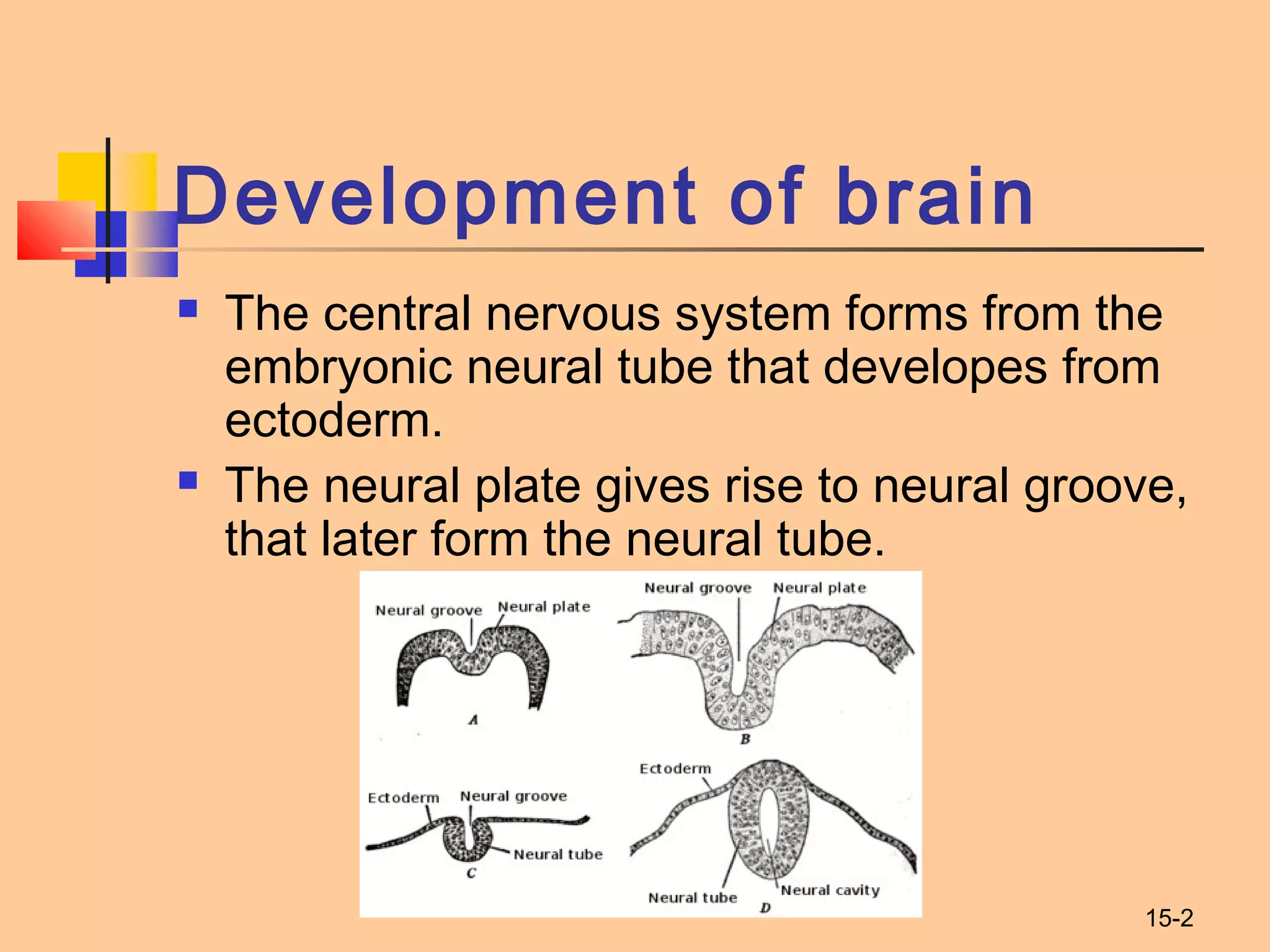
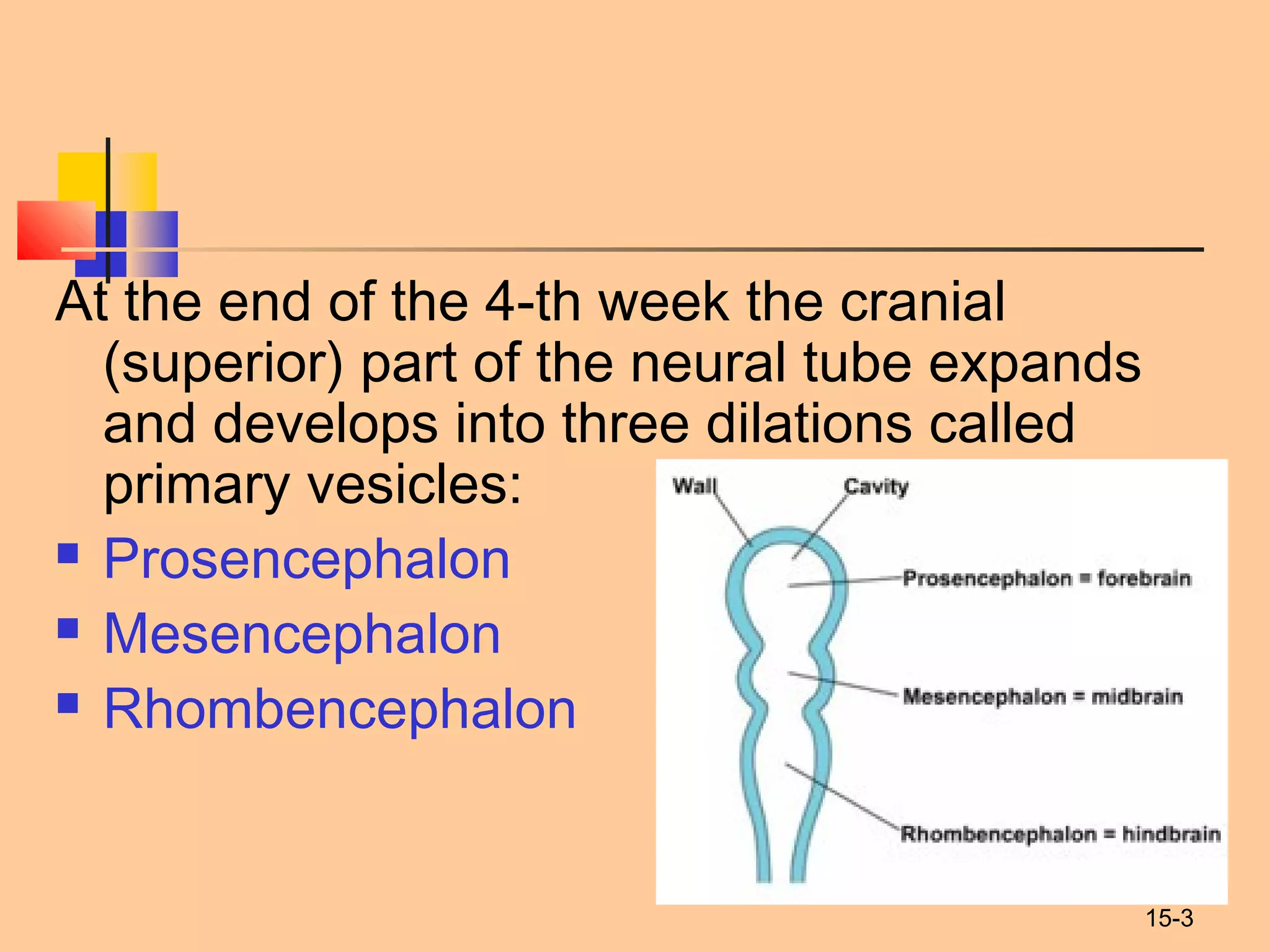
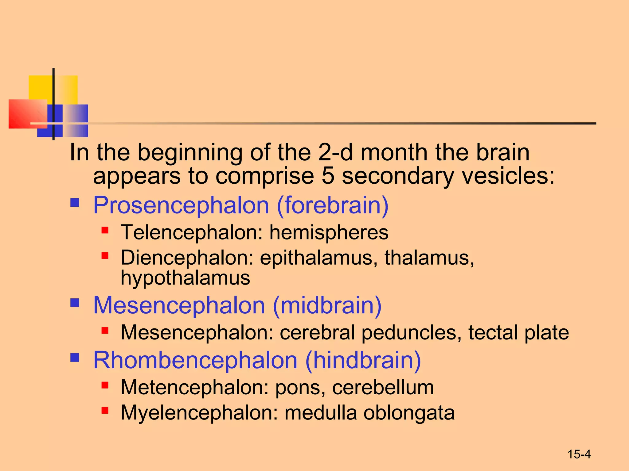
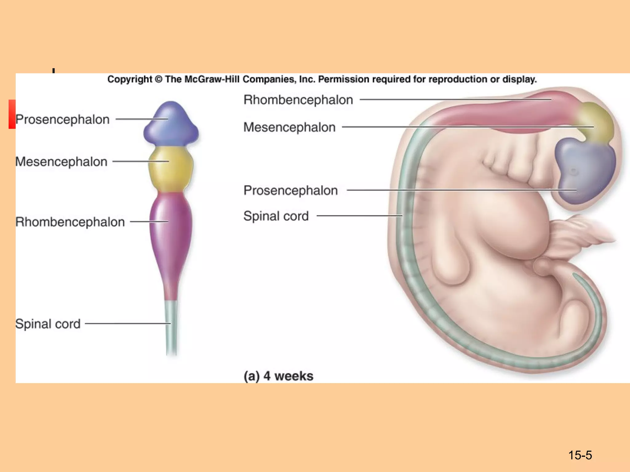
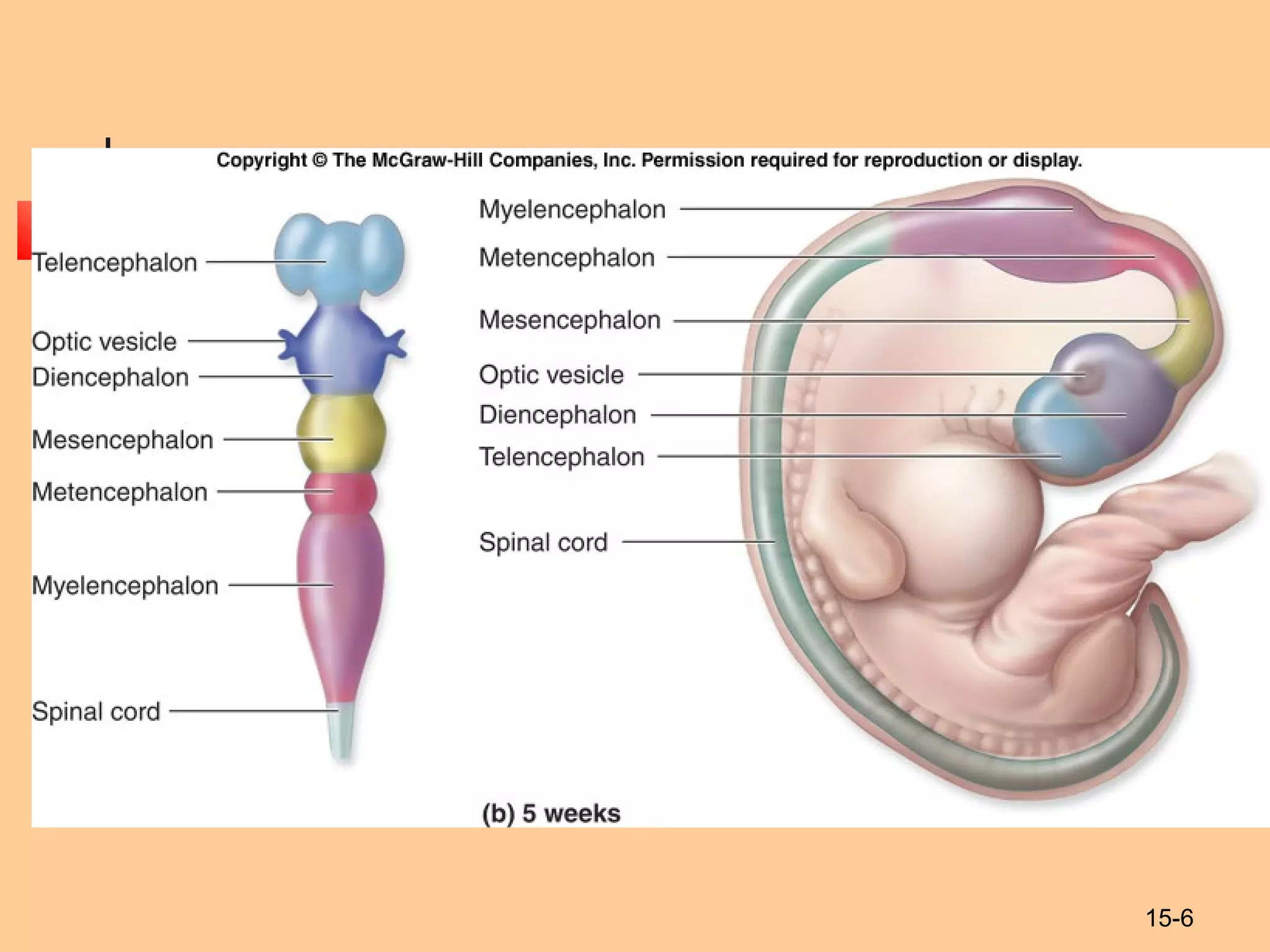
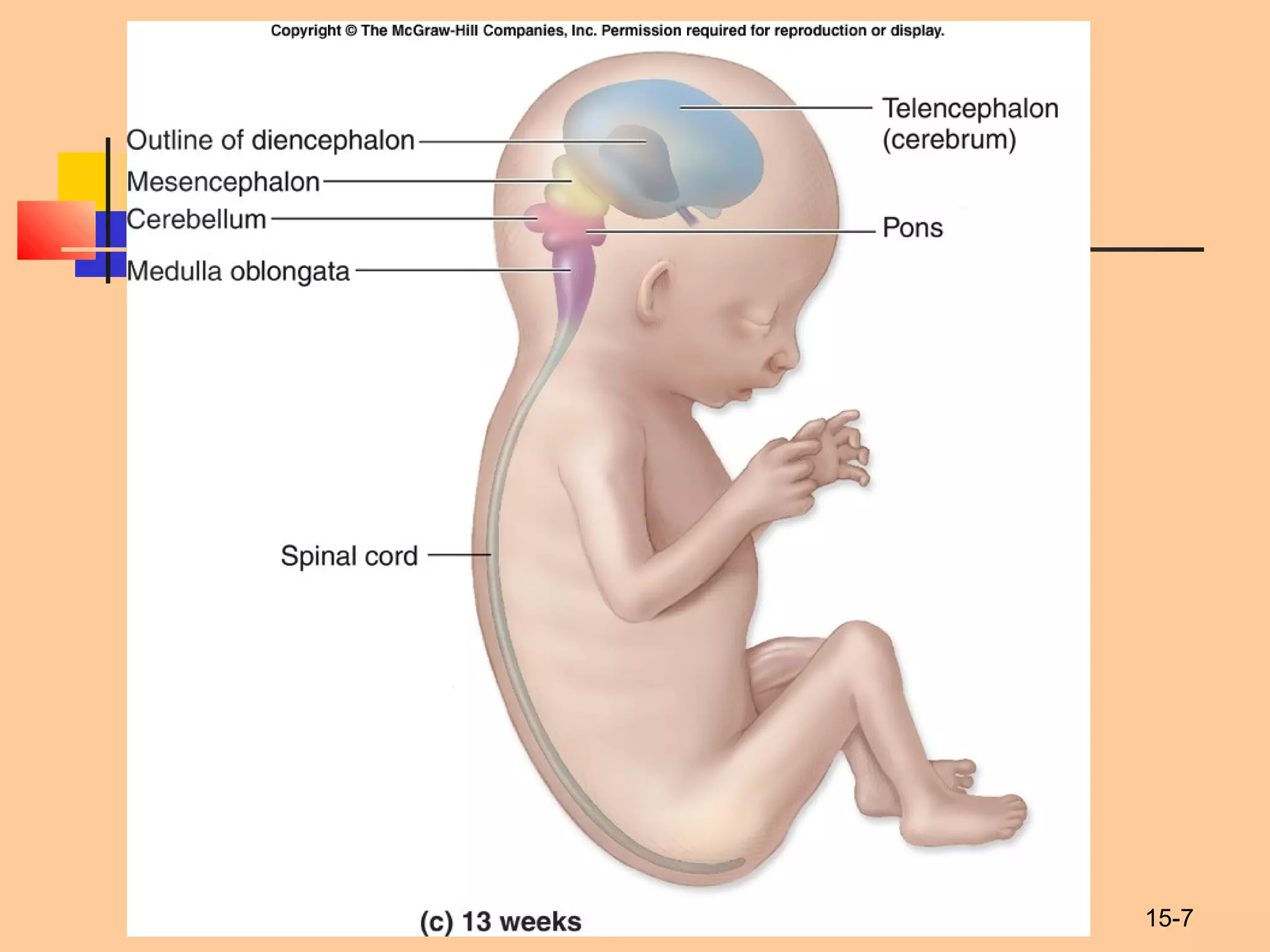
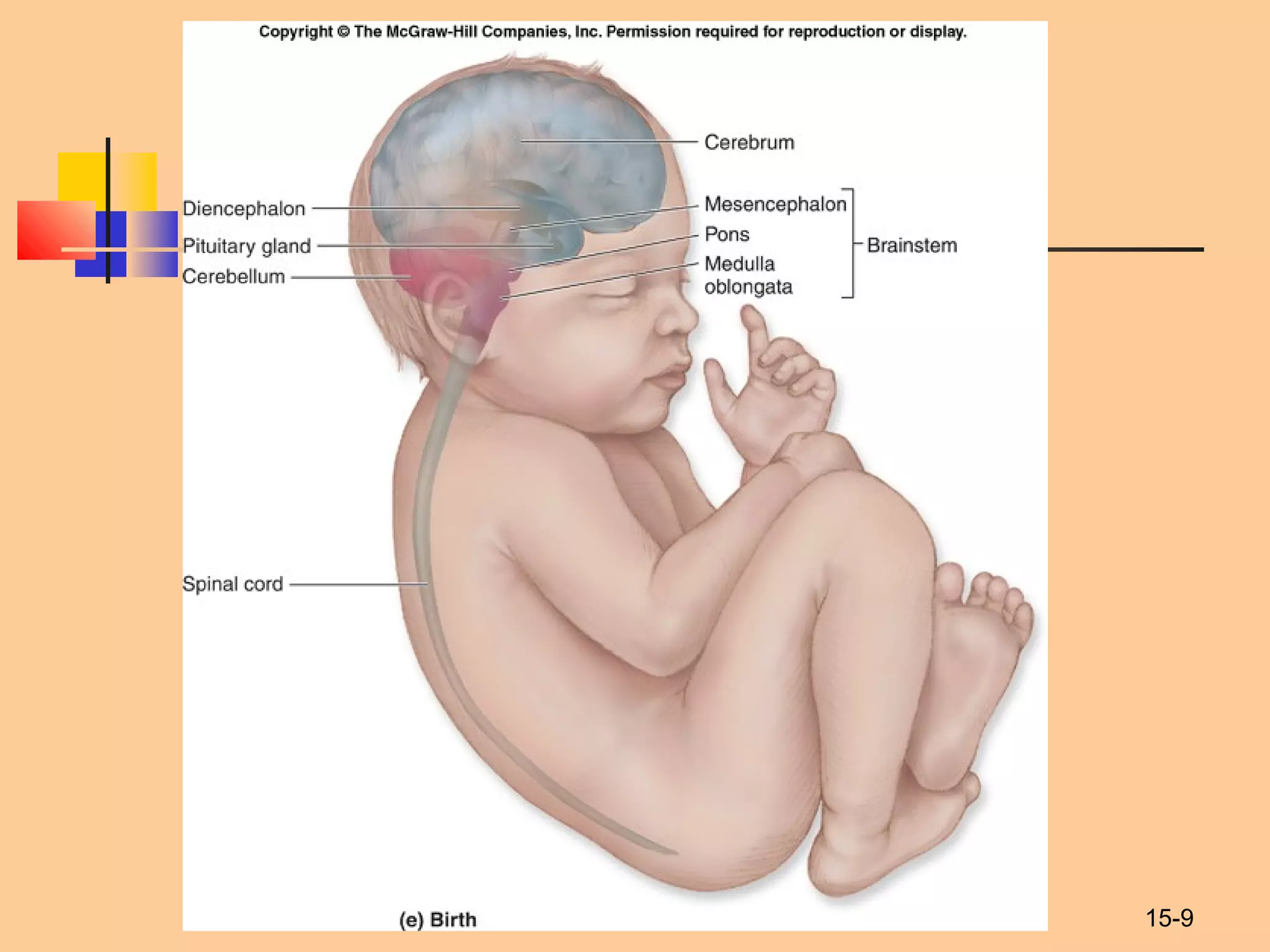
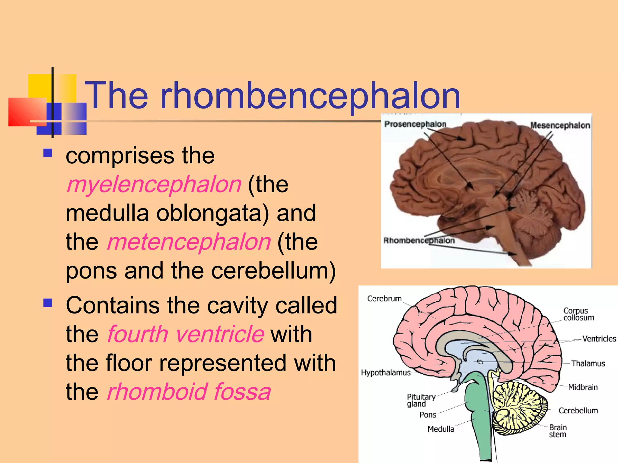
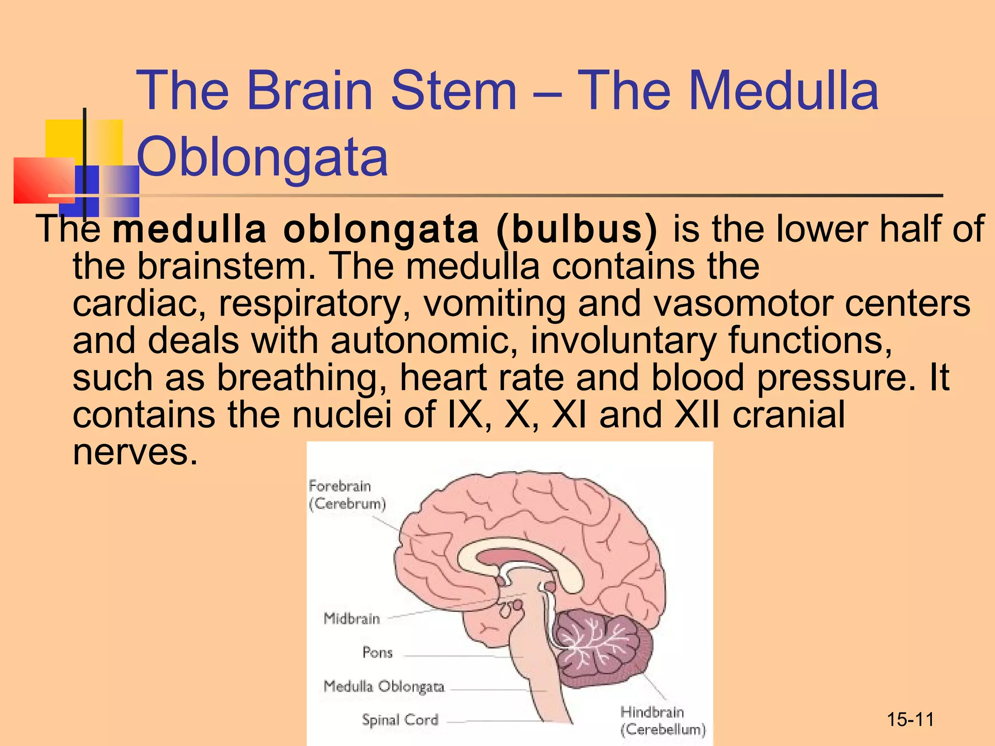
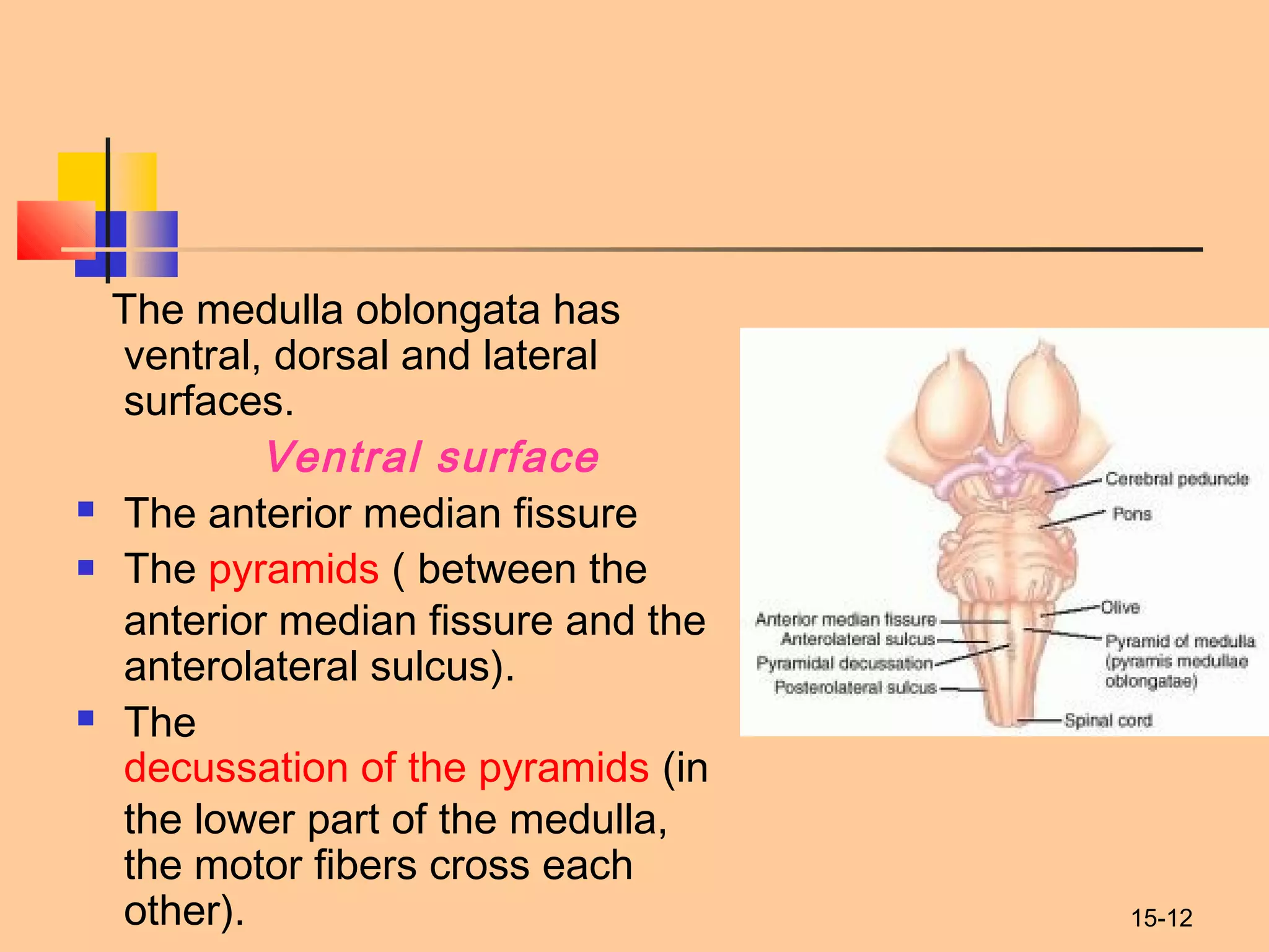
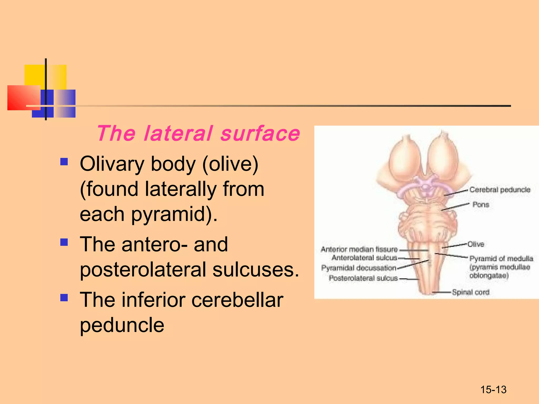
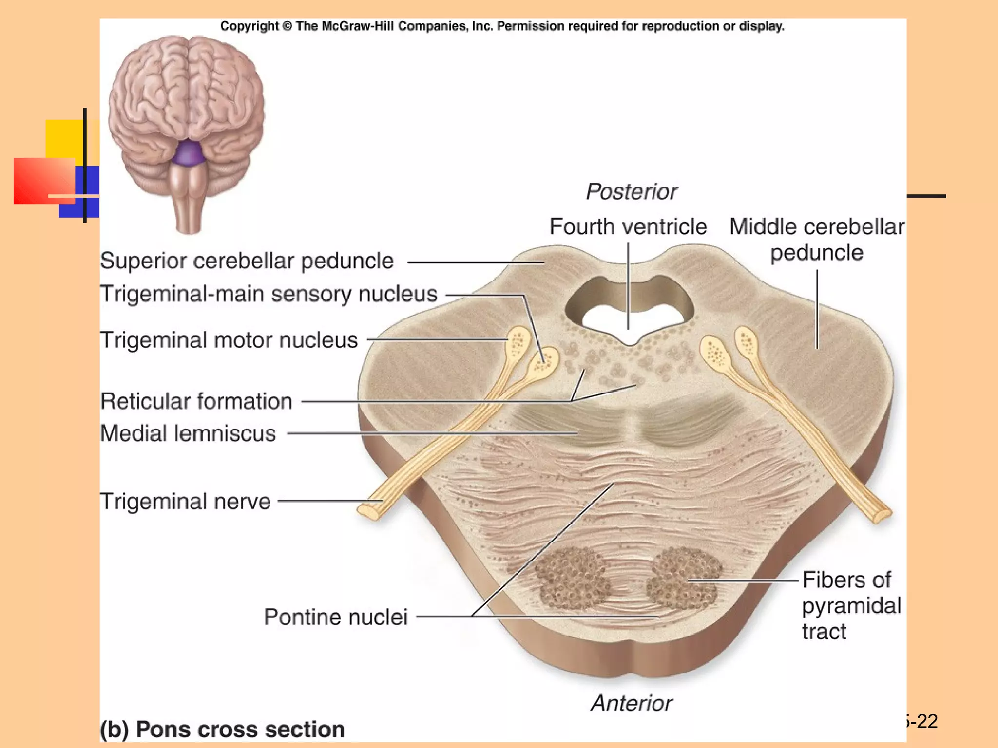
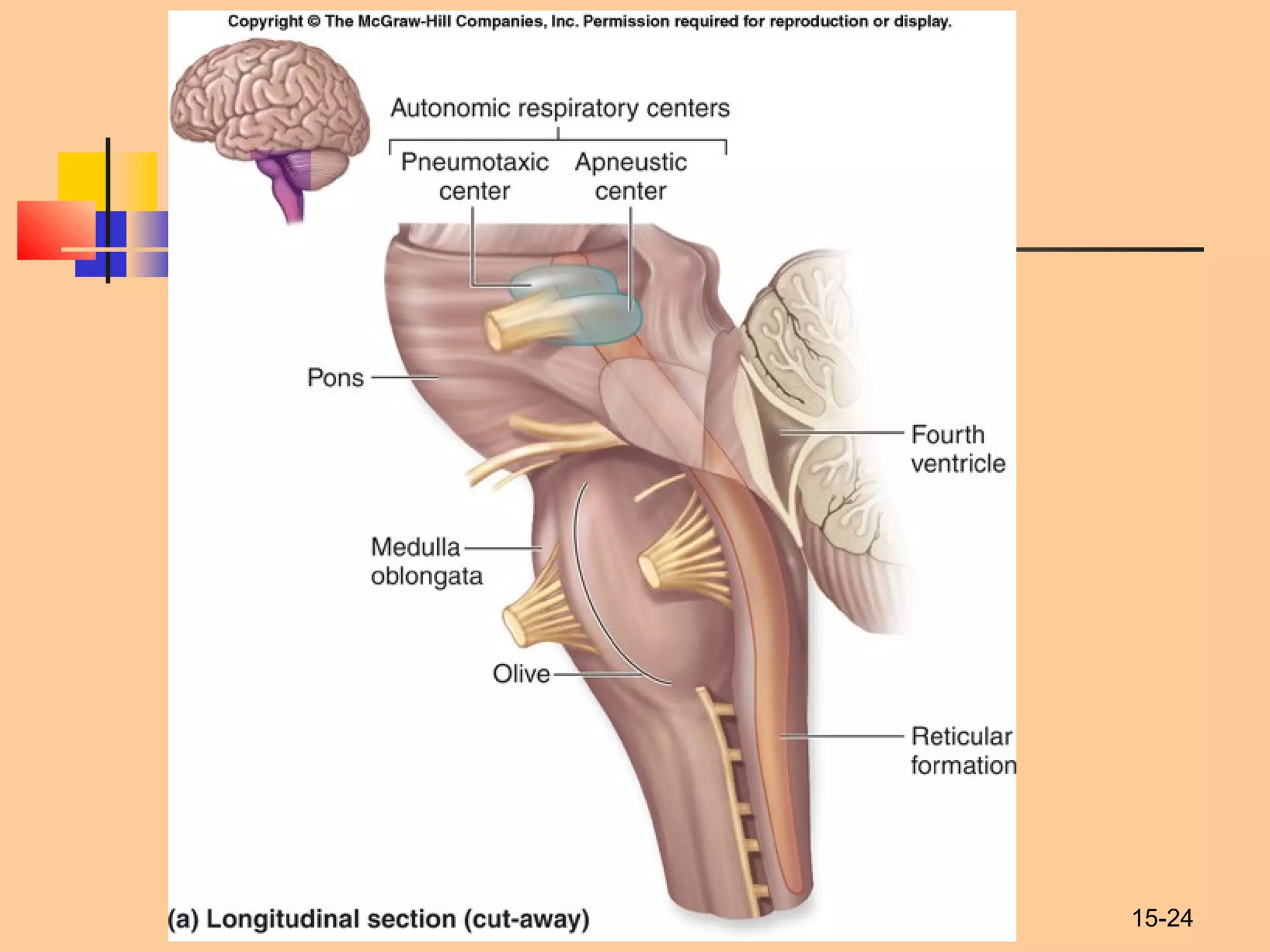
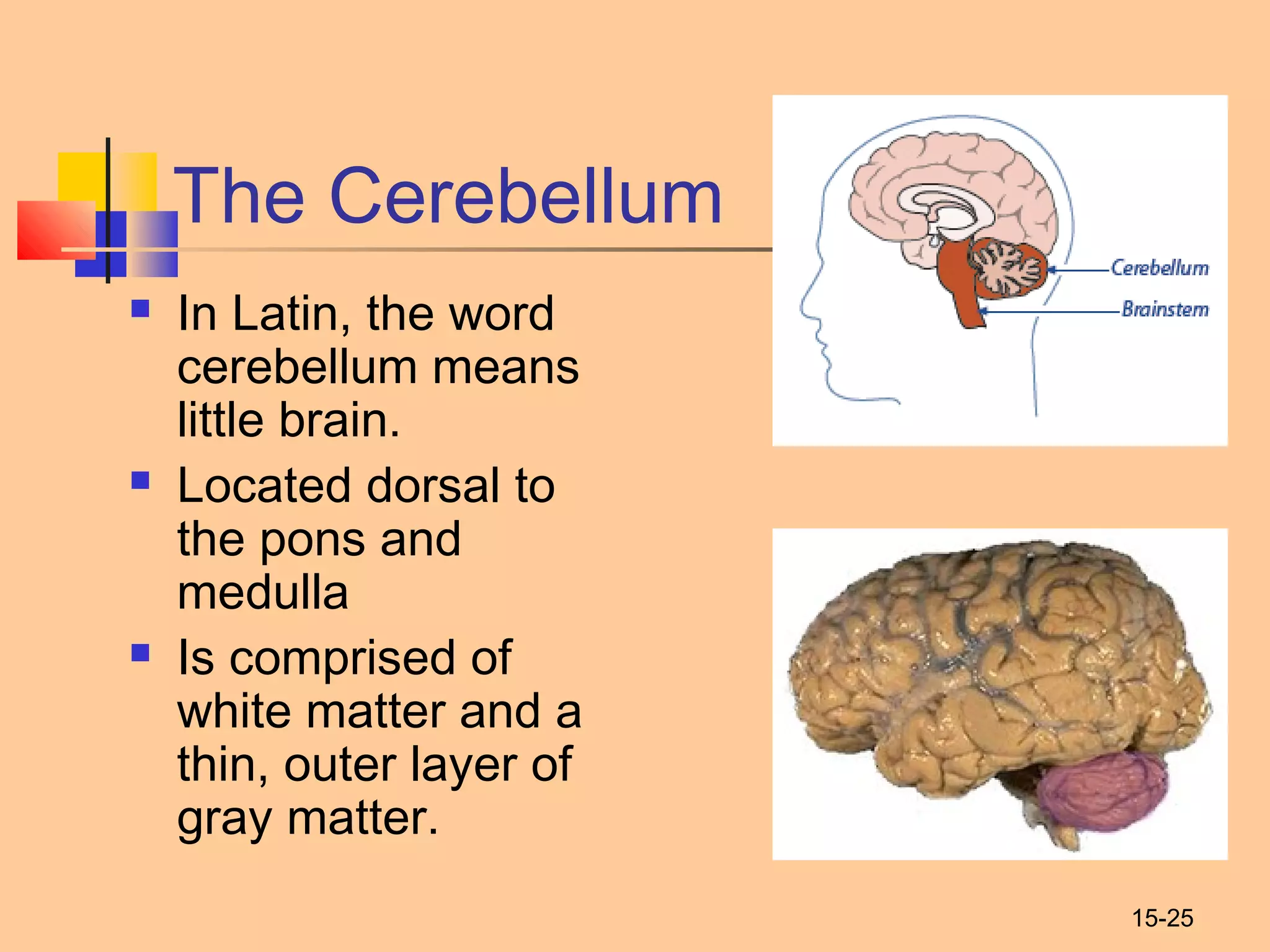
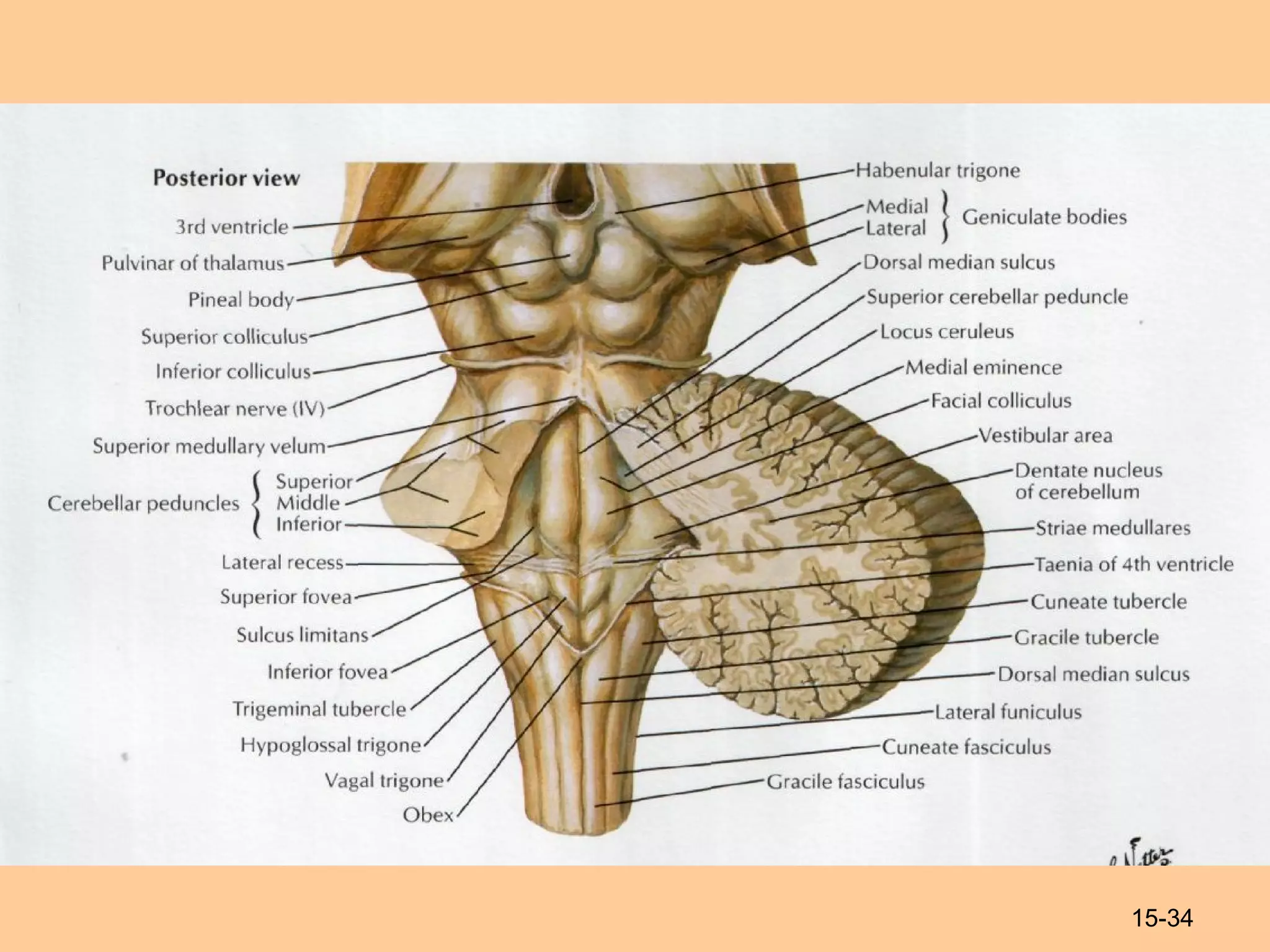
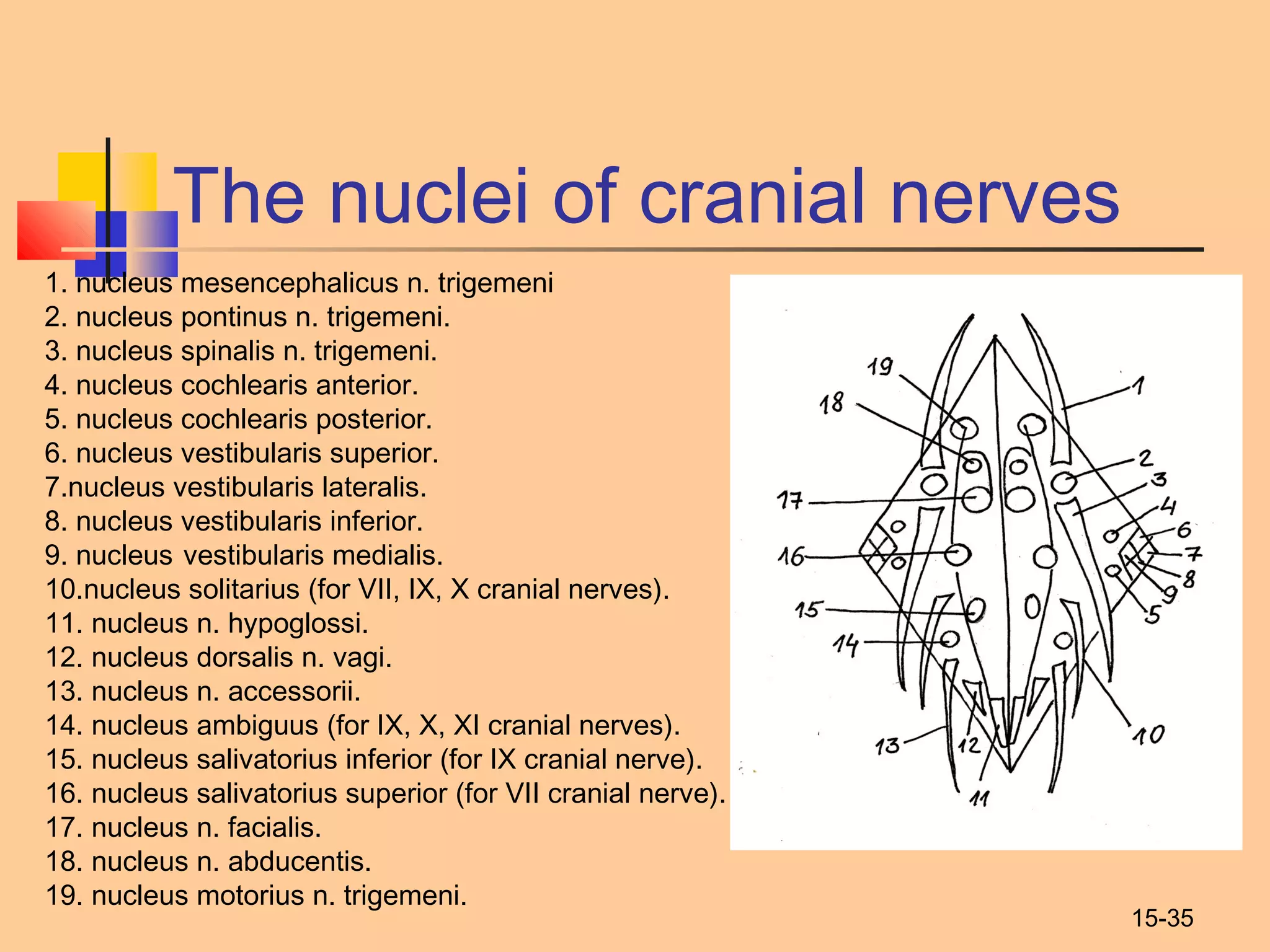
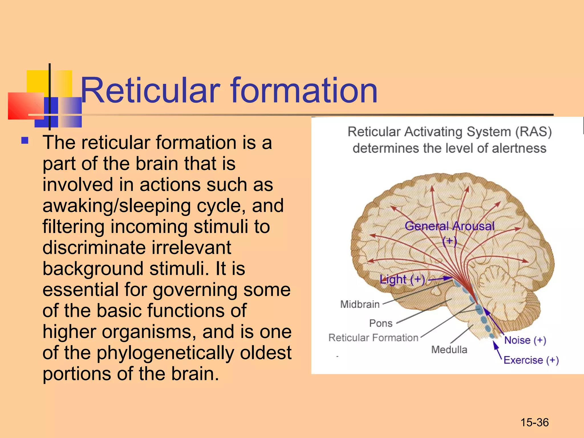
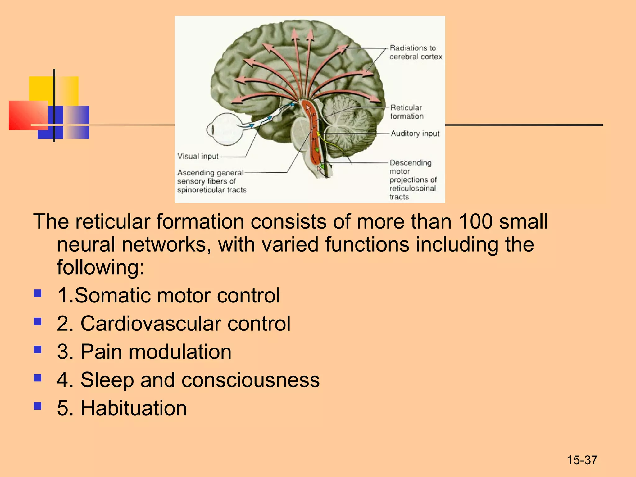
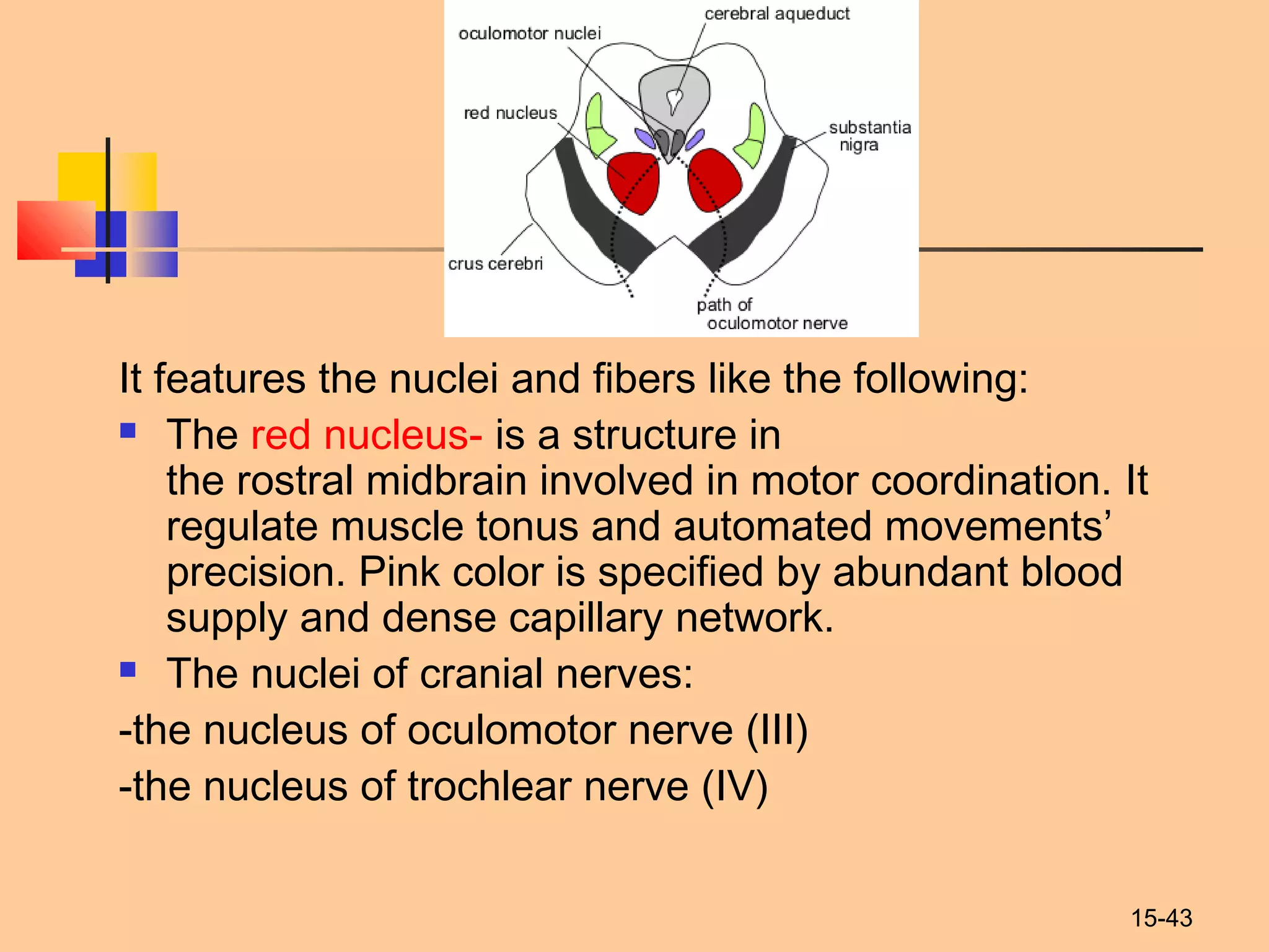
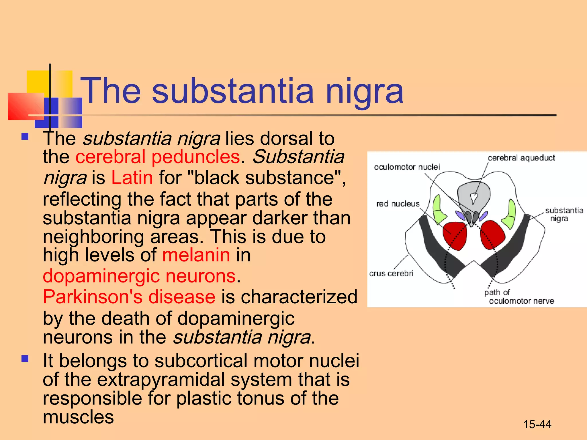
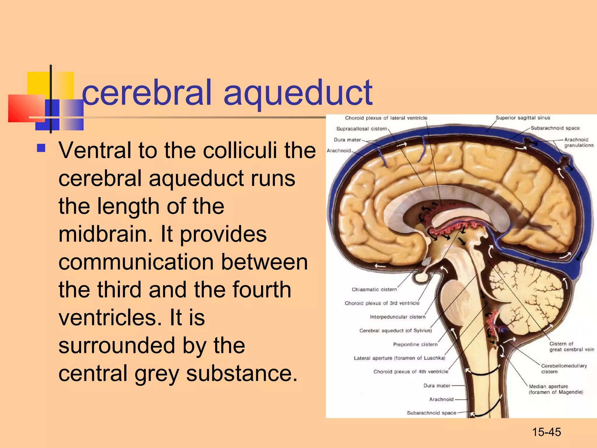
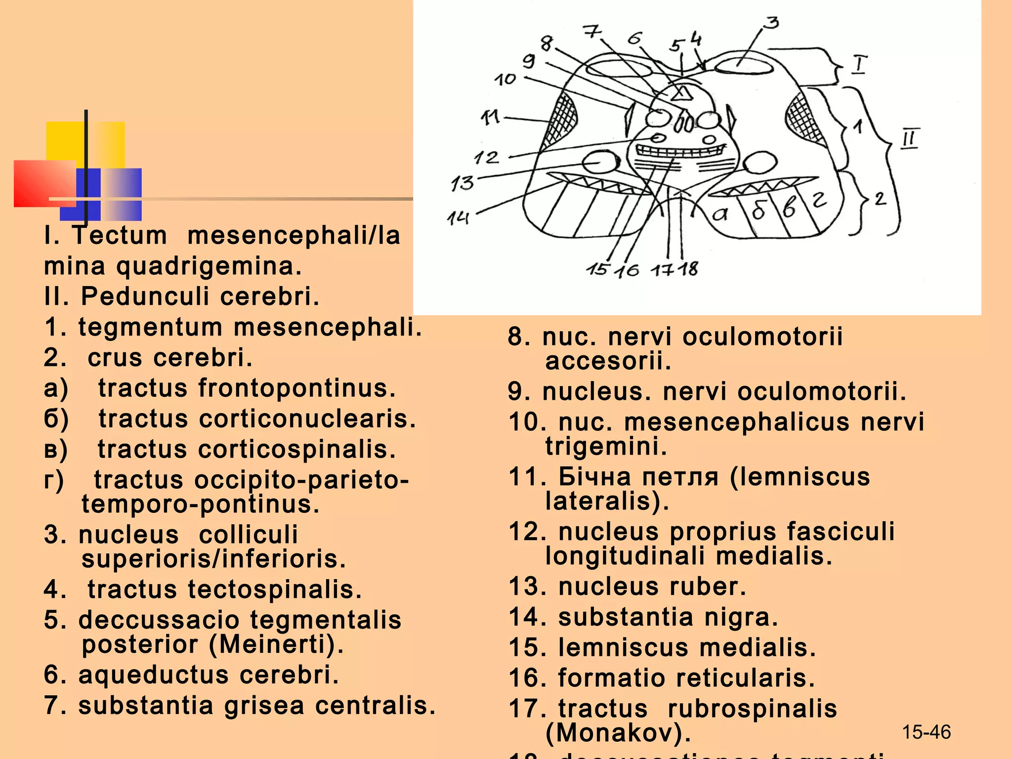
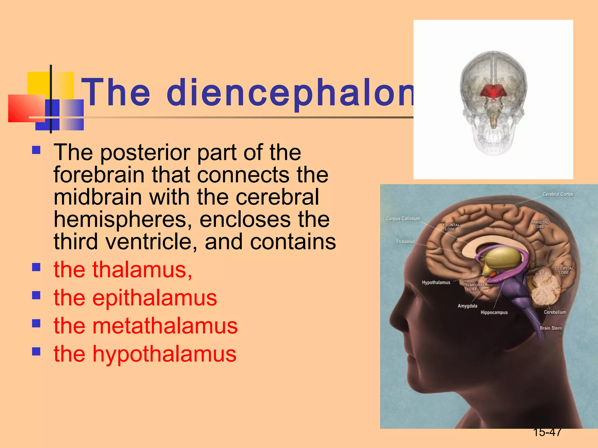
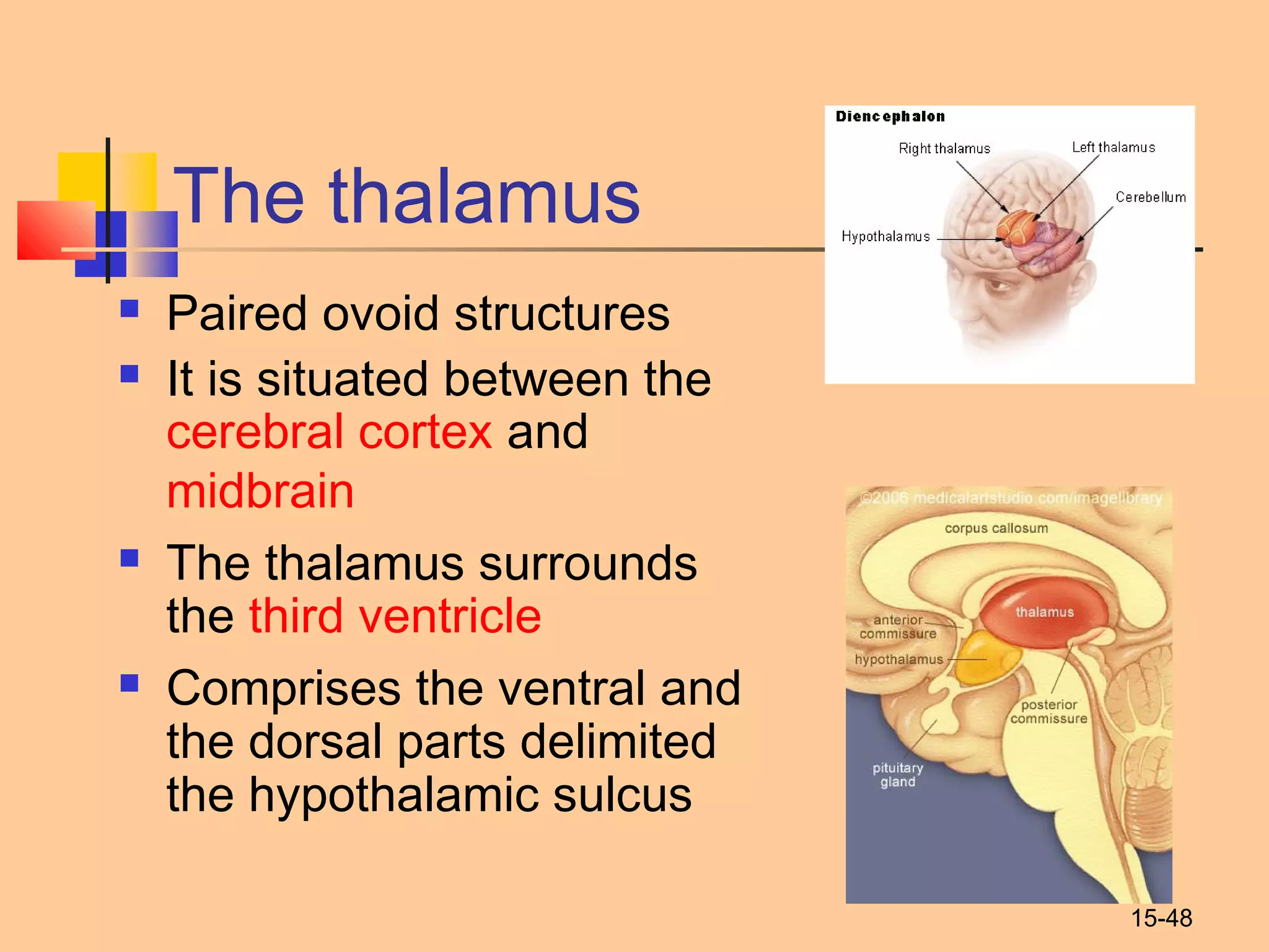
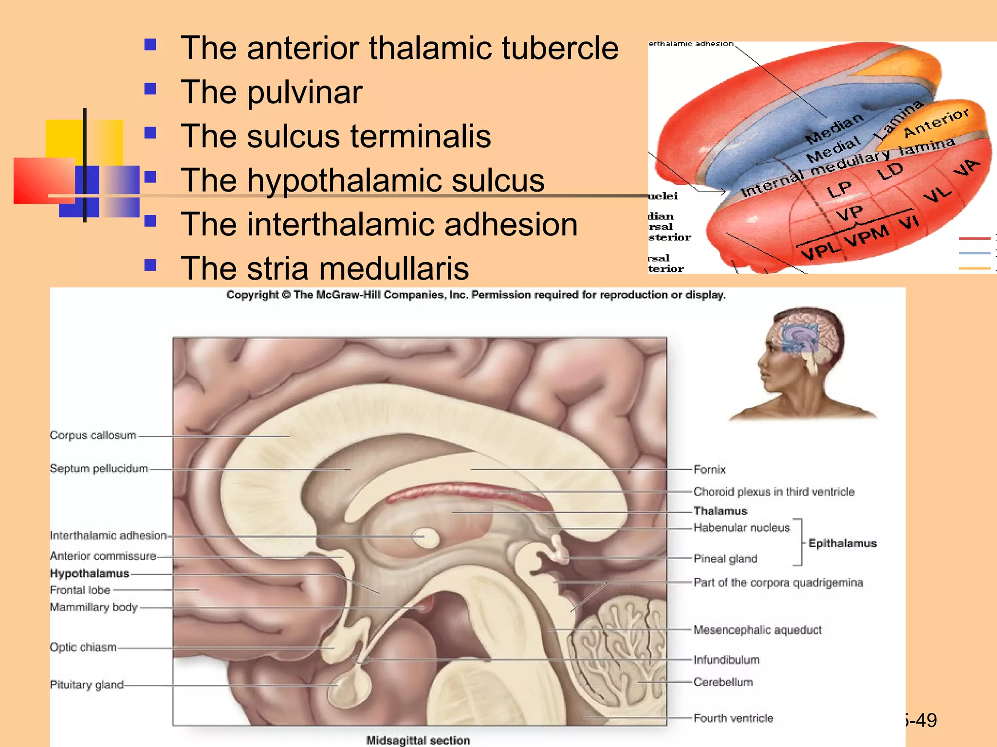
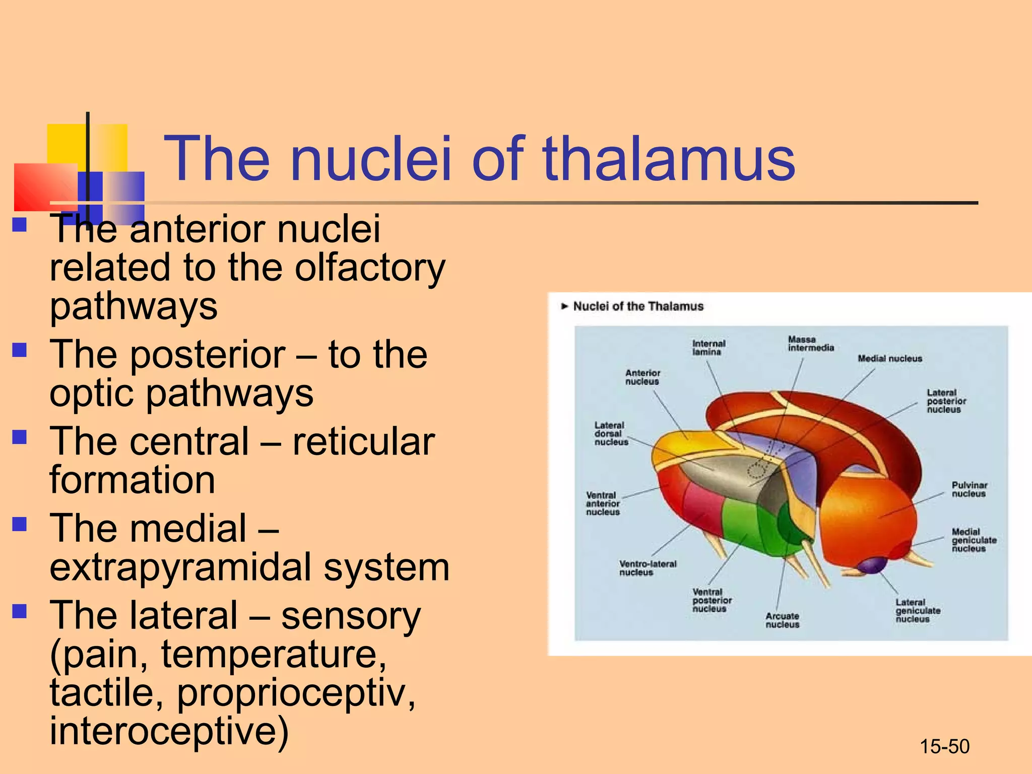
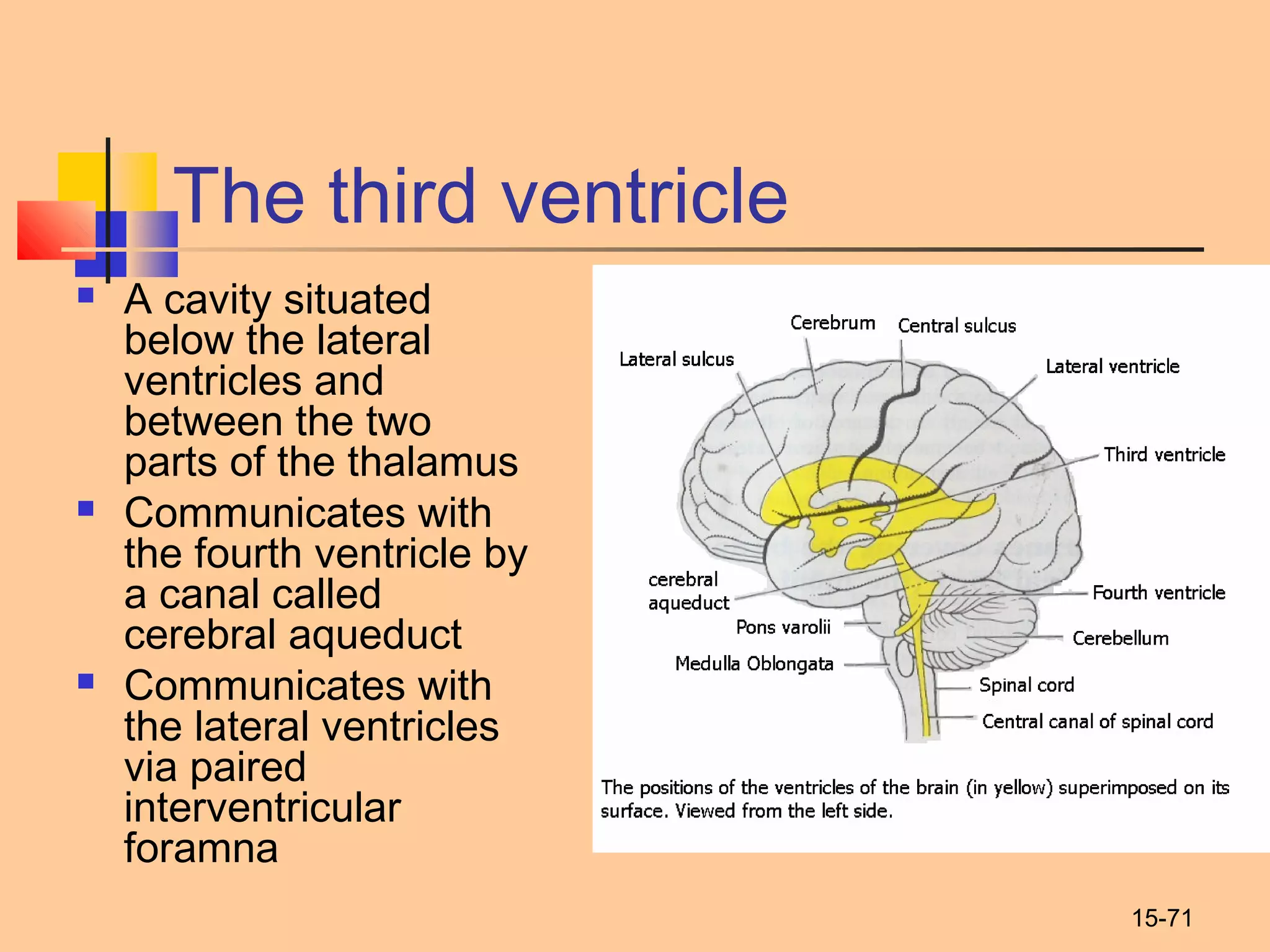
1
Ivano-Frankivsk National Medical
University
The Department of Human anatomy
Structure of rhombencephalon.
Development of brain.
Reticular formation.
Distribution of nuclei of cranial
nerves in rhomboid -shaped
fossa. Midbrain and
diencephalon. Vegetative
nuclei of hypothalamus. Notion
about neurosecretion.
Hypothalamo-hypophisial
system. Third ventricle.Prepared by PhDPrepared by PhD
Tetyana Knyazevych - ChornaTetyana Knyazevych - Chorna
15-2
 The central nervous system forms from the
embryonic neural tube that developes from
ectoderm.
 The neural plate gives rise to neural groove,
that later form the neural tube.
Development of brain
15-3
At the end of the 4-th week the cranial
(superior) part of the neural tube expands
and develops into three dilations called
primary vesicles:
 Prosencephalon
 Mesencephalon
 Rhombencephalon
15-4
In the beginning of the 2-d month the brain
appears to comprise 5 secondary vesicles:
 Prosencephalon (forebrain)
 Telencephalon: hemispheres
 Diencephalon: epithalamus, thalamus,
hypothalamus
 Mesencephalon (midbrain)
 Mesencephalon: cerebral peduncles, tectal plate
 Rhombencephalon (hindbrain)
 Metencephalon: pons, cerebellum
 Myelencephalon: medulla oblongata
15-5
15-6
15-7
15-8
15-9
15-10
The rhombencephalon
 comprises the
myelencephalon (the
medulla oblongata) and
the metencephalon (the
pons and the cerebellum)
 Contains the cavity called
the fourth ventricle with
the floor represented with
the rhomboid fossa
15-11
The Brain Stem – The Medulla
Oblongata
The medulla oblongata (bulbus) is the lower half of
the brainstem. The medulla contains the
cardiac, respiratory, vomiting and vasomotor centers
and deals with autonomic, involuntary functions,
such as breathing, heart rate and blood pressure. It
contains the nuclei of IX, X, XI and XII cranial
nerves.
15-12
The medulla oblongata has
ventral, dorsal and lateral
surfaces.
Ventral surface
 The anterior median fissure
 The pyramids ( between the
anterior median fissure and the
anterolateral sulcus).
 The 
decussation of the pyramids (in
the lower part of the medulla,
the motor fibers cross each
other).
15-13
The lateral surface
 Olivary body (olive)
(found laterally from
each pyramid).
 The antero- and
posterolateral sulcuses.
 The inferior cerebellar
peduncle
15-14
The dorsal surface
 The posterior median sulcus
 The fasciculus gracilis and the fasciculus cuneatus ( that
lying laterally).
 These fasciculi end in rounded elevations known as the
gracile and the cuneate tubercles. They are caused by
masses of gray matter known as the nucleus gracilis and
the nucleus cuneatus.
 Just above the tubercles, the posterior aspect of the
medulla is occupied by a triangular fossa, which forms the
lower part of the floor of the fourth ventricle.
15-15
15-16
Internal structure
 The cuneate and gracile nuclei that accept the
cuneate and gracile fasciculi (it transmit the impulses
of proprioceptive and tactile sensivity). The axons of
nuclei form the medial lemniscus.
 The pyramidal tract comprises the corticonuclear and
the corticospinal fibers.
 The inferior olivary nucleus (communicates with the
cerebellum and spinal cord)- control body equilibrium.
 The reticular formation.
 The nuclei of cranial nerves (IX-XII)
15-17
15-18
Functions
 The medulla oblongata controls autonomic functions,
and connects the higher levels of the brain to the 
spinal cord. It is also responsible for regulating
several basic functions of the 
autonomic nervous system which include:
 Respiration – chemoreceptors
 Cardiac center – sympathetic, parasympathetic
system
 Vasomotor center – baroreceptors
 Reflex centers of vomiting, coughing, sneezing, and
swallowing
 Destruction of the medulla causes instant death.
15-19
The Brain Stem – The Pons
(pons Varolii)
 The pons is about 2.5 cm in length. Most of it
appears as a broad anterior bulge rostral to
the medulla. Posteriorly, it consists mainly of
two pairs of thick stalks called
cerebellar peduncles. They connect the
cerebellum to the pons and midbrain.
 Contains the nuclei of cranial nerves V, VI, VII
and VIII.
15-20
The Pons
External
features
 The bulbopontine
sulcus
 The basilar sulcus
 The medullary stria
of fourth ventricle
 The middle
cerebellar peduncle
15-21
Internal structure of pons
The trapezoid body delimits two parts of pons:
The basilar part
 The pontine nuclei
 The pontocerebellar fibers
 The corticopontine fibers
 The pyramidal fibers
The tegmentum
 The nuclei of cranial nerves (V-VIII)
 The superior olivary nucleus (belongs to the auditory
nuclei group)
 Medial lemniscus
 Rubrospinal and tectospinal tracts
15-22
15-23
The functions of Pons
 The pons contains nuclei that relay
signals from the forebrain to the
cerebellum, along with nuclei that deal
primarily with sleep, respiration,
swallowing, bladder control, hearing,
equilibrium, taste, eye movement, facial
expressions, facial sensation, and
posture.
15-24
15-25
The Cerebellum
 In Latin, the word
cerebellum means
little brain.
 Located dorsal to
the pons and
medulla
 Is comprised of
white matter and a
thin, outer layer of
gray matter.
15-26
The Cerebellum
 Consists of two cerebellar
hemispheres and the vermis
 Surface feature various
cerebellar fissures delimit
lobes, lobules and folia
 The Cerebellum subdivided
into:
 Anterior lobe
 Posterior lobe
 Flocculonodular lobe
15-27
15-28
From the evolutional point the cerebellum has:
 The oldest part – archicerebellum (floculus
and nodule are associated with the vestibular
nuclei and are involved into body equilibrium
control).
 The ancient part – paleocerebellum (anterior
lobe) which control muscle tone, coordinates
the movement related to weight and inertia.
 The newest part - neocerebellum (posterior
lobe) which control voluntary and automated
movements.
15-29
The Cerebellum
Saggital section reveals
treelike arragment that call
the arbor vitae
The nuclei of cerebellum:
 The dentate (6)
 The emboliform (5)
 The globose (4)
 The fastigial (3)
15-30
The cerebellar peduncles
The inferior cerebellar peduncles:
 The posterior spinocerebellar tract
 The olivocerebellar tract
 The external arcuate fibers
 The vestibulocerebellar fibers
 The cerebellovestibular fibers
The middle cerebellar peduncles:
 The pontocerebellar fibers
The superior cerebellar peduncles:
 The cerebellorubral fibers
 The cerebellothalamic fibers
 The anterior spinocerebellar fibers
15-31
15-32
Functions of Cerebellum :
The cerebellum is involved in several functions of the body
including:
 Fine Movement Coordination
 Balance and Equilibrium
 Muscle Tone
 It may also be involved in some cognitive functions such as 
attention and language, and in regulating fear and pleasure
 responses
 The cerebellum does not initiate movement, but it
contributes to coordination, precision, and accurate timing.
It receives input from sensory systems and from other parts
of the brain and spinal cord, and integrates these inputs to
fine tune motor activity.
 Because of this fine-tuning function, damage to the
cerebellum does not cause paralysis, but instead produces
disorders in fine movement, equilibrium, posture, and
motor learning.
15-33
Rhomboid fossa
It resides on the dorsal surface of the medulla oblongata
and pons
Relief of the rhomboid fossa:
 Median sulcus
 Medial eminence
 Sulcus limitans
 Medullary stria
 Superior and inferior foveas
 Facial colliculus
 Vestibular area
 Locus caeruleus
 Hypoglossal and vagal
trigones
15-34
15-35
The nuclei of cranial nerves
1. nucleus mesencephalicus n. trigemeni
2. nucleus pontinus n. trigemeni.
3. nucleus spinalis n. trigemeni.
4. nucleus cochlearis anterior.
5. nucleus cochlearis posterior.
6. nucleus vestibularis superior.
7.nucleus vestibularis lateralis.
8. nucleus vestibularis inferior.
9. nucleus vestibularis medialis.
10.nucleus solitarius (for VII, IX, X cranial nerves).
11. nucleus n. hypoglossi.
12. nucleus dorsalis n. vagi.
13. nucleus n. accessorii.
14. nucleus ambiguus (for IX, X, XI cranial nerves).
15. nucleus salivatorius inferior (for IX cranial nerve).
16. nucleus salivatorius superior (for VII cranial nerve).
17. nucleus n. facialis.
18. nucleus n. abducentis.
19. nucleus motorius n. trigemeni.
15-36
Reticular formation
 The reticular formation is a
part of the brain that is
involved in actions such as
awaking/sleeping cycle, and
filtering incoming stimuli to
discriminate irrelevant
background stimuli. It is
essential for governing some
of the basic functions of
higher organisms, and is one
of the phylogenetically oldest
portions of the brain.
15-37
The reticular formation consists of more than 100 small
neural networks, with varied functions including the
following:
 1.Somatic motor control 
 2. Cardiovascular control
 3. Pain modulation
 4. Sleep and consciousness
 5. Habituation
15-38
 The midbrain or mesencephalon is
a portion of the central nervous
system associated with vision,
hearing, motor control, sleep/wake,
arousal (alertness), and temperature
regulation.
Midbrain
15-39
 Midbrain is formally divided into dorsal and
ventral parts at the level of cerebral aqueduct
 The dorsal portion is known as tectum (or
corpora quadrigemina), which largely consists
of inferior and superior colliculi
 The ventral portion is known as tegmentum
 Tegmentum is bounded ventrally by the massive
fibre system of the crus cerebri (
cerebral peduncles)
15-40
Tectum
(corpora quadrigemina)
 The superior colliculus is
involved with
saccadic eye movements
(subcortical visual centers);
while the inferior colliculus
is a synapsing point for
sound information
(subcortical auditory
centers). The nuclei of coliculi
are responsible for reflexes
associated with sudden sound
and visual stimuli by the
tectospinal tract.
15-41
Cerebral peduncle
 They are the most anterior structure
in the midbrain and contain the
large ascending and descending
tracts that run to and from the
cerebrum.
 Each Cerebral peduncle features
the ventral portion called the base
of peduncle and the dorsal portion
called the tegmentum of midbrain.
The two portions are delimited by
the substantia nigra.
 The base of peduncle contain the
pathways:
 The corticospinal fibers ant/lat
 The corticonuclear fibers
 The corticopontine fibers
15-42
The tegmentum
 The midbrain tegmentum
 is the part of the midbrain
 extending from the 
substantia nigra to the 
cerebral aqueduct in a
horizontal section. It forms
the floor of the midbrain that
surrounds the cerebral
aqueduct.
15-43
It features the nuclei and fibers like the following:
 The red nucleus- is a structure in
the rostral midbrain involved in motor coordination. It
regulate muscle tonus and automated movements’
precision. Pink color is specified by abundant blood
supply and dense capillary network.
 The nuclei of cranial nerves:
-the nucleus of oculomotor nerve (III)
-the nucleus of trochlear nerve (IV)
15-44
The substantia nigra
 The substantia nigra lies dorsal to
the cerebral peduncles. Substantia
nigra is Latin for "black substance",
reflecting the fact that parts of the
substantia nigra appear darker than
neighboring areas. This is due to
high levels of melanin in 
dopaminergic neurons. 
Parkinson's disease is characterized
by the death of dopaminergic
neurons in the substantia nigra.
 It belongs to subcortical motor nuclei
of the extrapyramidal system that is
responsible for plastic tonus of the
muscles
15-45
cerebral aqueduct
 Ventral to the colliculi the
cerebral aqueduct runs
the length of the
midbrain. It provides
communication between
the third and the fourth
ventricles. It is
surrounded by the
central grey substance.
15-46
I. Tectum mesencephali/la
mina quadrigemina.
II. Pedunculi cerebri.
1. tegmentum mesencephali.
2. crus cerebri.
а) tractus frontopontinus.
б) tractus corticonuclearis.
в) tractus corticospinalis.
г) tractus occipito-parieto-
temporo-pontinus.
3. nucleus colliculi
superioris/inferioris.
4. tractus tectospinalis.
5. deccussacio tegmentalis
posterior (Meinerti).
6. aqueductus cerebri.
7. substantia grisea centralis.
8. nuc. nervi oculomotorii
accesorii.
9. nucleus. nervi oculomotorii.
10. nuc. mesencephalicus nervi
trigemini.
11. Бічна петля (lemniscus
lateralis).
12. nucleus proprіus fasciculi
longitudinali medialis.
13. nucleus ruber.
14. substantia nigra.
15. lemniscus medialis.
16. formatio reticularis.
17. tractus rubrospinalis
(Monakov).
15-47
The diencephalon
 The posterior part of the
forebrain that connects the
midbrain with the cerebral
hemispheres, encloses the
third ventricle, and contains
 the thalamus,
 the epithalamus
 the metathalamus
 the hypothalamus
15-48
The thalamus
 Paired ovoid structures
 It is situated between the 
cerebral cortex and 
midbrain
 The thalamus surrounds
the third ventricle
 Comprises the ventral and
the dorsal parts delimited
the hypothalamic sulcus
15-49
 The anterior thalamic tubercle
 The pulvinar
 The sulcus terminalis
 The hypothalamic sulcus
 The interthalamic adhesion
 The stria medullaris
15-50
The nuclei of thalamus
 The anterior nuclei
related to the olfactory
pathways
 The posterior – to the
optic pathways
 The central – reticular
formation
 The medial –
extrapyramidal system
 The lateral – sensory
(pain, temperature,
tactile, proprioceptiv,
interoceptive)
15-51
The thalamus is involved in several
functions of the body including:
 Motor Control
 Receives Auditory, Somatosensory and
Visual Sensory Signals
 Relays Sensory Signals to the 
Cerebral Cortex
 Controls Sleep and Awake States
15-52
Clinical correlation
 Sensory loss

damage to the lateral
nucleus
 loss of all forms of
sensation

light touch

tactile localization
& discrimination

muscle joint sense
from the opposite
side of the body
 Thalamic pain

may be aroused by light
touch or by cold
 occurs on the
opposite side of the
body
 Thalamus – important
relay and integrative
center – lesions will
have profound effects
 3 common etiologies:
 Invasion of neoplasm
 Degeneration
following disease of
its arterial supply
 Damage by
Hemorrhage
15-53
 Abnormal involuntary movements
a. Chorea
 quick, jerky, irregular movements
 Ex. Grimaces and sudden movements of
the head and limbs
b. Athetosis
 slow, writhing movements commonly
involve the distal segments of the limbs
c. Ataxia
 muscle incoordination
15-54
The metathalamus
Presenting by:
the medial geniculate body that
by the inferior brachium
connects with the inferior
colliculi of midbrain (its
nucleus belong to the
subcortical auditory centers)
the lateral geniculate body that
by the superior brachium
connects with the superior
colliculi of midbrain  (its
nucleus belong to the
subcortical visual centers)
15-55
The epithalamus
The epithalamus is a major subdivision of the
diencephalon constituted by the habenular nuclei and
pineal complex:
 Pineal gland is (responsible
for secretion of melatonin
which is important in
the sleep/wakefulness cycle)
 Habenula ( is thought to be
important for regulating food
and water intake)
 Habenular commissure
 Habenular trigone
 The posterior commissure
15-56
15-57
Hypothalamus
 Lies within the floor and
ventral part of the walls of
third ventricle
 For maintenance of
homeostasis
 Hypothalamic sulcus
 Separates thalamus
from hypothalamus
 Subserves 3 systems:
 Autonomic NS
 Endocrine
 Limbic
 Subdivided into 3 areas:
 Supraoptic area

Below: Optic chiasm (1)

Optic tract
 Tuberal

Tuber cinereum (3)

Infundibulum
 Mamillary

Mamillary bodies (2)
(subcortical olfactory cenrers)
Hypothalamus
1
2
3
15-58
The Hypothalamus
 Functions include the following:
 Control of the autonomic nervous system
 Control of emotional responses
 Regulation of body temperature
 Regulation of hunger and thirst sensations
 Control of behavior
 Regulation of sleep-wake cycles
 Control of the endocrine system
 Formation of memory
15-59
The nuclei of Hypothalamus
All nuclei of Hypothalamus groups into four areas:
 The anterior hypothalamic area that houses the
supra-optic and paraventricular nuclei
 The posterior hypothalamic area that houses the
nuclei of mammillary bodies
 The dorsal hypothalamic area that comprises several
dorsal nuclei
 The intermediate hypothalamic area that houses
numerous small nuclei (infundibular, lateral tuberal,
dorsomedial, ventromedial ets)
15-60
releasing and inhibiting
hormones
 Hypothalamic releasing and inhibiting hormones are
carried directly ONLY to the anterior pituitary gland
via hypothalamic hypophyseal portal veins.
 Neurohormones from hypothalamus pass through
this portal system to stimulate or inhibit the
anterior pituitary
 Secretion is pulsatile
 They have short half lives so their actions on the
pituitary are of short duration.
 Their actions are limited by negative feedback
mechanisms.
15-61
Hypothalamic/hypoph
yseotropic nuclei
15-62
Hypothalamic
Neurohormones
Seven neurohormones are made in the hypothalamus
 Thyrotropin-releasing hormone (TRH)
 Corticotropin-releasing hormone (CRH)
 Gonadotropin-releasing hormone (GnRH)
 Growth hormone-releasing hormone (GHRH)
 Growth hormone-release inhibiting hormone
(GHIH) or somatostatin
 Prolactin-releasing factor (PRF)
 Prolactin-inhibiting hormone (PIH)
15-63
15-64
 The hormones secreted by the supraoptic and
paraventricular nucleus proceed to the
neurohypophysis via the hypothalamo-
hypophyseal tract (by means of axonal
transport)
 They synapase with capillaries in the posterior
pituitary
 Secrete the neurohormones oxytocin and
vasopressin ADH
 The antidiuretic hormone (ADH) is released in
response to solute concentration in the blood,
decreased blood volume or blood pressure. It
also causes vasoconstriction that helps
elevate blood pressure.
 Oxytocin is best known for its roles in sexual
reproduction, in particular during and after
childbirth. It is released in large amounts after 
distension of the cervix and uterus during
labor, facilitating birth, and facilitating 
breastfeeding.
15-65
Supraoptic and
paraventricular
neurons
15-66
I. Rregio hypothalamica
anterior:
1) nucleus supraopticus;
2) nucleus
paraventricularis
II. Regio hypothalamica
intermedia:
3) nucleus dorsomedialis
hypothalami;
4) nucleus
ventromedialis
hypothalami;
5) nucleus cinereum;
6) nucleus arcuatus;
7) nucleus infundibularis.
III. Regio hypothalamica
posterior:
8) nucleus corporis
mamillaris mediales et
lateralis;
9) nucleus posterior;
10) corpus subthalamicus
Luysi;
11. neurohypophysis;
12. adenohypophisis.
15-67
Pituitary Gland
(hypophysis )
 It lies in a depression of the
sphenoid bone called hypophyseal
fossa
 It consists of two parts:
 neurohypophysis of neural
origin developed as a
downgrowth from the floor of
the third ventricle (posterior
pituitary)
 Adenohypophysis of
ectodermal origin formed as an
up growth from the buccal
cavity (anterior pituitary)
15-68
 Neurohypophysis has three
parts: the median eminence
which is connected to the
neural lobe by the infundibular
stem.
 Adenohypophysis is also
divided into three parts: the
pars distalis, the pars
intermedia and pars tuberalis
15-69
Hypothalamic releasing & inhibiting hormones and
the Pituitary hormones they control
GnRH GHRH Somatostatin TRH PRH PIH CRH
GHIH Dopamine
Hypothalamus
FSH GH TSH Prolactin ACTH
LH
Ant
Pituitary
Gonads Liver Thyroid Breasts Adrenal
all cells Gland All Cells Cortex
Target
organ
+
-
+ +-+
+
-- ++
15-70
Condition Direction Hormone
Acromegaly
overprod
uction
growth hormone
Cushing's disease
overprod
uction
Adrenocorticotropic hormone
Growth hormone deficiency
underpro
duction
growth hormone
Syndrome of inappropriate
antidiuretic hormone
overprod
uction
vasopressin
Diabetes insipidus
(can also be nephrogenic)
underpro
duction
vasopressin
Sheehan syndrome
underpro
duction
any pituitary hormone
Pickardt-Fahlbusch-Syndrome
underpro
duction
any pituitary hormone,
exceptprolactin, which is
increased
Hyperpituitarism (most
commonlypituitary adenoma)
overprod
uction
any pituitary hormone
Hypopituitarism
underpro
any pituitary hormone
15-71
The third ventricle
 A cavity situated
below the lateral
ventricles and
between the two
parts of the thalamus
 Communicates with
the fourth ventricle by
a canal called
cerebral aqueduct
 Communicates with
the lateral ventricles
via paired
interventricular
foramna
15-72
 Lateral wall-medial
surfaces of the thalami
 Anterior wall- columns
of fornix, anterior
commissure, lamina
terminalis
 Posterior wall-posterior
commissure, habenular
commissure,
suprapineal recess
 Inferior wall-optic
chiasm, optic tract,
tuber cinereum,
infundibulum,
mammillary bodies,
supra-optic and
infundibular recess
 Superior wall-choroid
membrane
15-73
Thank you for attention!

Structure of rhombencephalon midbrain and diencephalon. vegetative nuclei of hypothalamus. notion about neurosecretion. hypothalamo hypophisial system.

  • 1.
    1 Ivano-Frankivsk National Medical University TheDepartment of Human anatomy Structure of rhombencephalon. Development of brain. Reticular formation. Distribution of nuclei of cranial nerves in rhomboid -shaped fossa. Midbrain and diencephalon. Vegetative nuclei of hypothalamus. Notion about neurosecretion. Hypothalamo-hypophisial system. Third ventricle.Prepared by PhDPrepared by PhD Tetyana Knyazevych - ChornaTetyana Knyazevych - Chorna
  • 2.
    15-2  The centralnervous system forms from the embryonic neural tube that developes from ectoderm.  The neural plate gives rise to neural groove, that later form the neural tube. Development of brain
  • 3.
    15-3 At the endof the 4-th week the cranial (superior) part of the neural tube expands and develops into three dilations called primary vesicles:  Prosencephalon  Mesencephalon  Rhombencephalon
  • 4.
    15-4 In the beginningof the 2-d month the brain appears to comprise 5 secondary vesicles:  Prosencephalon (forebrain)  Telencephalon: hemispheres  Diencephalon: epithalamus, thalamus, hypothalamus  Mesencephalon (midbrain)  Mesencephalon: cerebral peduncles, tectal plate  Rhombencephalon (hindbrain)  Metencephalon: pons, cerebellum  Myelencephalon: medulla oblongata
  • 5.
  • 6.
  • 7.
  • 8.
  • 9.
  • 10.
    15-10 The rhombencephalon  comprisesthe myelencephalon (the medulla oblongata) and the metencephalon (the pons and the cerebellum)  Contains the cavity called the fourth ventricle with the floor represented with the rhomboid fossa
  • 11.
    15-11 The Brain Stem– The Medulla Oblongata The medulla oblongata (bulbus) is the lower half of the brainstem. The medulla contains the cardiac, respiratory, vomiting and vasomotor centers and deals with autonomic, involuntary functions, such as breathing, heart rate and blood pressure. It contains the nuclei of IX, X, XI and XII cranial nerves.
  • 12.
    15-12 The medulla oblongatahas ventral, dorsal and lateral surfaces. Ventral surface  The anterior median fissure  The pyramids ( between the anterior median fissure and the anterolateral sulcus).  The  decussation of the pyramids (in the lower part of the medulla, the motor fibers cross each other).
  • 13.
    15-13 The lateral surface Olivary body (olive) (found laterally from each pyramid).  The antero- and posterolateral sulcuses.  The inferior cerebellar peduncle
  • 14.
    15-14 The dorsal surface The posterior median sulcus  The fasciculus gracilis and the fasciculus cuneatus ( that lying laterally).  These fasciculi end in rounded elevations known as the gracile and the cuneate tubercles. They are caused by masses of gray matter known as the nucleus gracilis and the nucleus cuneatus.  Just above the tubercles, the posterior aspect of the medulla is occupied by a triangular fossa, which forms the lower part of the floor of the fourth ventricle.
  • 15.
  • 16.
    15-16 Internal structure  Thecuneate and gracile nuclei that accept the cuneate and gracile fasciculi (it transmit the impulses of proprioceptive and tactile sensivity). The axons of nuclei form the medial lemniscus.  The pyramidal tract comprises the corticonuclear and the corticospinal fibers.  The inferior olivary nucleus (communicates with the cerebellum and spinal cord)- control body equilibrium.  The reticular formation.  The nuclei of cranial nerves (IX-XII)
  • 17.
  • 18.
    15-18 Functions  The medullaoblongata controls autonomic functions, and connects the higher levels of the brain to the  spinal cord. It is also responsible for regulating several basic functions of the  autonomic nervous system which include:  Respiration – chemoreceptors  Cardiac center – sympathetic, parasympathetic system  Vasomotor center – baroreceptors  Reflex centers of vomiting, coughing, sneezing, and swallowing  Destruction of the medulla causes instant death.
  • 19.
    15-19 The Brain Stem– The Pons (pons Varolii)  The pons is about 2.5 cm in length. Most of it appears as a broad anterior bulge rostral to the medulla. Posteriorly, it consists mainly of two pairs of thick stalks called cerebellar peduncles. They connect the cerebellum to the pons and midbrain.  Contains the nuclei of cranial nerves V, VI, VII and VIII.
  • 20.
    15-20 The Pons External features  Thebulbopontine sulcus  The basilar sulcus  The medullary stria of fourth ventricle  The middle cerebellar peduncle
  • 21.
    15-21 Internal structure ofpons The trapezoid body delimits two parts of pons: The basilar part  The pontine nuclei  The pontocerebellar fibers  The corticopontine fibers  The pyramidal fibers The tegmentum  The nuclei of cranial nerves (V-VIII)  The superior olivary nucleus (belongs to the auditory nuclei group)  Medial lemniscus  Rubrospinal and tectospinal tracts
  • 22.
  • 23.
    15-23 The functions ofPons  The pons contains nuclei that relay signals from the forebrain to the cerebellum, along with nuclei that deal primarily with sleep, respiration, swallowing, bladder control, hearing, equilibrium, taste, eye movement, facial expressions, facial sensation, and posture.
  • 24.
  • 25.
    15-25 The Cerebellum  InLatin, the word cerebellum means little brain.  Located dorsal to the pons and medulla  Is comprised of white matter and a thin, outer layer of gray matter.
  • 26.
    15-26 The Cerebellum  Consistsof two cerebellar hemispheres and the vermis  Surface feature various cerebellar fissures delimit lobes, lobules and folia  The Cerebellum subdivided into:  Anterior lobe  Posterior lobe  Flocculonodular lobe
  • 27.
  • 28.
    15-28 From the evolutionalpoint the cerebellum has:  The oldest part – archicerebellum (floculus and nodule are associated with the vestibular nuclei and are involved into body equilibrium control).  The ancient part – paleocerebellum (anterior lobe) which control muscle tone, coordinates the movement related to weight and inertia.  The newest part - neocerebellum (posterior lobe) which control voluntary and automated movements.
  • 29.
    15-29 The Cerebellum Saggital sectionreveals treelike arragment that call the arbor vitae The nuclei of cerebellum:  The dentate (6)  The emboliform (5)  The globose (4)  The fastigial (3)
  • 30.
    15-30 The cerebellar peduncles Theinferior cerebellar peduncles:  The posterior spinocerebellar tract  The olivocerebellar tract  The external arcuate fibers  The vestibulocerebellar fibers  The cerebellovestibular fibers The middle cerebellar peduncles:  The pontocerebellar fibers The superior cerebellar peduncles:  The cerebellorubral fibers  The cerebellothalamic fibers  The anterior spinocerebellar fibers
  • 31.
  • 32.
    15-32 Functions of Cerebellum: The cerebellum is involved in several functions of the body including:  Fine Movement Coordination  Balance and Equilibrium  Muscle Tone  It may also be involved in some cognitive functions such as  attention and language, and in regulating fear and pleasure  responses  The cerebellum does not initiate movement, but it contributes to coordination, precision, and accurate timing. It receives input from sensory systems and from other parts of the brain and spinal cord, and integrates these inputs to fine tune motor activity.  Because of this fine-tuning function, damage to the cerebellum does not cause paralysis, but instead produces disorders in fine movement, equilibrium, posture, and motor learning.
  • 33.
    15-33 Rhomboid fossa It resideson the dorsal surface of the medulla oblongata and pons Relief of the rhomboid fossa:  Median sulcus  Medial eminence  Sulcus limitans  Medullary stria  Superior and inferior foveas  Facial colliculus  Vestibular area  Locus caeruleus  Hypoglossal and vagal trigones
  • 34.
  • 35.
    15-35 The nuclei ofcranial nerves 1. nucleus mesencephalicus n. trigemeni 2. nucleus pontinus n. trigemeni. 3. nucleus spinalis n. trigemeni. 4. nucleus cochlearis anterior. 5. nucleus cochlearis posterior. 6. nucleus vestibularis superior. 7.nucleus vestibularis lateralis. 8. nucleus vestibularis inferior. 9. nucleus vestibularis medialis. 10.nucleus solitarius (for VII, IX, X cranial nerves). 11. nucleus n. hypoglossi. 12. nucleus dorsalis n. vagi. 13. nucleus n. accessorii. 14. nucleus ambiguus (for IX, X, XI cranial nerves). 15. nucleus salivatorius inferior (for IX cranial nerve). 16. nucleus salivatorius superior (for VII cranial nerve). 17. nucleus n. facialis. 18. nucleus n. abducentis. 19. nucleus motorius n. trigemeni.
  • 36.
    15-36 Reticular formation  Thereticular formation is a part of the brain that is involved in actions such as awaking/sleeping cycle, and filtering incoming stimuli to discriminate irrelevant background stimuli. It is essential for governing some of the basic functions of higher organisms, and is one of the phylogenetically oldest portions of the brain.
  • 37.
    15-37 The reticular formationconsists of more than 100 small neural networks, with varied functions including the following:  1.Somatic motor control   2. Cardiovascular control  3. Pain modulation  4. Sleep and consciousness  5. Habituation
  • 38.
    15-38  The midbrainor mesencephalon is a portion of the central nervous system associated with vision, hearing, motor control, sleep/wake, arousal (alertness), and temperature regulation. Midbrain
  • 39.
    15-39  Midbrain isformally divided into dorsal and ventral parts at the level of cerebral aqueduct  The dorsal portion is known as tectum (or corpora quadrigemina), which largely consists of inferior and superior colliculi  The ventral portion is known as tegmentum  Tegmentum is bounded ventrally by the massive fibre system of the crus cerebri ( cerebral peduncles)
  • 40.
    15-40 Tectum (corpora quadrigemina)  Thesuperior colliculus is involved with saccadic eye movements (subcortical visual centers); while the inferior colliculus is a synapsing point for sound information (subcortical auditory centers). The nuclei of coliculi are responsible for reflexes associated with sudden sound and visual stimuli by the tectospinal tract.
  • 41.
    15-41 Cerebral peduncle  Theyare the most anterior structure in the midbrain and contain the large ascending and descending tracts that run to and from the cerebrum.  Each Cerebral peduncle features the ventral portion called the base of peduncle and the dorsal portion called the tegmentum of midbrain. The two portions are delimited by the substantia nigra.  The base of peduncle contain the pathways:  The corticospinal fibers ant/lat  The corticonuclear fibers  The corticopontine fibers
  • 42.
    15-42 The tegmentum  The midbrain tegmentum  isthe part of the midbrain  extending from the  substantia nigra to the  cerebral aqueduct in a horizontal section. It forms the floor of the midbrain that surrounds the cerebral aqueduct.
  • 43.
    15-43 It features thenuclei and fibers like the following:  The red nucleus- is a structure in the rostral midbrain involved in motor coordination. It regulate muscle tonus and automated movements’ precision. Pink color is specified by abundant blood supply and dense capillary network.  The nuclei of cranial nerves: -the nucleus of oculomotor nerve (III) -the nucleus of trochlear nerve (IV)
  • 44.
    15-44 The substantia nigra The substantia nigra lies dorsal to the cerebral peduncles. Substantia nigra is Latin for "black substance", reflecting the fact that parts of the substantia nigra appear darker than neighboring areas. This is due to high levels of melanin in  dopaminergic neurons.  Parkinson's disease is characterized by the death of dopaminergic neurons in the substantia nigra.  It belongs to subcortical motor nuclei of the extrapyramidal system that is responsible for plastic tonus of the muscles
  • 45.
    15-45 cerebral aqueduct  Ventralto the colliculi the cerebral aqueduct runs the length of the midbrain. It provides communication between the third and the fourth ventricles. It is surrounded by the central grey substance.
  • 46.
    15-46 I. Tectum mesencephali/la minaquadrigemina. II. Pedunculi cerebri. 1. tegmentum mesencephali. 2. crus cerebri. а) tractus frontopontinus. б) tractus corticonuclearis. в) tractus corticospinalis. г) tractus occipito-parieto- temporo-pontinus. 3. nucleus colliculi superioris/inferioris. 4. tractus tectospinalis. 5. deccussacio tegmentalis posterior (Meinerti). 6. aqueductus cerebri. 7. substantia grisea centralis. 8. nuc. nervi oculomotorii accesorii. 9. nucleus. nervi oculomotorii. 10. nuc. mesencephalicus nervi trigemini. 11. Бічна петля (lemniscus lateralis). 12. nucleus proprіus fasciculi longitudinali medialis. 13. nucleus ruber. 14. substantia nigra. 15. lemniscus medialis. 16. formatio reticularis. 17. tractus rubrospinalis (Monakov).
  • 47.
    15-47 The diencephalon  Theposterior part of the forebrain that connects the midbrain with the cerebral hemispheres, encloses the third ventricle, and contains  the thalamus,  the epithalamus  the metathalamus  the hypothalamus
  • 48.
    15-48 The thalamus  Pairedovoid structures  It is situated between the  cerebral cortex and  midbrain  The thalamus surrounds the third ventricle  Comprises the ventral and the dorsal parts delimited the hypothalamic sulcus
  • 49.
    15-49  The anteriorthalamic tubercle  The pulvinar  The sulcus terminalis  The hypothalamic sulcus  The interthalamic adhesion  The stria medullaris
  • 50.
    15-50 The nuclei ofthalamus  The anterior nuclei related to the olfactory pathways  The posterior – to the optic pathways  The central – reticular formation  The medial – extrapyramidal system  The lateral – sensory (pain, temperature, tactile, proprioceptiv, interoceptive)
  • 51.
    15-51 The thalamus isinvolved in several functions of the body including:  Motor Control  Receives Auditory, Somatosensory and Visual Sensory Signals  Relays Sensory Signals to the  Cerebral Cortex  Controls Sleep and Awake States
  • 52.
    15-52 Clinical correlation  Sensoryloss  damage to the lateral nucleus  loss of all forms of sensation  light touch  tactile localization & discrimination  muscle joint sense from the opposite side of the body  Thalamic pain  may be aroused by light touch or by cold  occurs on the opposite side of the body  Thalamus – important relay and integrative center – lesions will have profound effects  3 common etiologies:  Invasion of neoplasm  Degeneration following disease of its arterial supply  Damage by Hemorrhage
  • 53.
    15-53  Abnormal involuntarymovements a. Chorea  quick, jerky, irregular movements  Ex. Grimaces and sudden movements of the head and limbs b. Athetosis  slow, writhing movements commonly involve the distal segments of the limbs c. Ataxia  muscle incoordination
  • 54.
    15-54 The metathalamus Presenting by: the medialgeniculate body that by the inferior brachium connects with the inferior colliculi of midbrain (its nucleus belong to the subcortical auditory centers) the lateral geniculate body that by the superior brachium connects with the superior colliculi of midbrain  (its nucleus belong to the subcortical visual centers)
  • 55.
    15-55 The epithalamus The epithalamusis a major subdivision of the diencephalon constituted by the habenular nuclei and pineal complex:  Pineal gland is (responsible for secretion of melatonin which is important in the sleep/wakefulness cycle)  Habenula ( is thought to be important for regulating food and water intake)  Habenular commissure  Habenular trigone  The posterior commissure
  • 56.
  • 57.
    15-57 Hypothalamus  Lies withinthe floor and ventral part of the walls of third ventricle  For maintenance of homeostasis  Hypothalamic sulcus  Separates thalamus from hypothalamus  Subserves 3 systems:  Autonomic NS  Endocrine  Limbic  Subdivided into 3 areas:  Supraoptic area  Below: Optic chiasm (1)  Optic tract  Tuberal  Tuber cinereum (3)  Infundibulum  Mamillary  Mamillary bodies (2) (subcortical olfactory cenrers) Hypothalamus 1 2 3
  • 58.
    15-58 The Hypothalamus  Functionsinclude the following:  Control of the autonomic nervous system  Control of emotional responses  Regulation of body temperature  Regulation of hunger and thirst sensations  Control of behavior  Regulation of sleep-wake cycles  Control of the endocrine system  Formation of memory
  • 59.
    15-59 The nuclei ofHypothalamus All nuclei of Hypothalamus groups into four areas:  The anterior hypothalamic area that houses the supra-optic and paraventricular nuclei  The posterior hypothalamic area that houses the nuclei of mammillary bodies  The dorsal hypothalamic area that comprises several dorsal nuclei  The intermediate hypothalamic area that houses numerous small nuclei (infundibular, lateral tuberal, dorsomedial, ventromedial ets)
  • 60.
    15-60 releasing and inhibiting hormones Hypothalamic releasing and inhibiting hormones are carried directly ONLY to the anterior pituitary gland via hypothalamic hypophyseal portal veins.  Neurohormones from hypothalamus pass through this portal system to stimulate or inhibit the anterior pituitary  Secretion is pulsatile  They have short half lives so their actions on the pituitary are of short duration.  Their actions are limited by negative feedback mechanisms.
  • 61.
  • 62.
    15-62 Hypothalamic Neurohormones Seven neurohormones aremade in the hypothalamus  Thyrotropin-releasing hormone (TRH)  Corticotropin-releasing hormone (CRH)  Gonadotropin-releasing hormone (GnRH)  Growth hormone-releasing hormone (GHRH)  Growth hormone-release inhibiting hormone (GHIH) or somatostatin  Prolactin-releasing factor (PRF)  Prolactin-inhibiting hormone (PIH)
  • 63.
  • 64.
    15-64  The hormonessecreted by the supraoptic and paraventricular nucleus proceed to the neurohypophysis via the hypothalamo- hypophyseal tract (by means of axonal transport)  They synapase with capillaries in the posterior pituitary  Secrete the neurohormones oxytocin and vasopressin ADH  The antidiuretic hormone (ADH) is released in response to solute concentration in the blood, decreased blood volume or blood pressure. It also causes vasoconstriction that helps elevate blood pressure.  Oxytocin is best known for its roles in sexual reproduction, in particular during and after childbirth. It is released in large amounts after  distension of the cervix and uterus during labor, facilitating birth, and facilitating  breastfeeding.
  • 65.
  • 66.
    15-66 I. Rregio hypothalamica anterior: 1)nucleus supraopticus; 2) nucleus paraventricularis II. Regio hypothalamica intermedia: 3) nucleus dorsomedialis hypothalami; 4) nucleus ventromedialis hypothalami; 5) nucleus cinereum; 6) nucleus arcuatus; 7) nucleus infundibularis. III. Regio hypothalamica posterior: 8) nucleus corporis mamillaris mediales et lateralis; 9) nucleus posterior; 10) corpus subthalamicus Luysi; 11. neurohypophysis; 12. adenohypophisis.
  • 67.
    15-67 Pituitary Gland (hypophysis ) It lies in a depression of the sphenoid bone called hypophyseal fossa  It consists of two parts:  neurohypophysis of neural origin developed as a downgrowth from the floor of the third ventricle (posterior pituitary)  Adenohypophysis of ectodermal origin formed as an up growth from the buccal cavity (anterior pituitary)
  • 68.
    15-68  Neurohypophysis hasthree parts: the median eminence which is connected to the neural lobe by the infundibular stem.  Adenohypophysis is also divided into three parts: the pars distalis, the pars intermedia and pars tuberalis
  • 69.
    15-69 Hypothalamic releasing &inhibiting hormones and the Pituitary hormones they control GnRH GHRH Somatostatin TRH PRH PIH CRH GHIH Dopamine Hypothalamus FSH GH TSH Prolactin ACTH LH Ant Pituitary Gonads Liver Thyroid Breasts Adrenal all cells Gland All Cells Cortex Target organ + - + +-+ + -- ++
  • 70.
    15-70 Condition Direction Hormone Acromegaly overprod uction growthhormone Cushing's disease overprod uction Adrenocorticotropic hormone Growth hormone deficiency underpro duction growth hormone Syndrome of inappropriate antidiuretic hormone overprod uction vasopressin Diabetes insipidus (can also be nephrogenic) underpro duction vasopressin Sheehan syndrome underpro duction any pituitary hormone Pickardt-Fahlbusch-Syndrome underpro duction any pituitary hormone, exceptprolactin, which is increased Hyperpituitarism (most commonlypituitary adenoma) overprod uction any pituitary hormone Hypopituitarism underpro any pituitary hormone
  • 71.
    15-71 The third ventricle A cavity situated below the lateral ventricles and between the two parts of the thalamus  Communicates with the fourth ventricle by a canal called cerebral aqueduct  Communicates with the lateral ventricles via paired interventricular foramna
  • 72.
    15-72  Lateral wall-medial surfacesof the thalami  Anterior wall- columns of fornix, anterior commissure, lamina terminalis  Posterior wall-posterior commissure, habenular commissure, suprapineal recess  Inferior wall-optic chiasm, optic tract, tuber cinereum, infundibulum, mammillary bodies, supra-optic and infundibular recess  Superior wall-choroid membrane
  • 73.