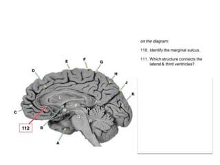
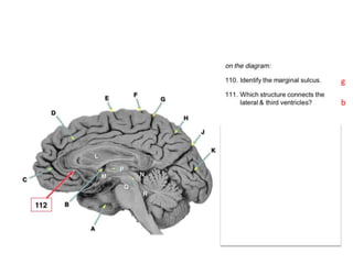
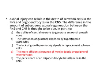
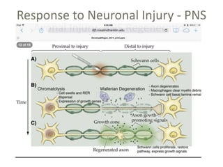
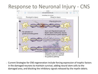
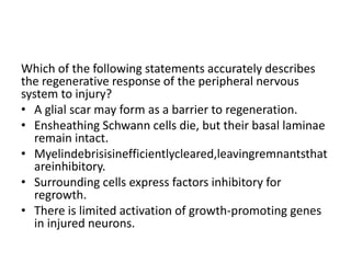
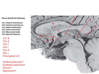
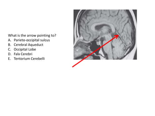
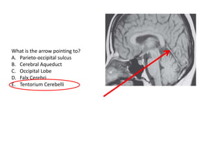
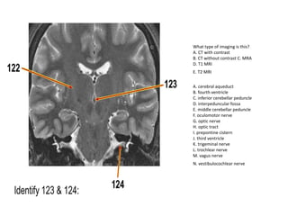
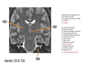
SlideShare a Scribd company logo
NEURO LARGE GROUP 1.1
• What type of image is
this?
a) T1 CT scan
b) T2 CT scan
c) T1 MRI scan
d) DTI
• What type of image is
this?
a) T1 CT scan
b) T2 CT scan
c) T1 MRI scan
d) DTI
T1/T2 are subsets of MRI
scans.
T(wh)one, white matter is
white.
• Which of the following brain imaging
modalities does NOT involve radiation?
a) CT
b) Invasive angiography
c) Nuclear medicine brain flow
d) MRI
e) SPECT
• Which of the following brain imaging
modalities does NOT involve radiation?
a) CT
b) Invasive angiography
c) Nuclear medicine brain flow
d) MRI
e) SPECT
• In CT which of these tissues appears darkest?
A) BLOOD
B) BONE
C) CSF
D) GRAY MATTER
E) WHITE MATTER
• In CT which of these tissues appears darkest?
A) BLOOD
B) BONE
C) CSF
D) GRAY MATTER
E) WHITE MATTER
• Functional MRI is based upon the fact that:
– blood flow is faster in more active brain regions
– Blood oxygenation is higher in more active brain
regions
– Deoxyhemoglobin levels rise in more active brain
regions
– glutamate reuptake is greater in more active brain
regions
– magnetic resonance is higher in more electrically
active neurons
• Which of the following structures is located in
the central nervous system?
a) DRG
b) CN 7
c) CN 3
d) Olfactory bulb
e) Trigeminal ganglion
• Which of the following structures is located in
the central nervous system?
a) DRG
b) CN 7
c) CN 3
d) Olfactory bulb
e) Trigeminal ganglion
• Which of the following structures is correctly
paired with its CNS level?
• A. facial colliculus : telencephalon
• B. mammillary bodies : midbrain
• C. optic chiasm : diencephalon
• D. pyramid : pons
• E. tectum : medulla
• Which of the following structures is correctly
paired with its CNS level?
• A. facial colliculus : telencephalon
• B. mammillary bodies : midbrain
• C. optic chiasm : diencephalon
• D. pyramid : pons
• E. tectum : medulla
Embryonic Origin of CNS
LV
3rd
4th
CA
Which one?
g
b
• A 23 YO male is brought, unconscious, to the
emergency department after a motorcycle accident.
Suspecting head trauma, the physician orders a CT scan
which reveals blood in the CSF, concentrated in the
quadrigeminal cistern. Which of the following
diagnoses is most appropriate for this patient?
a) Epidural hemorrhage
b) extracranial hemorrhage
c) Intraparenchymal hemorrhage
d) Interventricular hemorrhage
e) Subarachnoid hemorrhage
• You successfully administer an epidural anesthetic to a woman in labor.
You originally inserted the needle into the sacral hiatus, and correctly
placed the tip of the needle:
a) between the pia mater and subarachnoid space
b) between the dura mater and the periosteum of a vertebra
c) between the periosteum and the bone of a vertebra
d) into the subarachnoid space of the spinal cord
e) into the central canal of the spinal cord.
• A 20 YO student protestor is brought into the ED for
treatment of head injuries and bleeding after being
struck in the head by a rock. Neurological exam
indicates some disorientation and lethargy. A CT scan
reveals that the patient has suffered an epidural
hematoma, based upon which definitive observation?
a) Blood extending down into sulci
b) Lenticular shape of bleeding that does not cross suture
lines
c) Subfalcine herniation
d) Thin crescent of bleeding separating the dural layers
e) Ventricular distortion or obliteration due to elevated
intracranial pressure.
(not necessarily testable for this test,
but a useful teaching point)
• A 20 YO student protestor is brought into the ED for
treatment of head injuries and bleeding after being
struck in the head by a rock. Neurological exam
indicates some disorientation and lethargy. A CT scan
reveals that the patient has suffered an epidural
hematoma, based upon which definitive observation?
a) Blood extending down into sulci
b) Lenticular shape of bleeding that does not cross suture
lines
c) Subfalcine herniation
d) Thin crescent of bleeding separating the dural layers
e) Ventricular distortion or obliteration due to elevated
intracranial pressure.
• One of your patients has a neurological autoimmune
disease. Cells of her immune system are attempting to
migrate from the bloodstream into her brain. To do
this, an immune system cell must first cross the
endothelium of a brain capillary, and then cross a
second layer made of the cellular processes of
a) Astrocytes
b) Ependymal cells
c) Microglia
d) Neurons
e) Oligodendrocytes
Cell Types - Neurons
Key features: large cell body with nucleus, mitochondria, and Nissl Substance (RER);
dendrites for receiving NT input from neighboring neurons, axon hillock for
integration of dendritic input, axon for conduction of electrical impulses, terminal
branches for release of NT into synaptic cleft.
Several morphologies:
Bipolar: retinal, auditory, and olfactory sensory neurons
Pseudounipolar: peripheral sensory neurons of somatic and visceral nervous system
Multipolar: complex dendritic pattern, most CNS cells and motor neurons
Golgi Type I: Long projection neurons of the CNS
Golgi Type II: Short inhibitory interneurons
Cell Types - Astrocytes
Astrocytes are star-shaped glia with many thin processes radiating from a central cell body.
The processes wrap around capillaries in the CNS, preventing the free diffusion of soluble blood
components into the CNS tissues. This is called the blood brain barrier (BBB).
The area in between astrocyte processes and the capillary wall is called the Virchow-Robins space, and
these spaces are often enlarged in the elderly and people with neurodegenerative disease (inflammation).
Some processes also modulate synaptic activity.
During early development, astrocytes help guide migrating baby neurons to their proper location.
astrocytes that are near brain surfaces extend processes to those surfaces, forming a continuous covering
around the brain, the glia limitans which attaches to a basement membrane.
Laminin and dystroglycan mediate the connection between astrocytes and the basmement membrane
In cobblestone lissencephaly a mutation in one of the above proteins leads to an incomplete glia
limitans.
The Glial Fibrillary Acidic Protein (GFAP) stain is used to visualize astrocytes.
-Gliosis refers to the hypertrophy/hyperplasia of astrocytes in response to disease/injury. This helps
reinforce the integrity of the BBB and initiates a protective inflammatory response. The reactive astrocytes
that develop following an injury are called gemnistocytic astrocytes.
Crest Cells
The neural crest gives rise to the following neural elements (Figure 15):
• Sensory neurons of the cranial and spinal sensory ganglia.
• Postganglionic neurons of the sympathetic and
parasympathetic ganglia.
• Supporting cells of the PNS (Schwann cells and satellite cells
of the ganglia).
The neural crest also gives rise to non-neural elements:
• The cells of the pia mater and arachnoid mater.
• Some branchial cartilage and some cranial mesenchyme.
• Pigment-producing cells of the skin and subcutaneous
tissues (melanocytes).
• Chromaffin tissue (secretory cells of the adrenal medulla).
• The foot processes of CNS astrocytes do all of
the following EXCEPT:
– contribute to the blood-brain barrier
– Form the glia limitans that surrounds the brain
– Surround neuronal cell bodies and dendrites
– surround points of synaptic contact
– wrap around axons to form their myelin sheath
• The foot processes of CNS astrocytes do all of
the following EXCEPT:
– contribute to the blood-brain barrier
– Form the glia limitans that surrounds the brain
– Surround neuronal cell bodies and dendrites
– surround points of synaptic contact
– wrap around axons to form their myelin sheath
Cell Types-Other glia
Oligodendrocytes are egg-shaped glia with a clear cytoplasmic halo surrounding a dark nucleus. They
are abundant in white matter tracts. They extend several cellular processes that wrap several times
around multiple axons, creating myelin sheaths that greatly speed up signal conduction along the
axons.
-Oligodendrocytes do not respond to injury. Any damage to oligodendrocytes (i.e. demyelinating
disease) is irreversible.
Microglia are the resident macrophages of the CNS. They phagocytose particulate matter and cellular
debris, as well as help initiate inflammatory and repair pathways following injury via cytokine release.
The microglia aggressively proliferate in response to local injury, promoting an inflammatory response and clearing any necrotic
tissue.
Ependymal cells are the cells of the choroid plexus which continuously filter the blood and secrete
CSF into the ventricles. Choroid plexus is a specialized version. No basement membrane.
• Which of the following is NOT present in CNS
white matter?
– astrocyte cell bodies
– Microglial cell bodies
– myelin
– neuronal cell bodies
– oligodendrocyte cell bodies
• Which of the following is NOT present in CNS
white matter?
– astrocyte cell bodies
– MicrogliaL cell bodies
– myelin
– neuronal cell bodies
– oligodendrocyte cell bodies
BEWARE THE CNS DEVELOPMENT
LECTURE!
• Deletions or missense mutations of sonic
hedgehog (SHH) can produce incomplete
separation of forebrain structures during
development, including the formation of a single
midline cyclopean eye. This most common
developmental forebrain malformation is called:
– anencephaly
– Dandy-Walkersyndrome
– holoprosencephaly
– lissencephaly
– Waardenburg syndrome
BEWARE THE CNS DEVELOPMENT
LECTURE!
• In contrast to the classical radial migration, wehre
newly generated neuroblasts stay within the brain
regions where they were generated and mature into
variosu types of neurons, newly generated neuroblasts
from the medial and lateral ganglionic eminences
migrate long distances tangentially to mature into what
type of cell?
a) Astrocytes
b) Cholinergic neurons
c) Dopaminergic neurons
d) GABAergic interneurons
e) Glutaminergic neurons
BEWARE THE CNS DEVELOPMENT
LECTURE!
• In contrast to the classical radial migration, where
newly generated neuroblasts stay within the brain
regions where they were generated and mature into
various types of neurons, newly generated neuroblasts
from the medial and lateral ganglionic eminences
migrate long distances tangentially to mature into what
type of cell?
a) Astrocytes
b) Cholinergic neurons
c) Dopaminergic neurons
d) GABAergic interneurons
e) Glutaminergic neurons
Not all migration occurs radially. Recent
work has determined that cortical
interneurons arise from proliferative
centers along the ventricles, the lateral
ganglionic eminence (LGE) and the medial
ganglionic eminence (MGE). Cells from
these two regions migrate throughout the
cerebral cortex. See Box 22F for more
information about this long-distance
migration.!
• During cell fate specification, the concept of
competence refers to:
a) Gradient of signaling molecules that generates
multiple cell types
b) A transcriptional switch that permits a cell to secrete
inducing factors
c) The ability of a cell to maintain an uncommitted
state almost indefinitely
d) The capacity of a cell to respond to inducing factors
e) The signaling that instructs a cell to end its
migraiton.
Understand dorsal/ventral functions
NRG = neureglin (seen in migration slide)
• Axonal injury can result in the death of schwann cells in the
PNS and oligodendrocytes in the CNS. The difference in the
amount of subsequent axonal regeneration between the
PNS and CNS is thought to be due, in part, to:
a) the ability of central neurons to generate an axonal growth
cone
b) The formation of guidance channels by hypertrophic
astrocytes
c) The lack of growth promoting signals in replacement schwann
cells
d) The more efficient clearance of myelin debris by peripheral
macrophages
e) The persistence of an oligodendrocyte basal lamina in the
CNS.
Response to Neuronal Injury - PNS
Response to Neuronal Injury - CNS
Current Strategies for CNS regeneration include forcing expression of trophic factors
in the damaged neurons to maintain survival, adding neural stem cells to the
damaged area, and blocking the inhibitory signals released by the myelin debris.
Which of the following statements accurately describes
the regenerative response of the peripheral nervous
system to injury?
• A glial scar may form as a barrier to regeneration.
• Ensheathing Schwann cells die, but their basal laminae
remain intact.
• Myelindebrisisinefficientlycleared,leavingremnantsthat
areinhibitory.
• Surrounding cells express factors inhibitory for
regrowth.
• There is limited activation of growth-promoting genes
in injured neurons.
Identify these structures
A. Central Sulcus
B. Temporal Lobe
C. Pons
D. Medulla
E. Corpus Callosum
F. Uncus
G. Cerebellum
H. Parieto-occpital sulcus
I. Spinal Cord
J. Midbrain
1.
2.
Identify these structures
A. Central Sulcus
B. Temporal Lobe
C. Pons
D. Medulla
E. Corpus Callosum
F. Uncus
G. Cerebellum
H. Parieto-occpital sulcus (1)
I. Spinal Cord
J. Midbrain (2)
1.
2.
Identify the following:
But first…Plane of Section?
A
B
C
D
Image from: http://www.neuroanatomy.ca/cross_sections/sections_horizontal.html
From Natalie Popenko’s practice question cohort
Mammillary Body
Crus Cerebri aka Cerebral
Peduncle
Inferior horn of
Lateral Ventricle
Cerebral Aqueduct
Image from: http://www.neuroanatomy.ca/cross_sections/sections_horizontal.html
From Natalie Popenko’s practice question cohort
102.
103
104
101.
Numbers 101, 102, and 103
Correspond with which of the
Following Structures?
A. Fornix
B. Pons
C. Optic Nerve
D. Infundibular Stalk
(Infundibulum)
E. Oculomotor Nerve
F. Thalamus
G. Pineal Gland
H. Cingulate Gyrus
I. Central Sulcus
102.
103
104
101.
Numbers 101, 102, and 103
Correspond with which of the
Following Structures?
A. Fornix
B. Pons
C. Optic Nerve
D. Infundibular Stalk
(Infundibulum)
E. Oculomotor Nerve
F. Thalamus
G. Pineal Gland
H. Cingulate Gyrus
I. Central Sulcus
101: H) Cingulate Gyrus
102. D)Infundibular stalk
103: G) Pineal Gland
102.
103
104
101.
Structure 103 is associated
With which level of the CNS?
A. Telencephalon
B. Diencephalon
C. Metencephalon
D. Rhombencephalon
E. Spinal Cord
What the heck is #104?
A. Pituitary tumor
B. Pons
C. Mammillary body
D. Oculomotor Nerve
E. Uncus
Which ventricle is #104
Associated with?
A. Left Lateral Ventricle
B. Third Ventricle
C. Cerebral Aqueduct
D. Fourth Ventricle
Structure 103 is associated
With which level of the CNS?
A. Telencephalon
B. Diencephalon
C. Metencephalon
D. Rhombencephalon
E. Spinal Cord
What the heck is #104?
A. Pituitary tumor
B. Pons
C. Mammillary body
D. Oculomotor Nerve
E. Uncus
Which ventricle is #104
Associated with?
A. Left Lateral Ventricle
B. Third Ventricle
C. Cerebral Aqueduct
D. Fourth Ventricle 102.
103
104
101.
What view of the brain is this (BE SPECIFIC)
Please match the following structures with
The appropriate letters:
Crus Cerebri:
Trochlear Nerve:
Superior Colliculus:
Inferior Colliculus:
Pineal Gland:
C
B
E
A
D
What view of the brain is this (BE SPECIFIC)
Posterior view of the brainstem
Please match the following structures with
The appropriate letters:
Crus Cerebri: A
Trochlear Nerve: D
Superior Colliculus: E
Inferior Colliculus: B
Pineal Gland: C
C
B
E
A
D
C
F
D
B
G
A
H
EI
J
Please identify the following:
111. Anterior Commissure
112. Posterior Commissure
113. Septum Pellucidum
114. Lamina Terminalis
115. Massa Intermedia
116. (Dorsal) Thalamus
C
F
D
B
G
A
H
EI
J
Please identify the following:
111. Anterior Commissure
112. Posterior Commissure
113. Septum Pellucidum
114. Lamina Terminalis
115. Massa Intermedia
116. (Dorsal) Thalamus
111. B
112. A
113. F
114. I
115. H
116. J
Pineal gland is G
Cerebral peduncles?
Cerebellar peduncles?
Tectum?
Tegmentum?
What is the arrow pointing to?
A. Parieto-occipital sulcus
B. Cerebral Aqueduct
C. Occipital Lobe
D. Falx Cerebri
E. Tentorium Cerebelli
What is the arrow pointing to?
A. Parieto-occipital sulcus
B. Cerebral Aqueduct
C. Occipital Lobe
D. Falx Cerebri
E. Tentorium Cerebelli
A. cerebral aqueduct
B. fourth ventricle
C. inferior cerebellar peduncle
D. interpeduncular fossa
E. middle cerebellar peduncle
F. oculomotor nerve
G. optic nerve
H. optic tract
I. prepontine cistern
J. third ventricle
K. trigeminal nerve
L. trochlear nerve
M. vagus nerve
N. vestibulocochlear nerve
What type of imaging is this?
A. CT with contrast
B. CT without contrast C. MRA
D. T1 MRI
E. T2 MRI
• A multipolar neuron resides in the gray matter of
the spinal cord, at T4. In order to send a message
to its target neuron, which resides in the gray
matter of S2, it must send a long efferent fiber
through the spinal cord. Which statement is NOT
correct?
a) It conducts an action potential
b) It contains intermediate filaments
c) It contains microtubules
d) It contains rough ER
e) It is myelinated
END OF SESSION 1
• Charcot Marie-Tooth disease results from a
defect in:
a) Myelination of peripheral axons
b) Nicotinic acetylcholine receptors
c) SNARE proteins
d) Synaptic vesicle recycling
e) Voltage-gated calcium channels.
• Charcot Marie-Tooth disease results from a
defect in:
a) Myelination of peripheral axons
b) Nicotinic acetylcholine receptors
c) SNARE proteins
d) Synaptic vesicle recycling
e) Voltage-gated calcium channels.
(can affect motor and sensory nerves of PNS)
• Review Chemical Synaptic Transmission
– Slower than electrical transmission
– How to Terminate transmitter action:
• Diffusion
• Reuptake
– Transporters (drug targets, cocaine affects dopamine
reuptake)
• Degradation
• Which statement about small molecular
(“classical”) transmitters is true?
– They do not activate G-protein coupled receptors.
– Substance P is a member of this family.
– They are synthesized in the cell body.
– They are synthesized from larger precursor
proteins.
– For many of these, synaptic action is terminated
by transporter proteins.
• Which of the following correctly describes the
relationship between the size of the EPP and the
MEPP at the neuromuscular junction?
– In low calcium solution, the MEPP will be larger than
the EPP.
– The EPP cannot be more than twice the magnitude of
the MEPP.
– The EPP is always a whole number multiple of the
MEPP.
– The EPP is always the same size as the MEPP.
– The MEPP is always larger than the EPP.
MYASTHENIA GRAVIS
• Muscle weakness
– Drooping eyelids (ptosis)
– Double vision (diplopia)
– Facial expression, chewing, swallowing
– TRIAD! (think uncal, tonsillar herniation)
• Treat with neostigmine (what kind of molecule
is this?)
MYASTHENIA GRAVIS
• Muscle weakness
– Drooping eyelids (ptosis)
– Double vision (diplopia)
– Facial expression, chewing, swallowing
– TRIAD! (think uncal, tonsillar herniation)
• Treat with
– Immunosuppressants
– Thymectomy
– plasmapheresis
– neostigmine (what kind of molecule is this?)
acetylcholinesterase inhibitor.
• What type of receptor is being affected?
MYASTHENIA GRAVIS
• Muscle weakness
– Drooping eyelids (ptosis)
– Double vision (diplopia)
– Facial expression, chewing, swallowing
– TRIAD! (think uncal, tonsillar herniation)
• Treat with neostigmine (what kind of molecule is
this?) acetylcholinesterase inhibitor.
• What type of receptor is being affected?
– Antibodies are binding to NICOTINIC Ach receptor.
• Dendritic spines are expected to contain:
– acetylcholinesterase
– dopamine transporters
– glutamate receptors
– SNARE proteins
– synaptic vesicles
• Dendritic spines are expected to contain:
– acetylcholinesterase
– dopamine transporters
– glutamate receptors
– SNARE proteins
– synaptic vesicles
• Neuromodulators influence the way a cell will respond
to another transmitter that produces an EPSP or IPSP.
In the example considered in class (and many other
instances), the neuromodulator does this by activating
a signaling pathway that alters the function of:
• synaptic vesicle proteins
• A transcription factor
• A voltage-gated ion channel
• an enzyme that synthesizes a neurotransmitter
• a plasma membrane transporter
MYASTHENIA SYNDROMES
• POSTSYNAPTIC:
– Myasthenia gravis
• Presynaptic:
– Congenital myasthenic syndromes
• Insufficient Ach release (defects in vesicle recycling)
– Lambert-Eaton myasthenic syndrome
• Autoimmune, response to voltage gated Ca2+ channels
• Associated with small cell lung cancer
TOXINS
• Affect presynaptic terminal:
– A-latrotoxin (black widow venom)
• Ca2+ release
• massive transmitter release
– Clostridium (makes botulinim and tetanus toxins)
• Proteases that cleave SNARE proteins
• Prevent release
• Which statement about botulinum toxin is
NOT true:
– It disrupts synaptic vesicle fusion.
– It is a protease that cleaves SNARE proteins.
– It is approved by the FDA for several uses.
– It is taken up by inhibitory interneurons in the
spinal cord.
– It results in paralysis of neuromuscular synapses.
Neuro 1.1

More Related Content

What's hot

Neuroimaging basics
Neuroimaging basicsNeuroimaging basics
Neuroimaging basics
Sujit Kumar Kar
 
CT brain in emergencies ... for ER doctors
CT brain in emergencies ... for ER doctorsCT brain in emergencies ... for ER doctors
CT brain in emergencies ... for ER doctors
Mohamed Shaaban
 
Epilepsy getting the most out of neuroimaging 2019
Epilepsy getting the most out of neuroimaging 2019Epilepsy getting the most out of neuroimaging 2019
Epilepsy getting the most out of neuroimaging 2019
Felice D'Arco
 
CT Brain
CT BrainCT Brain
CT Brain
Ajay Nagisetti
 
Presentation1.pptx, radiological imaging of peri natal acute ischemia and hyp...
Presentation1.pptx, radiological imaging of peri natal acute ischemia and hyp...Presentation1.pptx, radiological imaging of peri natal acute ischemia and hyp...
Presentation1.pptx, radiological imaging of peri natal acute ischemia and hyp...
Abdellah Nazeer
 
Diagnostic Neuroradiology
Diagnostic NeuroradiologyDiagnostic Neuroradiology
Diagnostic Neuroradiology
Sumit Prajapati
 
Brain ct and mri in icu
Brain ct and mri in icuBrain ct and mri in icu
Brain ct and mri in icu
samirelansary
 
101 ct neuroimaging
101 ct neuroimaging101 ct neuroimaging
101 ct neuroimaging
Ahmad Shahir
 
Neuroradiology for the ED
Neuroradiology for the EDNeuroradiology for the ED
Neuroradiology for the ED
SCGH ED CME
 
Diagnostic Imaging of Bilateral Abnormalities of the Basal Ganglia & Thalamus
Diagnostic Imaging of Bilateral Abnormalities of the Basal Ganglia & ThalamusDiagnostic Imaging of Bilateral Abnormalities of the Basal Ganglia & Thalamus
Diagnostic Imaging of Bilateral Abnormalities of the Basal Ganglia & Thalamus
Mohamed M.A. Zaitoun
 
Radiology of central nervous system
Radiology of central nervous systemRadiology of central nervous system
Radiology of central nervous system
WEEKLYMEDIC
 
Ct and mri interpretation
Ct and mri interpretationCt and mri interpretation
Ct and mri interpretation
Dr. Mohamed Maged Kharabish
 
Basics of ct mri
Basics of ct mriBasics of ct mri
Basics of ct mri
Omkar Singh
 
recent investigation in epilepsy
recent investigation in epilepsyrecent investigation in epilepsy
recent investigation in epilepsy
Osama Ragab
 
Ct brain presentation
Ct brain presentationCt brain presentation
Ct brain presentation
Qamar Zaman
 
BASICS OF MRI
BASICS OF MRIBASICS OF MRI
BASICS OF MRI
Krishna Kiran Karanth
 
Integrated Diagnostics – A Unique Epilepsy Approach
Integrated Diagnostics – A Unique Epilepsy ApproachIntegrated Diagnostics – A Unique Epilepsy Approach
Integrated Diagnostics – A Unique Epilepsy Approach
Neurokrish - the neuropsychiatry centre
 
Brain tumours seminar by dr pushpendra patel
Brain tumours seminar by dr pushpendra patelBrain tumours seminar by dr pushpendra patel
Brain tumours seminar by dr pushpendra patel
DrPushpendraPatel
 
Cns path congenital, edema
Cns path congenital, edemaCns path congenital, edema
Cns path congenital, edema
raj kumar
 
12 b. ct brain
12 b. ct brain12 b. ct brain
12 b. ct brain
Yerragunta Tirumal
 

What's hot (20)

Neuroimaging basics
Neuroimaging basicsNeuroimaging basics
Neuroimaging basics
 
CT brain in emergencies ... for ER doctors
CT brain in emergencies ... for ER doctorsCT brain in emergencies ... for ER doctors
CT brain in emergencies ... for ER doctors
 
Epilepsy getting the most out of neuroimaging 2019
Epilepsy getting the most out of neuroimaging 2019Epilepsy getting the most out of neuroimaging 2019
Epilepsy getting the most out of neuroimaging 2019
 
CT Brain
CT BrainCT Brain
CT Brain
 
Presentation1.pptx, radiological imaging of peri natal acute ischemia and hyp...
Presentation1.pptx, radiological imaging of peri natal acute ischemia and hyp...Presentation1.pptx, radiological imaging of peri natal acute ischemia and hyp...
Presentation1.pptx, radiological imaging of peri natal acute ischemia and hyp...
 
Diagnostic Neuroradiology
Diagnostic NeuroradiologyDiagnostic Neuroradiology
Diagnostic Neuroradiology
 
Brain ct and mri in icu
Brain ct and mri in icuBrain ct and mri in icu
Brain ct and mri in icu
 
101 ct neuroimaging
101 ct neuroimaging101 ct neuroimaging
101 ct neuroimaging
 
Neuroradiology for the ED
Neuroradiology for the EDNeuroradiology for the ED
Neuroradiology for the ED
 
Diagnostic Imaging of Bilateral Abnormalities of the Basal Ganglia & Thalamus
Diagnostic Imaging of Bilateral Abnormalities of the Basal Ganglia & ThalamusDiagnostic Imaging of Bilateral Abnormalities of the Basal Ganglia & Thalamus
Diagnostic Imaging of Bilateral Abnormalities of the Basal Ganglia & Thalamus
 
Radiology of central nervous system
Radiology of central nervous systemRadiology of central nervous system
Radiology of central nervous system
 
Ct and mri interpretation
Ct and mri interpretationCt and mri interpretation
Ct and mri interpretation
 
Basics of ct mri
Basics of ct mriBasics of ct mri
Basics of ct mri
 
recent investigation in epilepsy
recent investigation in epilepsyrecent investigation in epilepsy
recent investigation in epilepsy
 
Ct brain presentation
Ct brain presentationCt brain presentation
Ct brain presentation
 
BASICS OF MRI
BASICS OF MRIBASICS OF MRI
BASICS OF MRI
 
Integrated Diagnostics – A Unique Epilepsy Approach
Integrated Diagnostics – A Unique Epilepsy ApproachIntegrated Diagnostics – A Unique Epilepsy Approach
Integrated Diagnostics – A Unique Epilepsy Approach
 
Brain tumours seminar by dr pushpendra patel
Brain tumours seminar by dr pushpendra patelBrain tumours seminar by dr pushpendra patel
Brain tumours seminar by dr pushpendra patel
 
Cns path congenital, edema
Cns path congenital, edemaCns path congenital, edema
Cns path congenital, edema
 
12 b. ct brain
12 b. ct brain12 b. ct brain
12 b. ct brain
 

Viewers also liked

Vijay Achar_11+ Yrs of Exp in Testing+TM
Vijay Achar_11+ Yrs of Exp in Testing+TMVijay Achar_11+ Yrs of Exp in Testing+TM
Vijay Achar_11+ Yrs of Exp in Testing+TM
Achari Vijaya
 
Càtaleg productes.odt 2
Càtaleg productes.odt 2Càtaleg productes.odt 2
Càtaleg productes.odt 2
doloreteslore
 
Zno 2015
Zno 2015Zno 2015
Food Drive
Food DriveFood Drive
Food Drive
John Albert
 
Michele_Brun-Final
Michele_Brun-FinalMichele_Brun-Final
Michele_Brun-Final
Michele Brun
 
Indepencia del peru
Indepencia del peruIndepencia del peru
Indepencia del peru
SofiaAranguren
 
Easy Serve, an Online Services Marketplace Startup in India
Easy Serve, an Online Services Marketplace Startup in IndiaEasy Serve, an Online Services Marketplace Startup in India
Easy Serve, an Online Services Marketplace Startup in India
Upamanyu Acharya
 
Verbrevf3 (2)
Verbrevf3 (2)Verbrevf3 (2)
Verbrevf3 (2)
leexa mishaal
 
Domain registration and protection in vietnam
Domain registration and protection in vietnamDomain registration and protection in vietnam
Domain registration and protection in vietnam
PATON CO., LTD.
 

Viewers also liked (9)

Vijay Achar_11+ Yrs of Exp in Testing+TM
Vijay Achar_11+ Yrs of Exp in Testing+TMVijay Achar_11+ Yrs of Exp in Testing+TM
Vijay Achar_11+ Yrs of Exp in Testing+TM
 
Càtaleg productes.odt 2
Càtaleg productes.odt 2Càtaleg productes.odt 2
Càtaleg productes.odt 2
 
Zno 2015
Zno 2015Zno 2015
Zno 2015
 
Food Drive
Food DriveFood Drive
Food Drive
 
Michele_Brun-Final
Michele_Brun-FinalMichele_Brun-Final
Michele_Brun-Final
 
Indepencia del peru
Indepencia del peruIndepencia del peru
Indepencia del peru
 
Easy Serve, an Online Services Marketplace Startup in India
Easy Serve, an Online Services Marketplace Startup in IndiaEasy Serve, an Online Services Marketplace Startup in India
Easy Serve, an Online Services Marketplace Startup in India
 
Verbrevf3 (2)
Verbrevf3 (2)Verbrevf3 (2)
Verbrevf3 (2)
 
Domain registration and protection in vietnam
Domain registration and protection in vietnamDomain registration and protection in vietnam
Domain registration and protection in vietnam
 

Similar to Neuro 1.1

Meningitis
MeningitisMeningitis
Meningitis
Anil Regmi
 
Revision of neuroanatomy
Revision of neuroanatomyRevision of neuroanatomy
Revision of neuroanatomy
Abdul Ansari
 
ESSENTIALS_OF_CT_BRAIN_For_Undergraduate.pptx
ESSENTIALS_OF_CT_BRAIN_For_Undergraduate.pptxESSENTIALS_OF_CT_BRAIN_For_Undergraduate.pptx
ESSENTIALS_OF_CT_BRAIN_For_Undergraduate.pptx
ssuser39be96
 
Essentials of CT brain (For Undergraduates)
Essentials of CT brain (For Undergraduates)Essentials of CT brain (For Undergraduates)
Essentials of CT brain (For Undergraduates)
Department of Health & Family Welfare, Government of West Bengal
 
Neuroimaging in Psychiatry
Neuroimaging in PsychiatryNeuroimaging in Psychiatry
Neuroimaging in Psychiatry
Enoch R G
 
RAH Med 4 MHU - Brain CT 1
RAH Med 4 MHU - Brain CT 1RAH Med 4 MHU - Brain CT 1
RAH Med 4 MHU - Brain CT 1
Luke Oakden-Rayner
 
MRI evaluation of developing brain and its role in congenital brain malformat...
MRI evaluation of developing brain and its role in congenital brain malformat...MRI evaluation of developing brain and its role in congenital brain malformat...
MRI evaluation of developing brain and its role in congenital brain malformat...
Shaurya Agarwal
 
Ct scan brain lecture by rashimul haque rimon
Ct scan brain lecture by rashimul haque rimonCt scan brain lecture by rashimul haque rimon
Ct scan brain lecture by rashimul haque rimon
Rashimul haque Rimon
 
Neonatal MRI Brain
Neonatal MRI BrainNeonatal MRI Brain
Neonatal MRI Brain
Vinayak Kodur
 
Neurosurgery 1.pptx
Neurosurgery 1.pptxNeurosurgery 1.pptx
Neurosurgery 1.pptx
joendesh
 
pediatric neuroradiology essentials
pediatric neuroradiology essentialspediatric neuroradiology essentials
pediatric neuroradiology essentials
dr jyoti prajapati
 
Cortical dysplasia and epilepsy
Cortical dysplasia and epilepsyCortical dysplasia and epilepsy
Cortical dysplasia and epilepsy
Dr-Ashraf Abdou
 
Head injury and CNS infection.pdf
Head injury and CNS infection.pdfHead injury and CNS infection.pdf
Head injury and CNS infection.pdf
gp9dprrjvx
 
Decompressive craniectomy in Traumatic Brain Injury
Decompressive craniectomy in Traumatic Brain InjuryDecompressive craniectomy in Traumatic Brain Injury
Decompressive craniectomy in Traumatic Brain Injury
joemdas
 
Cns infections perfect
Cns infections perfectCns infections perfect
Cns infections perfect
Ali Jiwani
 
MRI BRAIN(AIIMS DELHI).pptx
MRI BRAIN(AIIMS DELHI).pptxMRI BRAIN(AIIMS DELHI).pptx
MRI BRAIN(AIIMS DELHI).pptx
Gyandeep28
 
USMLE GENERAL EMBRYOLOGY 012 Fourth week development A embryo .pdf
USMLE   GENERAL EMBRYOLOGY  012 Fourth week development A embryo .pdfUSMLE   GENERAL EMBRYOLOGY  012 Fourth week development A embryo .pdf
USMLE GENERAL EMBRYOLOGY 012 Fourth week development A embryo .pdf
AHMED ASHOUR
 
Peripheral nerve blocks
Peripheral nerve blocksPeripheral nerve blocks
Peripheral nerve blocks
sarmistha panigrahi
 
BMS2-K13 Pemeriksaan Radiologi pada Sistem Saraf.pptx
BMS2-K13 Pemeriksaan Radiologi pada Sistem Saraf.pptxBMS2-K13 Pemeriksaan Radiologi pada Sistem Saraf.pptx
BMS2-K13 Pemeriksaan Radiologi pada Sistem Saraf.pptx
ssuser144901
 
Bedside Ultrasound in Neurosurgery Part 1/3
Bedside Ultrasound in Neurosurgery Part 1/3Bedside Ultrasound in Neurosurgery Part 1/3
Bedside Ultrasound in Neurosurgery Part 1/3
Liew Boon Seng
 

Similar to Neuro 1.1 (20)

Meningitis
MeningitisMeningitis
Meningitis
 
Revision of neuroanatomy
Revision of neuroanatomyRevision of neuroanatomy
Revision of neuroanatomy
 
ESSENTIALS_OF_CT_BRAIN_For_Undergraduate.pptx
ESSENTIALS_OF_CT_BRAIN_For_Undergraduate.pptxESSENTIALS_OF_CT_BRAIN_For_Undergraduate.pptx
ESSENTIALS_OF_CT_BRAIN_For_Undergraduate.pptx
 
Essentials of CT brain (For Undergraduates)
Essentials of CT brain (For Undergraduates)Essentials of CT brain (For Undergraduates)
Essentials of CT brain (For Undergraduates)
 
Neuroimaging in Psychiatry
Neuroimaging in PsychiatryNeuroimaging in Psychiatry
Neuroimaging in Psychiatry
 
RAH Med 4 MHU - Brain CT 1
RAH Med 4 MHU - Brain CT 1RAH Med 4 MHU - Brain CT 1
RAH Med 4 MHU - Brain CT 1
 
MRI evaluation of developing brain and its role in congenital brain malformat...
MRI evaluation of developing brain and its role in congenital brain malformat...MRI evaluation of developing brain and its role in congenital brain malformat...
MRI evaluation of developing brain and its role in congenital brain malformat...
 
Ct scan brain lecture by rashimul haque rimon
Ct scan brain lecture by rashimul haque rimonCt scan brain lecture by rashimul haque rimon
Ct scan brain lecture by rashimul haque rimon
 
Neonatal MRI Brain
Neonatal MRI BrainNeonatal MRI Brain
Neonatal MRI Brain
 
Neurosurgery 1.pptx
Neurosurgery 1.pptxNeurosurgery 1.pptx
Neurosurgery 1.pptx
 
pediatric neuroradiology essentials
pediatric neuroradiology essentialspediatric neuroradiology essentials
pediatric neuroradiology essentials
 
Cortical dysplasia and epilepsy
Cortical dysplasia and epilepsyCortical dysplasia and epilepsy
Cortical dysplasia and epilepsy
 
Head injury and CNS infection.pdf
Head injury and CNS infection.pdfHead injury and CNS infection.pdf
Head injury and CNS infection.pdf
 
Decompressive craniectomy in Traumatic Brain Injury
Decompressive craniectomy in Traumatic Brain InjuryDecompressive craniectomy in Traumatic Brain Injury
Decompressive craniectomy in Traumatic Brain Injury
 
Cns infections perfect
Cns infections perfectCns infections perfect
Cns infections perfect
 
MRI BRAIN(AIIMS DELHI).pptx
MRI BRAIN(AIIMS DELHI).pptxMRI BRAIN(AIIMS DELHI).pptx
MRI BRAIN(AIIMS DELHI).pptx
 
USMLE GENERAL EMBRYOLOGY 012 Fourth week development A embryo .pdf
USMLE   GENERAL EMBRYOLOGY  012 Fourth week development A embryo .pdfUSMLE   GENERAL EMBRYOLOGY  012 Fourth week development A embryo .pdf
USMLE GENERAL EMBRYOLOGY 012 Fourth week development A embryo .pdf
 
Peripheral nerve blocks
Peripheral nerve blocksPeripheral nerve blocks
Peripheral nerve blocks
 
BMS2-K13 Pemeriksaan Radiologi pada Sistem Saraf.pptx
BMS2-K13 Pemeriksaan Radiologi pada Sistem Saraf.pptxBMS2-K13 Pemeriksaan Radiologi pada Sistem Saraf.pptx
BMS2-K13 Pemeriksaan Radiologi pada Sistem Saraf.pptx
 
Bedside Ultrasound in Neurosurgery Part 1/3
Bedside Ultrasound in Neurosurgery Part 1/3Bedside Ultrasound in Neurosurgery Part 1/3
Bedside Ultrasound in Neurosurgery Part 1/3
 

Recently uploaded

Pests of Storage_Identification_Dr.UPR.pdf
Pests of Storage_Identification_Dr.UPR.pdfPests of Storage_Identification_Dr.UPR.pdf
Pests of Storage_Identification_Dr.UPR.pdf
PirithiRaju
 
Mending Clothing to Support Sustainable Fashion_CIMaR 2024.pdf
Mending Clothing to Support Sustainable Fashion_CIMaR 2024.pdfMending Clothing to Support Sustainable Fashion_CIMaR 2024.pdf
Mending Clothing to Support Sustainable Fashion_CIMaR 2024.pdf
Selcen Ozturkcan
 
11.1 Role of physical biological in deterioration of grains.pdf
11.1 Role of physical biological in deterioration of grains.pdf11.1 Role of physical biological in deterioration of grains.pdf
11.1 Role of physical biological in deterioration of grains.pdf
PirithiRaju
 
8.Isolation of pure cultures and preservation of cultures.pdf
8.Isolation of pure cultures and preservation of cultures.pdf8.Isolation of pure cultures and preservation of cultures.pdf
8.Isolation of pure cultures and preservation of cultures.pdf
by6843629
 
The cost of acquiring information by natural selection
The cost of acquiring information by natural selectionThe cost of acquiring information by natural selection
The cost of acquiring information by natural selection
Carl Bergstrom
 
Eukaryotic Transcription Presentation.pptx
Eukaryotic Transcription Presentation.pptxEukaryotic Transcription Presentation.pptx
Eukaryotic Transcription Presentation.pptx
RitabrataSarkar3
 
The binding of cosmological structures by massless topological defects
The binding of cosmological structures by massless topological defectsThe binding of cosmological structures by massless topological defects
The binding of cosmological structures by massless topological defects
Sérgio Sacani
 
NuGOweek 2024 Ghent programme overview flyer
NuGOweek 2024 Ghent programme overview flyerNuGOweek 2024 Ghent programme overview flyer
NuGOweek 2024 Ghent programme overview flyer
pablovgd
 
Direct Seeded Rice - Climate Smart Agriculture
Direct Seeded Rice - Climate Smart AgricultureDirect Seeded Rice - Climate Smart Agriculture
Direct Seeded Rice - Climate Smart Agriculture
International Food Policy Research Institute- South Asia Office
 
Compexometric titration/Chelatorphy titration/chelating titration
Compexometric titration/Chelatorphy titration/chelating titrationCompexometric titration/Chelatorphy titration/chelating titration
Compexometric titration/Chelatorphy titration/chelating titration
Vandana Devesh Sharma
 
ESR spectroscopy in liquid food and beverages.pptx
ESR spectroscopy in liquid food and beverages.pptxESR spectroscopy in liquid food and beverages.pptx
ESR spectroscopy in liquid food and beverages.pptx
PRIYANKA PATEL
 
在线办理(salfor毕业证书)索尔福德大学毕业证毕业完成信一模一样
在线办理(salfor毕业证书)索尔福德大学毕业证毕业完成信一模一样在线办理(salfor毕业证书)索尔福德大学毕业证毕业完成信一模一样
在线办理(salfor毕业证书)索尔福德大学毕业证毕业完成信一模一样
vluwdy49
 
molar-distalization in orthodontics-seminar.pptx
molar-distalization in orthodontics-seminar.pptxmolar-distalization in orthodontics-seminar.pptx
molar-distalization in orthodontics-seminar.pptx
Anagha Prasad
 
Sharlene Leurig - Enabling Onsite Water Use with Net Zero Water
Sharlene Leurig - Enabling Onsite Water Use with Net Zero WaterSharlene Leurig - Enabling Onsite Water Use with Net Zero Water
Sharlene Leurig - Enabling Onsite Water Use with Net Zero Water
Texas Alliance of Groundwater Districts
 
SAR of Medicinal Chemistry 1st by dk.pdf
SAR of Medicinal Chemistry 1st by dk.pdfSAR of Medicinal Chemistry 1st by dk.pdf
SAR of Medicinal Chemistry 1st by dk.pdf
KrushnaDarade1
 
Authoring a personal GPT for your research and practice: How we created the Q...
Authoring a personal GPT for your research and practice: How we created the Q...Authoring a personal GPT for your research and practice: How we created the Q...
Authoring a personal GPT for your research and practice: How we created the Q...
Leonel Morgado
 
Applied Science: Thermodynamics, Laws & Methodology.pdf
Applied Science: Thermodynamics, Laws & Methodology.pdfApplied Science: Thermodynamics, Laws & Methodology.pdf
Applied Science: Thermodynamics, Laws & Methodology.pdf
University of Hertfordshire
 
20240520 Planning a Circuit Simulator in JavaScript.pptx
20240520 Planning a Circuit Simulator in JavaScript.pptx20240520 Planning a Circuit Simulator in JavaScript.pptx
20240520 Planning a Circuit Simulator in JavaScript.pptx
Sharon Liu
 
GBSN - Biochemistry (Unit 6) Chemistry of Proteins
GBSN - Biochemistry (Unit 6) Chemistry of ProteinsGBSN - Biochemistry (Unit 6) Chemistry of Proteins
GBSN - Biochemistry (Unit 6) Chemistry of Proteins
Areesha Ahmad
 
Immersive Learning That Works: Research Grounding and Paths Forward
Immersive Learning That Works: Research Grounding and Paths ForwardImmersive Learning That Works: Research Grounding and Paths Forward
Immersive Learning That Works: Research Grounding and Paths Forward
Leonel Morgado
 

Recently uploaded (20)

Pests of Storage_Identification_Dr.UPR.pdf
Pests of Storage_Identification_Dr.UPR.pdfPests of Storage_Identification_Dr.UPR.pdf
Pests of Storage_Identification_Dr.UPR.pdf
 
Mending Clothing to Support Sustainable Fashion_CIMaR 2024.pdf
Mending Clothing to Support Sustainable Fashion_CIMaR 2024.pdfMending Clothing to Support Sustainable Fashion_CIMaR 2024.pdf
Mending Clothing to Support Sustainable Fashion_CIMaR 2024.pdf
 
11.1 Role of physical biological in deterioration of grains.pdf
11.1 Role of physical biological in deterioration of grains.pdf11.1 Role of physical biological in deterioration of grains.pdf
11.1 Role of physical biological in deterioration of grains.pdf
 
8.Isolation of pure cultures and preservation of cultures.pdf
8.Isolation of pure cultures and preservation of cultures.pdf8.Isolation of pure cultures and preservation of cultures.pdf
8.Isolation of pure cultures and preservation of cultures.pdf
 
The cost of acquiring information by natural selection
The cost of acquiring information by natural selectionThe cost of acquiring information by natural selection
The cost of acquiring information by natural selection
 
Eukaryotic Transcription Presentation.pptx
Eukaryotic Transcription Presentation.pptxEukaryotic Transcription Presentation.pptx
Eukaryotic Transcription Presentation.pptx
 
The binding of cosmological structures by massless topological defects
The binding of cosmological structures by massless topological defectsThe binding of cosmological structures by massless topological defects
The binding of cosmological structures by massless topological defects
 
NuGOweek 2024 Ghent programme overview flyer
NuGOweek 2024 Ghent programme overview flyerNuGOweek 2024 Ghent programme overview flyer
NuGOweek 2024 Ghent programme overview flyer
 
Direct Seeded Rice - Climate Smart Agriculture
Direct Seeded Rice - Climate Smart AgricultureDirect Seeded Rice - Climate Smart Agriculture
Direct Seeded Rice - Climate Smart Agriculture
 
Compexometric titration/Chelatorphy titration/chelating titration
Compexometric titration/Chelatorphy titration/chelating titrationCompexometric titration/Chelatorphy titration/chelating titration
Compexometric titration/Chelatorphy titration/chelating titration
 
ESR spectroscopy in liquid food and beverages.pptx
ESR spectroscopy in liquid food and beverages.pptxESR spectroscopy in liquid food and beverages.pptx
ESR spectroscopy in liquid food and beverages.pptx
 
在线办理(salfor毕业证书)索尔福德大学毕业证毕业完成信一模一样
在线办理(salfor毕业证书)索尔福德大学毕业证毕业完成信一模一样在线办理(salfor毕业证书)索尔福德大学毕业证毕业完成信一模一样
在线办理(salfor毕业证书)索尔福德大学毕业证毕业完成信一模一样
 
molar-distalization in orthodontics-seminar.pptx
molar-distalization in orthodontics-seminar.pptxmolar-distalization in orthodontics-seminar.pptx
molar-distalization in orthodontics-seminar.pptx
 
Sharlene Leurig - Enabling Onsite Water Use with Net Zero Water
Sharlene Leurig - Enabling Onsite Water Use with Net Zero WaterSharlene Leurig - Enabling Onsite Water Use with Net Zero Water
Sharlene Leurig - Enabling Onsite Water Use with Net Zero Water
 
SAR of Medicinal Chemistry 1st by dk.pdf
SAR of Medicinal Chemistry 1st by dk.pdfSAR of Medicinal Chemistry 1st by dk.pdf
SAR of Medicinal Chemistry 1st by dk.pdf
 
Authoring a personal GPT for your research and practice: How we created the Q...
Authoring a personal GPT for your research and practice: How we created the Q...Authoring a personal GPT for your research and practice: How we created the Q...
Authoring a personal GPT for your research and practice: How we created the Q...
 
Applied Science: Thermodynamics, Laws & Methodology.pdf
Applied Science: Thermodynamics, Laws & Methodology.pdfApplied Science: Thermodynamics, Laws & Methodology.pdf
Applied Science: Thermodynamics, Laws & Methodology.pdf
 
20240520 Planning a Circuit Simulator in JavaScript.pptx
20240520 Planning a Circuit Simulator in JavaScript.pptx20240520 Planning a Circuit Simulator in JavaScript.pptx
20240520 Planning a Circuit Simulator in JavaScript.pptx
 
GBSN - Biochemistry (Unit 6) Chemistry of Proteins
GBSN - Biochemistry (Unit 6) Chemistry of ProteinsGBSN - Biochemistry (Unit 6) Chemistry of Proteins
GBSN - Biochemistry (Unit 6) Chemistry of Proteins
 
Immersive Learning That Works: Research Grounding and Paths Forward
Immersive Learning That Works: Research Grounding and Paths ForwardImmersive Learning That Works: Research Grounding and Paths Forward
Immersive Learning That Works: Research Grounding and Paths Forward
 

Neuro 1.1

  • 2.
  • 3. • What type of image is this? a) T1 CT scan b) T2 CT scan c) T1 MRI scan d) DTI
  • 4. • What type of image is this? a) T1 CT scan b) T2 CT scan c) T1 MRI scan d) DTI T1/T2 are subsets of MRI scans. T(wh)one, white matter is white.
  • 5. • Which of the following brain imaging modalities does NOT involve radiation? a) CT b) Invasive angiography c) Nuclear medicine brain flow d) MRI e) SPECT
  • 6. • Which of the following brain imaging modalities does NOT involve radiation? a) CT b) Invasive angiography c) Nuclear medicine brain flow d) MRI e) SPECT
  • 7. • In CT which of these tissues appears darkest? A) BLOOD B) BONE C) CSF D) GRAY MATTER E) WHITE MATTER
  • 8. • In CT which of these tissues appears darkest? A) BLOOD B) BONE C) CSF D) GRAY MATTER E) WHITE MATTER
  • 9. • Functional MRI is based upon the fact that: – blood flow is faster in more active brain regions – Blood oxygenation is higher in more active brain regions – Deoxyhemoglobin levels rise in more active brain regions – glutamate reuptake is greater in more active brain regions – magnetic resonance is higher in more electrically active neurons
  • 10. • Functional MRI is based upon the fact that: – blood flow is faster in more active brain regions – Blood oxygenation is higher in more active brain regions – Deoxyhemoglobin levels rise in more active brain regions – glutamate reuptake is greater in more active brain regions – magnetic resonance is higher in more electrically active neurons
  • 11. • Which of the following structures is located in the central nervous system? a) DRG b) CN 7 c) CN 3 d) Olfactory bulb e) Trigeminal ganglion
  • 12. • Which of the following structures is located in the central nervous system? a) DRG b) CN 7 c) CN 3 d) Olfactory bulb e) Trigeminal ganglion
  • 13.
  • 14. • Which of the following structures is correctly paired with its CNS level? • A. facial colliculus : telencephalon • B. mammillary bodies : midbrain • C. optic chiasm : diencephalon • D. pyramid : pons • E. tectum : medulla
  • 15. • Which of the following structures is correctly paired with its CNS level? • A. facial colliculus : telencephalon • B. mammillary bodies : midbrain • C. optic chiasm : diencephalon • D. pyramid : pons • E. tectum : medulla
  • 16. Embryonic Origin of CNS LV 3rd 4th CA
  • 17.
  • 18.
  • 19.
  • 20.
  • 22.
  • 23. g b
  • 24. • A 23 YO male is brought, unconscious, to the emergency department after a motorcycle accident. Suspecting head trauma, the physician orders a CT scan which reveals blood in the CSF, concentrated in the quadrigeminal cistern. Which of the following diagnoses is most appropriate for this patient? a) Epidural hemorrhage b) extracranial hemorrhage c) Intraparenchymal hemorrhage d) Interventricular hemorrhage e) Subarachnoid hemorrhage
  • 25. • A 23 YO male is brought, unconscious, to the emergency department after a motorcycle accident. Suspecting head trauma, the physician orders a CT scan which reveals blood in the CSF, concentrated in the quadrigeminal cistern. Which of the following diagnoses is most appropriate for this patient? a) Epidural hemorrhage b) extracranial hemorrhage c) Intraparenchymal hemorrhage d) Interventricular hemorrhage e) Subarachnoid hemorrhage
  • 26.
  • 27.
  • 28. • You successfully administer an epidural anesthetic to a woman in labor. You originally inserted the needle into the sacral hiatus, and correctly placed the tip of the needle: a) between the pia mater and subarachnoid space b) between the dura mater and the periosteum of a vertebra c) between the periosteum and the bone of a vertebra d) into the subarachnoid space of the spinal cord e) into the central canal of the spinal cord.
  • 29. • You successfully administer an epidural anesthetic to a woman in labor. You originally inserted the needle into the sacral hiatus, and correctly placed the tip of the needle: a) between the pia mater and subarachnoid space b) between the dura mater and the periosteum of a vertebra c) between the periosteum and the bone of a vertebra d) into the subarachnoid space of the spinal cord e) into the central canal of the spinal cord.
  • 30. • A 20 YO student protestor is brought into the ED for treatment of head injuries and bleeding after being struck in the head by a rock. Neurological exam indicates some disorientation and lethargy. A CT scan reveals that the patient has suffered an epidural hematoma, based upon which definitive observation? a) Blood extending down into sulci b) Lenticular shape of bleeding that does not cross suture lines c) Subfalcine herniation d) Thin crescent of bleeding separating the dural layers e) Ventricular distortion or obliteration due to elevated intracranial pressure.
  • 31. (not necessarily testable for this test, but a useful teaching point) • A 20 YO student protestor is brought into the ED for treatment of head injuries and bleeding after being struck in the head by a rock. Neurological exam indicates some disorientation and lethargy. A CT scan reveals that the patient has suffered an epidural hematoma, based upon which definitive observation? a) Blood extending down into sulci b) Lenticular shape of bleeding that does not cross suture lines c) Subfalcine herniation d) Thin crescent of bleeding separating the dural layers e) Ventricular distortion or obliteration due to elevated intracranial pressure.
  • 32. • One of your patients has a neurological autoimmune disease. Cells of her immune system are attempting to migrate from the bloodstream into her brain. To do this, an immune system cell must first cross the endothelium of a brain capillary, and then cross a second layer made of the cellular processes of a) Astrocytes b) Ependymal cells c) Microglia d) Neurons e) Oligodendrocytes
  • 33. • One of your patients has a neurological autoimmune disease. Cells of her immune system are attempting to migrate from the bloodstream into her brain. To do this, an immune system cell must first cross the endothelium of a brain capillary, and then cross a second layer made of the cellular processes of a) Astrocytes b) Ependymal cells c) Microglia d) Neurons e) Oligodendrocytes
  • 34. Cell Types - Neurons Key features: large cell body with nucleus, mitochondria, and Nissl Substance (RER); dendrites for receiving NT input from neighboring neurons, axon hillock for integration of dendritic input, axon for conduction of electrical impulses, terminal branches for release of NT into synaptic cleft. Several morphologies: Bipolar: retinal, auditory, and olfactory sensory neurons Pseudounipolar: peripheral sensory neurons of somatic and visceral nervous system Multipolar: complex dendritic pattern, most CNS cells and motor neurons Golgi Type I: Long projection neurons of the CNS Golgi Type II: Short inhibitory interneurons
  • 35. Cell Types - Astrocytes Astrocytes are star-shaped glia with many thin processes radiating from a central cell body. The processes wrap around capillaries in the CNS, preventing the free diffusion of soluble blood components into the CNS tissues. This is called the blood brain barrier (BBB). The area in between astrocyte processes and the capillary wall is called the Virchow-Robins space, and these spaces are often enlarged in the elderly and people with neurodegenerative disease (inflammation). Some processes also modulate synaptic activity. During early development, astrocytes help guide migrating baby neurons to their proper location. astrocytes that are near brain surfaces extend processes to those surfaces, forming a continuous covering around the brain, the glia limitans which attaches to a basement membrane. Laminin and dystroglycan mediate the connection between astrocytes and the basmement membrane In cobblestone lissencephaly a mutation in one of the above proteins leads to an incomplete glia limitans. The Glial Fibrillary Acidic Protein (GFAP) stain is used to visualize astrocytes. -Gliosis refers to the hypertrophy/hyperplasia of astrocytes in response to disease/injury. This helps reinforce the integrity of the BBB and initiates a protective inflammatory response. The reactive astrocytes that develop following an injury are called gemnistocytic astrocytes.
  • 36.
  • 37. Crest Cells The neural crest gives rise to the following neural elements (Figure 15): • Sensory neurons of the cranial and spinal sensory ganglia. • Postganglionic neurons of the sympathetic and parasympathetic ganglia. • Supporting cells of the PNS (Schwann cells and satellite cells of the ganglia). The neural crest also gives rise to non-neural elements: • The cells of the pia mater and arachnoid mater. • Some branchial cartilage and some cranial mesenchyme. • Pigment-producing cells of the skin and subcutaneous tissues (melanocytes). • Chromaffin tissue (secretory cells of the adrenal medulla).
  • 38. • The foot processes of CNS astrocytes do all of the following EXCEPT: – contribute to the blood-brain barrier – Form the glia limitans that surrounds the brain – Surround neuronal cell bodies and dendrites – surround points of synaptic contact – wrap around axons to form their myelin sheath
  • 39. • The foot processes of CNS astrocytes do all of the following EXCEPT: – contribute to the blood-brain barrier – Form the glia limitans that surrounds the brain – Surround neuronal cell bodies and dendrites – surround points of synaptic contact – wrap around axons to form their myelin sheath
  • 40. Cell Types-Other glia Oligodendrocytes are egg-shaped glia with a clear cytoplasmic halo surrounding a dark nucleus. They are abundant in white matter tracts. They extend several cellular processes that wrap several times around multiple axons, creating myelin sheaths that greatly speed up signal conduction along the axons. -Oligodendrocytes do not respond to injury. Any damage to oligodendrocytes (i.e. demyelinating disease) is irreversible. Microglia are the resident macrophages of the CNS. They phagocytose particulate matter and cellular debris, as well as help initiate inflammatory and repair pathways following injury via cytokine release. The microglia aggressively proliferate in response to local injury, promoting an inflammatory response and clearing any necrotic tissue. Ependymal cells are the cells of the choroid plexus which continuously filter the blood and secrete CSF into the ventricles. Choroid plexus is a specialized version. No basement membrane.
  • 41.
  • 42.
  • 43. • Which of the following is NOT present in CNS white matter? – astrocyte cell bodies – Microglial cell bodies – myelin – neuronal cell bodies – oligodendrocyte cell bodies
  • 44. • Which of the following is NOT present in CNS white matter? – astrocyte cell bodies – MicrogliaL cell bodies – myelin – neuronal cell bodies – oligodendrocyte cell bodies
  • 45. BEWARE THE CNS DEVELOPMENT LECTURE!
  • 46. • Deletions or missense mutations of sonic hedgehog (SHH) can produce incomplete separation of forebrain structures during development, including the formation of a single midline cyclopean eye. This most common developmental forebrain malformation is called: – anencephaly – Dandy-Walkersyndrome – holoprosencephaly – lissencephaly – Waardenburg syndrome
  • 47. • Deletions or missense mutations of sonic hedgehog (SHH) can produce incomplete separation of forebrain structures during development, including the formation of a single midline cyclopean eye. This most common developmental forebrain malformation is called: – anencephaly – Dandy-Walkersyndrome – holoprosencephaly – lissencephaly – Waardenburg syndrome
  • 48. BEWARE THE CNS DEVELOPMENT LECTURE! • In contrast to the classical radial migration, wehre newly generated neuroblasts stay within the brain regions where they were generated and mature into variosu types of neurons, newly generated neuroblasts from the medial and lateral ganglionic eminences migrate long distances tangentially to mature into what type of cell? a) Astrocytes b) Cholinergic neurons c) Dopaminergic neurons d) GABAergic interneurons e) Glutaminergic neurons
  • 49. BEWARE THE CNS DEVELOPMENT LECTURE! • In contrast to the classical radial migration, where newly generated neuroblasts stay within the brain regions where they were generated and mature into various types of neurons, newly generated neuroblasts from the medial and lateral ganglionic eminences migrate long distances tangentially to mature into what type of cell? a) Astrocytes b) Cholinergic neurons c) Dopaminergic neurons d) GABAergic interneurons e) Glutaminergic neurons Not all migration occurs radially. Recent work has determined that cortical interneurons arise from proliferative centers along the ventricles, the lateral ganglionic eminence (LGE) and the medial ganglionic eminence (MGE). Cells from these two regions migrate throughout the cerebral cortex. See Box 22F for more information about this long-distance migration.!
  • 50. • During cell fate specification, the concept of competence refers to: a) Gradient of signaling molecules that generates multiple cell types b) A transcriptional switch that permits a cell to secrete inducing factors c) The ability of a cell to maintain an uncommitted state almost indefinitely d) The capacity of a cell to respond to inducing factors e) The signaling that instructs a cell to end its migraiton.
  • 51. • During cell fate specification, the concept of competence refers to: a) Gradient of signaling molecules that generates multiple cell types b) A transcriptional switch that permits a cell to secrete inducing factors c) The ability of a cell to maintain an uncommitted state almost indefinitely d) The capacity of a cell to respond to inducing factors e) The signaling that instructs a cell to end its migraiton.
  • 52.
  • 53. Understand dorsal/ventral functions NRG = neureglin (seen in migration slide)
  • 54.
  • 55. • Axonal injury can result in the death of schwann cells in the PNS and oligodendrocytes in the CNS. The difference in the amount of subsequent axonal regeneration between the PNS and CNS is thought to be due, in part, to: a) the ability of central neurons to generate an axonal growth cone b) The formation of guidance channels by hypertrophic astrocytes c) The lack of growth promoting signals in replacement schwann cells d) The more efficient clearance of myelin debris by peripheral macrophages e) The persistence of an oligodendrocyte basal lamina in the CNS.
  • 56. • Axonal injury can result in the death of schwann cells in the PNS and oligodendrocytes in the CNS. The difference in the amount of subsequent axonal regeneration between the PNS and CNS is thought to be due, in part, to: a) the ability of central neurons to generate an axonal growth cone b) The formation of guidance channels by hypertrophic astrocytes c) The lack of growth promoting signals in replacement schwann cells d) The more efficient clearance of myelin debris by peripheral macrophages e) The persistence of an oligodendrocyte basal lamina in the CNS.
  • 57. Response to Neuronal Injury - PNS
  • 58. Response to Neuronal Injury - CNS Current Strategies for CNS regeneration include forcing expression of trophic factors in the damaged neurons to maintain survival, adding neural stem cells to the damaged area, and blocking the inhibitory signals released by the myelin debris.
  • 59. Which of the following statements accurately describes the regenerative response of the peripheral nervous system to injury? • A glial scar may form as a barrier to regeneration. • Ensheathing Schwann cells die, but their basal laminae remain intact. • Myelindebrisisinefficientlycleared,leavingremnantsthat areinhibitory. • Surrounding cells express factors inhibitory for regrowth. • There is limited activation of growth-promoting genes in injured neurons.
  • 60. Which of the following statements accurately describes the regenerative response of the peripheral nervous system to injury? • A glial scar may form as a barrier to regeneration. • Ensheathing Schwann cells die, but their basal laminae remain intact. • Myelindebrisisinefficientlycleared,leavingremnantsthat areinhibitory. • Surrounding cells express factors inhibitory for regrowth. • There is limited activation of growth-promoting genes in injured neurons.
  • 61. Identify these structures A. Central Sulcus B. Temporal Lobe C. Pons D. Medulla E. Corpus Callosum F. Uncus G. Cerebellum H. Parieto-occpital sulcus I. Spinal Cord J. Midbrain 1. 2.
  • 62. Identify these structures A. Central Sulcus B. Temporal Lobe C. Pons D. Medulla E. Corpus Callosum F. Uncus G. Cerebellum H. Parieto-occpital sulcus (1) I. Spinal Cord J. Midbrain (2) 1. 2.
  • 63. Identify the following: But first…Plane of Section? A B C D Image from: http://www.neuroanatomy.ca/cross_sections/sections_horizontal.html From Natalie Popenko’s practice question cohort
  • 64. Mammillary Body Crus Cerebri aka Cerebral Peduncle Inferior horn of Lateral Ventricle Cerebral Aqueduct Image from: http://www.neuroanatomy.ca/cross_sections/sections_horizontal.html From Natalie Popenko’s practice question cohort
  • 65. 102. 103 104 101. Numbers 101, 102, and 103 Correspond with which of the Following Structures? A. Fornix B. Pons C. Optic Nerve D. Infundibular Stalk (Infundibulum) E. Oculomotor Nerve F. Thalamus G. Pineal Gland H. Cingulate Gyrus I. Central Sulcus
  • 66. 102. 103 104 101. Numbers 101, 102, and 103 Correspond with which of the Following Structures? A. Fornix B. Pons C. Optic Nerve D. Infundibular Stalk (Infundibulum) E. Oculomotor Nerve F. Thalamus G. Pineal Gland H. Cingulate Gyrus I. Central Sulcus 101: H) Cingulate Gyrus 102. D)Infundibular stalk 103: G) Pineal Gland
  • 67. 102. 103 104 101. Structure 103 is associated With which level of the CNS? A. Telencephalon B. Diencephalon C. Metencephalon D. Rhombencephalon E. Spinal Cord What the heck is #104? A. Pituitary tumor B. Pons C. Mammillary body D. Oculomotor Nerve E. Uncus Which ventricle is #104 Associated with? A. Left Lateral Ventricle B. Third Ventricle C. Cerebral Aqueduct D. Fourth Ventricle
  • 68. Structure 103 is associated With which level of the CNS? A. Telencephalon B. Diencephalon C. Metencephalon D. Rhombencephalon E. Spinal Cord What the heck is #104? A. Pituitary tumor B. Pons C. Mammillary body D. Oculomotor Nerve E. Uncus Which ventricle is #104 Associated with? A. Left Lateral Ventricle B. Third Ventricle C. Cerebral Aqueduct D. Fourth Ventricle 102. 103 104 101.
  • 69. What view of the brain is this (BE SPECIFIC) Please match the following structures with The appropriate letters: Crus Cerebri: Trochlear Nerve: Superior Colliculus: Inferior Colliculus: Pineal Gland: C B E A D
  • 70. What view of the brain is this (BE SPECIFIC) Posterior view of the brainstem Please match the following structures with The appropriate letters: Crus Cerebri: A Trochlear Nerve: D Superior Colliculus: E Inferior Colliculus: B Pineal Gland: C C B E A D
  • 71. C F D B G A H EI J Please identify the following: 111. Anterior Commissure 112. Posterior Commissure 113. Septum Pellucidum 114. Lamina Terminalis 115. Massa Intermedia 116. (Dorsal) Thalamus
  • 72. C F D B G A H EI J Please identify the following: 111. Anterior Commissure 112. Posterior Commissure 113. Septum Pellucidum 114. Lamina Terminalis 115. Massa Intermedia 116. (Dorsal) Thalamus 111. B 112. A 113. F 114. I 115. H 116. J Pineal gland is G Cerebral peduncles? Cerebellar peduncles? Tectum? Tegmentum?
  • 73. What is the arrow pointing to? A. Parieto-occipital sulcus B. Cerebral Aqueduct C. Occipital Lobe D. Falx Cerebri E. Tentorium Cerebelli
  • 74. What is the arrow pointing to? A. Parieto-occipital sulcus B. Cerebral Aqueduct C. Occipital Lobe D. Falx Cerebri E. Tentorium Cerebelli
  • 75. A. cerebral aqueduct B. fourth ventricle C. inferior cerebellar peduncle D. interpeduncular fossa E. middle cerebellar peduncle F. oculomotor nerve G. optic nerve H. optic tract I. prepontine cistern J. third ventricle K. trigeminal nerve L. trochlear nerve M. vagus nerve N. vestibulocochlear nerve What type of imaging is this? A. CT with contrast B. CT without contrast C. MRA D. T1 MRI E. T2 MRI
  • 76. A. cerebral aqueduct B. fourth ventricle C. inferior cerebellar peduncle D. interpeduncular fossa E. middle cerebellar peduncle F. oculomotor nerve G. optic nerve H. optic tract I. prepontine cistern J. third ventricle K. trigeminal nerve L. trochlear nerve M. vagus nerve N. vestibulocochlear nerve What type of imaging is this? A. CT with contrast B. CT without contrast C. MRA D. T1 MRI E. T2 MRI
  • 77. • A multipolar neuron resides in the gray matter of the spinal cord, at T4. In order to send a message to its target neuron, which resides in the gray matter of S2, it must send a long efferent fiber through the spinal cord. Which statement is NOT correct? a) It conducts an action potential b) It contains intermediate filaments c) It contains microtubules d) It contains rough ER e) It is myelinated
  • 78. • A multipolar neuron resides in the gray matter of the spinal cord, at T4. In order to send a message to its target neuron, which resides in the gray matter of S2, it must send a long efferent fiber through the spinal cord. Which statement is NOT correct? a) It conducts an action potential b) It contains intermediate filaments c) It contains microtubules d) It contains rough ER e) It is myelinated
  • 80. • Charcot Marie-Tooth disease results from a defect in: a) Myelination of peripheral axons b) Nicotinic acetylcholine receptors c) SNARE proteins d) Synaptic vesicle recycling e) Voltage-gated calcium channels.
  • 81. • Charcot Marie-Tooth disease results from a defect in: a) Myelination of peripheral axons b) Nicotinic acetylcholine receptors c) SNARE proteins d) Synaptic vesicle recycling e) Voltage-gated calcium channels. (can affect motor and sensory nerves of PNS)
  • 82. • Review Chemical Synaptic Transmission – Slower than electrical transmission – How to Terminate transmitter action: • Diffusion • Reuptake – Transporters (drug targets, cocaine affects dopamine reuptake) • Degradation
  • 83. • Which statement about small molecular (“classical”) transmitters is true? – They do not activate G-protein coupled receptors. – Substance P is a member of this family. – They are synthesized in the cell body. – They are synthesized from larger precursor proteins. – For many of these, synaptic action is terminated by transporter proteins.
  • 84. • Which statement about small molecular (“classical”) transmitters is true? – They do not activate G-protein coupled receptors. – Substance P is a member of this family. – They are synthesized in the cell body. – They are synthesized from larger precursor proteins. – For many of these, synaptic action is terminated by transporter proteins.
  • 85.
  • 86. • Which of the following correctly describes the relationship between the size of the EPP and the MEPP at the neuromuscular junction? – In low calcium solution, the MEPP will be larger than the EPP. – The EPP cannot be more than twice the magnitude of the MEPP. – The EPP is always a whole number multiple of the MEPP. – The EPP is always the same size as the MEPP. – The MEPP is always larger than the EPP.
  • 87. • Which of the following correctly describes the relationship between the size of the EPP and the MEPP at the neuromuscular junction? – In low calcium solution, the MEPP will be larger than the EPP. – The EPP cannot be more than twice the magnitude of the MEPP. – The EPP is always a whole number multiple of the MEPP. – The EPP is always the same size as the MEPP. – The MEPP is always larger than the EPP.
  • 88.
  • 89. MYASTHENIA GRAVIS • Muscle weakness – Drooping eyelids (ptosis) – Double vision (diplopia) – Facial expression, chewing, swallowing – TRIAD! (think uncal, tonsillar herniation) • Treat with neostigmine (what kind of molecule is this?)
  • 90. MYASTHENIA GRAVIS • Muscle weakness – Drooping eyelids (ptosis) – Double vision (diplopia) – Facial expression, chewing, swallowing – TRIAD! (think uncal, tonsillar herniation) • Treat with – Immunosuppressants – Thymectomy – plasmapheresis – neostigmine (what kind of molecule is this?) acetylcholinesterase inhibitor. • What type of receptor is being affected?
  • 91. MYASTHENIA GRAVIS • Muscle weakness – Drooping eyelids (ptosis) – Double vision (diplopia) – Facial expression, chewing, swallowing – TRIAD! (think uncal, tonsillar herniation) • Treat with neostigmine (what kind of molecule is this?) acetylcholinesterase inhibitor. • What type of receptor is being affected? – Antibodies are binding to NICOTINIC Ach receptor.
  • 92. • Dendritic spines are expected to contain: – acetylcholinesterase – dopamine transporters – glutamate receptors – SNARE proteins – synaptic vesicles
  • 93. • Dendritic spines are expected to contain: – acetylcholinesterase – dopamine transporters – glutamate receptors – SNARE proteins – synaptic vesicles
  • 94. • Neuromodulators influence the way a cell will respond to another transmitter that produces an EPSP or IPSP. In the example considered in class (and many other instances), the neuromodulator does this by activating a signaling pathway that alters the function of: • synaptic vesicle proteins • A transcription factor • A voltage-gated ion channel • an enzyme that synthesizes a neurotransmitter • a plasma membrane transporter
  • 95. • Neuromodulators influence the way a cell will respond to another transmitter that produces an EPSP or IPSP. In the example considered in class (and many other instances), the neuromodulator does this by activating a signaling pathway that alters the function of: • synaptic vesicle proteins • A transcription factor • A voltage-gated ion channel • an enzyme that synthesizes a neurotransmitter • a plasma membrane transporter
  • 96.
  • 97.
  • 98. MYASTHENIA SYNDROMES • POSTSYNAPTIC: – Myasthenia gravis • Presynaptic: – Congenital myasthenic syndromes • Insufficient Ach release (defects in vesicle recycling) – Lambert-Eaton myasthenic syndrome • Autoimmune, response to voltage gated Ca2+ channels • Associated with small cell lung cancer
  • 99. TOXINS • Affect presynaptic terminal: – A-latrotoxin (black widow venom) • Ca2+ release • massive transmitter release – Clostridium (makes botulinim and tetanus toxins) • Proteases that cleave SNARE proteins • Prevent release
  • 100. • Which statement about botulinum toxin is NOT true: – It disrupts synaptic vesicle fusion. – It is a protease that cleaves SNARE proteins. – It is approved by the FDA for several uses. – It is taken up by inhibitory interneurons in the spinal cord. – It results in paralysis of neuromuscular synapses.
  • 101. • Which statement about botulinum toxin is NOT true: – It disrupts synaptic vesicle fusion. – It is a protease that cleaves SNARE proteins. – It is approved by the FDA for several uses. – It is taken up by inhibitory interneurons in the spinal cord. – It results in paralysis of neuromuscular synapses.

Editor's Notes

  1. Skipping embryo, ask later… Start as pseudostratified cells Usually cortex develops radially out from the ventricles. But we have another method. Reelin- expressed for exit sign. Know that neureguglin, DCX LIS 1 are helping with process of migration. Lissencephaly when no lis1. no gyri Pachygyri – weird gyri.