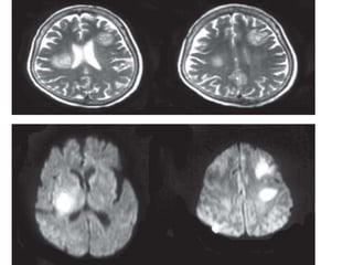
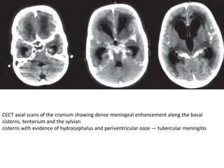
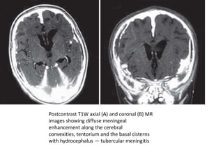
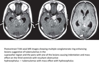
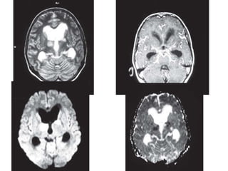
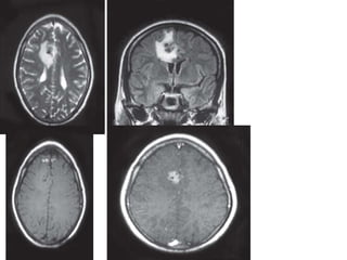
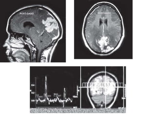
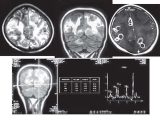
CNS INFECTIONS
DR ALI JIWANI
JNMC WARDHA
BACTERIAL/PYOGENIC INFECTIONS
PYOGENIC MENINGITIS
• Bacteria reach the meninges through one of
the following pathways:
• Hematogenous spread
• Local extension
• Direct implantation of microorganisms
• By transmission along the cranial nerves.
Clinical and Pathophysiologic Features
• Fever, irritation and variable neurologic
impairment
• headache, photophobia,clouding of
consciousness and neck stiffness
Imaging in Meningitis
• In early meningitis, the CT or MR findings may
be normal
• The characteristic positive finding on
noncontrast CT scans secondary to the
presence of inflammatory exudate and brain
swelling is obliteration of the basal
subarachnoid cisterns, fissures and cerebral
and cerebellar sulci.
• On contrast enhanced CT scan, there is
enhancement of the inflammatory exudate in
the involved basal cisterns, fissures or sulci
• The abnormal leptomeningeal contrast
enhancement is typically more readily
apparent and more intense on MR imaging
rather than on CT scanning.
Postcontrast T1W axial (A) and coronal (B) MR images
showing intense abnormal leptomeningeal enhancement along the fissures,
cerebral sulci, tentorium, over the cerebral convexities and around the
brainstem. Note is made of dilatation of the ventricular system suggestive
of hydrocephalus — pyogenic meningitis with hydrocephalous
COMPLICATIONS OR ALTERNATIVE
MANIFESTATIONS OF BACTERIAL MENINGITIS
Hydrocephalus
• Communicating hydrocephalus is the most
common complication associated with meningitis
• It is a complication more frequently seen in
children than adults
• It is related to inflammatory debris blocking the
flow and resorption of cerebrospinal fluid
particularly at the level of the arachnoid villi. An
exudative obstruction may also occur within the
cerebral aqueduct or along the foraminal outflow
of the lateral or fourth ventricles
Extra-axial Fluid and Pus Collections
Subdural Effusion and Hygroma
• Extra-axial collections associated with
meningitis may be infected or sterile.’
• Presumably, such subdural effusions are
secondary to irritation of the dura mater by
the infectious agent or its products.
Alternately, effusions can occur secondary to
inflammation
• These collections are typically isodense to
cerebrospinal fluid on CT scans and isointense
to cerebrospinal fluid on all MR imaging
sequences
Subdural Abscess (Empyema)
• The most common locations of subdural
empyemas are the cerebral convexities and
the interhemispheric fissure
• Unenhanced CT scanning reveals a crescentic
or lentiform extra-axial fluid collection that is
slightly denser than cerebrospinal fluid.
• Which may show peripheral rim enhancement
on post contrast study
• This rim represents an inflammatory membrane
of granulation tissue on the leptomeningeal
surface bordering the empyema
• Hypodensity or contrast enhancement of the
adjacent brain parenchyma may also be seen
secondary to thrombophlebitis of the bridging
draining veins crossing the subdural space
resulting in venous occlusion and venous
infarction of the involved brain.
Cerebritis and Abscess
• Cerebritis and abscess formation constitute a
continuum
• The majority of patients with brain abscess
demonstrate a contiguous focus of infection usually
sinusitis or otitis media
• Hematogenous brain abscesses have the following
characteristics (i) a distant focus of infection (ii)
location in the distribution of the middle cerebral
artery (iii) initial location at the grey-white matter
junction (iv) poor encapsulation (v) high mortality.
evolution from cerebritis to abscess
• The evolution from cerebritis to abscess has been
categorized into four stages:
• Early cerebritis, late cerebritis, early capsule
formation and late capsule formation
• In the early cerebritis (the first 4 to 5 days) stage,
the organism grows in the parenchyma. Acute
inflammatory cells, particularly
polymorphonuclear leucocytes, migrate into the
parenchyma to ingest or destroy bacteria.
Opening of the blood-brain barrier produces
edema
Noncontrast axial scan of the cranium (A)
showing an
ill-defined hypodensity in the right parietal
region with evidence of
postcontrast gyriform enhancement (B and C)
— cerebritis
• Microscopic hemorrhage may be seen during
the acute cerebritis stage but is unusual later.
In the late cerebritis stage (7 to 10 days), the
small areas of necrosis coalesce into one
• large focus filled with necrotic debris. Edema
and small foci of cerebritis are seen
surrounding this area.
• These small foci form satellite lesions adjacent
to the large abscess
• In the early capsule stage (10 to 14 days), the
body attempts to wall off the infection by forming
a fibrous capsule.The central necrotic area is
liquefied and the surrounding edema persists
• The late capsule stage (> 14 days) is characterized
by a decrease in the surrounding edema. A gliotic
reaction develops at the outer margin of the
abscess capsule
Imaging of Brain Abscess
• Typically the centre of a mature abscess
contains necrotic material hypointense on T1-
weighted images and hyperintense to brain on
T2-weighted images.
• Edema surrounding an abscess may be greater
in volume than the abscess itself and causes
much of the associated mass effect .
CECT axial scans of the cranium showing a large ring
enhancing lesion with smooth thin walls in a left parafalcine location with
extensive vasogenic edema causing effacement of the ipsilateral frontal
horn and midline shift to the contralateral side. Adjacent smaller enhancing
rings are also seen in another case—brain abscess with daughter
abscesses
• The collagenous capsule of the abscess can be
seen on unenhanced scans as a thin walled
rim that is isointense to slightly hyperintense
to brain on T1-weighted images and is
hypointense on T2-weighted images.
T1W sagittal (A) and T2W axial (B) MR images showing
multiple lesions having a hypointense center on T1 and hyperintense
center on T2W images with perilesional edema. The collagenous capsule
of the lesions is iso to slightly hyperintense on T1 and hypointense on
T2W images. Mass effect on the ipsilateral ventricular system with
dilatation of the contralateral temporal horn is seen — multiple brain
abscesses
Proton MR spectroscopy and diffusion
weighted imaging
• In vivo proton MR spectroscopy has been found
to be useful in the differentiation of brain abscess
from other cystic mass lesions
• In an untreated abscess, the usual metabolites
observed are succinate (2.4 ppm), acetate (1.92
ppm), lactate/lipids (1.3 ppm),
leucine/isoleucine/valine (0.85 ppm), glycine (3.5
ppm), alanine (1.5 ppm) and other amino acids
• Demonstration of these metabolites is considered
specific of an abscess and has not been observed
in a cystic neoplasm.
• Recently diffusion-weighted MR imaging has been used in
differentiation of cerebral abscess from intracerebral
necrotic tumors.
• The abscesses appear bright on diffusion-weighted images
and show a very low apparent diffusion coefficient (ADC)
while the necrotic tumors appear hypointense on
diffusionweighted images and have a very high ADC.
• The pus structure itself is responsible for low ADC values
and the heavily impeded water mobility of pus may be
related to its high cellularity and viscosity and this is useful
in differentiating abscess from cystic necrotic tumors25
Multiple Brain Abscesses Secondary
to Septic Emboli
• Patients with a history of cyanotic heart
disease, rheumatic heart disease, bacterial
endocarditis and intravenous drug abuse are
at risk
• Depending on the size of the emboli, the MR
findings may vary from major arterial branch
infarction to multiple small abscesses at the
grey-white matter junction secondary to
occlusion of small arteries and arterioles
• The small abscesses are accompanied by
surrounding edema and mass effect well
demonstrated on MR
• Mycotic aneurysms may occur as a result of
septic embolism and involve intermediate to
small cerebral arteries.
• MR angiography may be useful in detecting
the mycotic aneurysms
Central Nervous System Infarction
• Central nervous system infarction associated
with meningitis occurs because of
inflammation-induced arterial spasm or
because of direct inflammation of the walls of
arteries and arterioles resulting in an
infectious arteritis
• Most infarcts occur in the basal ganglia
secondary to involvement of the penetrating
lenticulostriate arteries. Large arterial branch
occlusions seen in some instances can result in
cortical infarction
CECT axial scans of the cranium showing intense
leptomeningeal enhancement with an area of hypodensity involving both
the grey and white matter in the left parietal region — meningitis with
cortical infarct
Ventriculitis
• Ventriculitis is an uncommon but potentially
serious infectious process involving the
cerebral ependyma
• Ventriculitis may be seen in direct association
with meningitis, secondary to rupture of a
parenchymal abscess into the ventricular
system or after ventricular catheter placement
• On unenhanced CT scans there may be subtle
areas of low density along the ventricular
margins. After intravenous contrast
administration there is diffuse enhancement
along the ventricular walls
• On T2-weighted MR images, ventriculomegaly
is often present accompanied by abnormal
increased signal intensity along periventricular
margins
CECT axial scans of the cranium showing massive
dilatation of the ventricular system with transependymal edema with
diffuse enhancement along the ventricular walls — ventriculitis
GRANULOMATOUS INFECTION
TUBERCULOSIS
• M. tuberculosis is responsible for almost all
cases of the tuberculous infections of the CNS
Clinical Manifestations:
• Malaise, lassitude, low-grade fever and
intermittent headache characterize the
insidious prodrome of tuberculous meningitis.
• The most common clinical manifestation
occurs with fever, headache, nausea,
vomiting, photophobia and neck stiffness.
Imaging:
CT
• Computed tomography shows obliteration by
isoattenuating or marginally hyperattenuating exudate
at the basal cisterns and plaque like dural thickening
• On intravenous contrast administration, there is dense
homogeneous enhancement of the basal cisterns
which may extend to the convexities, tentorium and
sylvian fissures
• ependymitis is seen as a linear enhancement along the
ventricular margins.
• The presence of hydrocephalus is thought to be related
to poor prognosis.
CECT axial scans of the cranium showing dense meningeal enhancement along the basal
cisterns, tentorium and the sylvian
cisterns with evidence of hydrocephalus and periventricular ooze — tubercular meningitis
MRI
• MRI may be normal in early stage
• Later, there may be widening of the
subarachnoid spaces with associated T1 and
T2 shortening of the CSF.
• Postcontrast T1-weighted MR images show
diffuse meningeal enhancement, mainly at the
basal cisterns and the sylvian fissures
Postcontrast T1W axial (A) and coronal (B) MR
images showing diffuse meningeal
enhancement along the cerebral
convexities, tentorium and the basal cisterns
with hydrocephalus — tubercular meningitis
COMPLICATIONS
Hydrocephalus
• This is a common complication and the
hydrocephalus produced by tuberculous
meningitis is usually persistent and progressive
leading to transependymal edema
• The inflammatory exudate in tuberculosis
becomes thick, gelatinous, adhesive and partly
fibrotic blocking the basal cisterns, impeding the
cerebrospinal fluid circulation and resorption
causing communicating hydrocephalus
Postcontrast T1W axial MR images showing multiple conglomerate ring enhancing
lesions suggestive of tuberculomas in the
suprasellar region and the pons with one of the lesions causing indentation and mass
effect on the third ventricle with resultant obstructive
hydrocephalus — tuberculomas with mass effect with hydrocephalus
Vasculitis
• Vasculitis is initiated by direct invasion of vessel
wall by mycobacteria or may result from
secondary extension of adjacent arachnoiditis
• On CT scan, infarctions are seen as areas of
hypoattenuation whereas on
• MR images, they are seen as areas of high signal
intensities on T2-weighted images.
• Diffusion weighted images are the gold standard
for the diagnosis of acute infarctions seen as
increasing hyperintensity with increasing b values
GRANULOMATOUS TUBERCULOUS
MENINGITIS
PACHYMENINGITIS• Common sites of dural involvement are cavernous sinus,
floor of middle cranial fossa, cerebral convexity and the
tentorium.
• On noncontrast CT scanning, it exhibits hyperattenuating
densities because of calcium deposition.
• On MR imaging, it appears hypointense to brain
parenchyma on T1- and T2-weighted images.
• On postcontrast study, intense homogeneous
enhancement of the thickened meninges is seen
• The differential diagnosis of the causative factors of
pachymeningitis includes meningioma, lymphoma and
sarcoidosis
PARENCHYMAL TUBERCULOMAS
• Intracranial tuberculoma is thought to be
secondary to an infective focus elsewhere in the
body
• The incidence of intracranial tuberculoma is
higher in the pediatric age group because of their
greater susceptibility to infections
• Tuberculomas usually involve the
corticomedullary junction and periventricular
location as expected for hematogenous
dissemination
• CT, the noncaseating granulomas usually
appear solid, are hyperdense or isodense and
enhance homogeneously while the caseating
granulomas enhance peripherally. The “target
sign” seen on CT is characterized by a central
area of calcification surrounded by rim of
enhancement and is considered to be typical
of tuberculous lesions but is not
pathognomonic
• The MR features of the individual
tuberculoma will depend on whether the
granuloma is noncaseating with a solid center
or caseating with liquid center
• Noncaseating Granuloma is usually
iso/hypointense on T1- and hyperintense on
T2- weighted images. These show
homogeneous enhancement with gadolinium
Caseating Solid Granuloma
• The caseating solid granuloma usually is
hypointense relative to brain on T1W and
strikingly hypointense on T2W images
• This relative hypointensity is attributed to the
granulation tissue and compressed glial tissue
in the central core resulting in its greater
cellular density than the brain parenchyma
V
Axial T2W MR (A) and FLAIR Coronal image (B)
showing multiple conglomerate lesions
hypointense on T2W in the right parietal
region with surrounding edema. Mass effect
noted on the ipsilateral right lateral ventricle
with shift of the midline to the contralateral
side. On contrast
enhancement (C) conglomeration and ring
enhancement of the lesions seen—Multiple
tuberculomas.
MR spectroscopy
• shows prominent lipid peaks in tuberculomas
• Caseous material typical of tuberculomas has
a high lipid content .This spectral pattern is
highly specific for tuberculomas
TUBERCULOUS ABSCESSES
• They may be solitary or multiple and are more
frequent in the elderly or in the
immunocompromised patients.
• On imaging studies, a TB abscess may be
indistinguishable from a caseating tuberculoma
or a pyogenic abscess. However, a TB abscess has
thinner and smoother walls, is commonally
multiloculated and is larger (more than 3 cm in
diameter), its walls enhance and it has peripheral
edema and mass effect
NEUROSARCOIDOSIS
• Sarcoidosis is an idiopathic systemic disease
characterized histologically by the formation
of noncaseating granulomas
• Two major patterns of intracranial
involvement are:
• 1. Meningeal or ependymal
• 2. Parenchymal.
• Granulomatous leptomeningitis may occur
diffusely or as a circumscribed process at the skull
base involving the optic chiasma, the pituitary
gland, floor of the third ventricle and
hypothalamus
• This usually appears as thickening and
enhancement of the leptomeninges on
contrastenhanced T1-weighted MR images. The
enhancement may be diffuse or focal/multifocal
and there is a predilection for the basilar
meninges.
• Dural involvement by sarcoidosis can present
as diffuse thickening or focal masses. Lesions
typically enhance homogenously on contrast-
enhanced T1-weighted images.
• They are commonly hypointense on T2-
weighted images which can serve as a clue to
the diagnosis but still may be indistinguishable
from calcified meningiomas or very cellular
dural metastases
• The parenchymal pattern of CNS sarcoid
consists of noncaseating granulomas scattered
diffusely in the brain parenchyma or occurring
as a single large mass which may mimic a
brain neoplasm.
• On MR, sarcoid granulomas are iso to
hypointense on T1W and hyperintense on
T2W images. They enhance homogeneously
with contrast.
Leptomeningeal Involvement. (a) Pre-contrast
T1, and (b) post-contrast axial, and (c) coronal
sequences show widespread leptomeningeal
thickening and enhancement along the
convexities of the brain (arrowheads). Both
diffuse and nodular patterns are evident
FUNGAL INFECTIONS OF THE CENTRAL
NERVOUS SYSTEM
ASPERGILLOSIS
• It is an opportunistic infection affecting only
those with immunodeficiency
• MRI reveals a hyperintense lesion with
hypointense rim on T2-weight images. The
lesions show vasogenic edema and contrast
enhancement
• The presence of ring or nodular enhancement
consistent with granuloma or abscess formation
indicates that the host defence system is able to
encapsulate the offending organism
• diffusion weighted MR imaging where
conventional CT and MRI are not of much
help. Diffusion weighted images are of value
in identifying early lesions as these lesions
represent septic infarction
T2W axial (A) postcontrast T1W coronal (B) and postcontrast axial (C) MR images showing
multiple ring or nodular enhancing
lesions in the right basal ganglia, right parietal and left temporal regions. Extensive
perilesional edema is seen around the lesions causing mass effect
and shift of the midline of the contralateral side — CNS aspergillosis
CRYPTOCOCCAL INFECTION
• Cryptococcal infection is the most common
fungal infection to involve the CNS.
• Cryptococcosis is nearly always an
opportunistic organism and is only rarely
found in immunocompetent patients.
• The portal of entry into the human body is
through inhalation.
Imaging findings in cryptococcal
meningitis are non-specific
• The positive CT findings may include cerebral atrophy
and communicating hydrocephalus.
• Meningeal enhancement on contrast enhanced CT is
very uncommon as cryptococcus produces only mild
inflammatory reaction in the subarachnoid space.
• Also in immunocompromised patients hydrocephalus
is uncommonly seen due to the lack of inflammatory
leptomeningeal reaction and hence paucity of
adhesions within the basal cisterns.
• The MR appearances of CNS cryptococcosis in
AIDS patients can be (1) Dilated Virchow-Robin
spaces (2) Cryptococcoma (3) Miliary enhancing
nodules (4) Mixed pattern.
• On ct it apperas as Hypoattenuating
nonenhancing lesions within the basal ganglia
• On MR images, these areas are isointense to
cerebrospinal fluid, are less than 3 mm in size, are
bilateral and symmetric with a propensity for the
basal ganglia and the brainstem.
• These foci do not enhance with contrast and
are not associated with mass effect or edema.
• These spaces may become voluminous as the
yeasts which are present in these areas,
produce mucoid material that distends these
areas.
• Other patterns of involvement include
parenchymal mass lesions also known as
cryptococcoma which may be of low or high
attenuation on CT scan.
• Cryptococcomas represent a collection of
organisms, inflammatory cells and gelatinous
mucoid material
Axial T2W MR images (A, B) showing dilated
Virchow-Robin spaces in a bilateral and
symmetric manner involving the basal ganglia.
Also seen are round hyperintense lesion in
periventricular region and the left cerebellar
hemisphere representing cryptococcomas (C,
D)—
Cryptococcosis in an immunocompromised
patient
• MR Spectroscopy in Cryptococcomas:
• The large amount of trehalose relative to other
MR visible compounds on the spectra of C
neoformans defines trehalose as one marker that
can be used to distinguish C neoformans from
other fungi.
• The high level of MR visible α trehalose recorded
from cryptococcomas provides a basis for the
pathologic diagnosis of cerebral cryptococcomas
CANDIDIASIS
• Candida is present as part of the normal
intestinal flora.
• Superinfection from Candida results from
disturbances in the equilibrium that normally
exists within the intestinal flora, usually
resulting from chemotherapy, antibiotic
therapy, and hematologic abnormalities such
as thrombocytopenia
Imaging Findings
• On CT scan, the microabscesses and
granulomas typically show enhancement.
• On MR imaging, the hypointense to
isointense signal on T2-weighted images may
be helpful in distinguishing fungal granuloma.
A target appearance has also been described
in candidal abscess, which on T2-weighted
images shows a well-delineated hypointense
area surrounded by a hyperintense rim.
Axial T2W MR image (A) and postcontrast T1W
axial (B) and postcontrast coronal T1W axial (C)
MR images showing multiple
microabscesses seen as nodular enhancing
lesions scattered in bilateral cerebral
hemispheres — candidiasis
PARASITIC INFECTIONS
NEUROCYSTICERCOSIS
• Cysticercosis is a disease transmitted by the
ingestion of the eggs of the worm Taenia
solium
• It is the most common parasitic disease in
humans.
• Clinical Features:seizures, headaches,
syncope, dementia, diplopia, visual field
defects, arachnoiditis, hydrocephalus, focal
neurologic deficits and stroke
• Pathology:
• Infection which leads to extra-intestinal disease
(including neurocysticercosis) usually occurs as a
result of eating food or drinking water
contaminated by human faeces containing T.
solium eggs. This is distinct from the 'normal' life
cycle in which the undercooked pork is eaten and
the larval cysts contained within, mature into
adult intestinal tapeworm
• Stages
• There are four main stages (also known as Escobar's pathological
stages):
• Vesicular: viable parasite with intact membrane and therefore no
host reaction.
• Colloidal vesicular: parasite dies within 4-5 years 1 untreated, or
earlier with treatment and the cyst fluid becomes turbid. As the
membrane becomes leaky oedema surrounds the cyst. This is the
most symptomatic stage.
• Granular nodular: oedema decreases as the cyst retracts further;
enhancement persists.
• Nodular calcified: end-stage quiescent calcified cyst remnant; no
oedema.
imaging
Vesicular
• cyst with dot sign
• CSF density/intensity
• hyperintense scolex on T1 can sometimes be
seen
• no enhancement is typical, although very faint
enhancement of the wall and enhancement of
the scolex may be seen
Colloidal vesicular
• cyst fluid becomes turbid
• CT: hyperattenuating to CSF
• MRI T1: hyperintense to CSF 2
• surrounding oedema
• cyst and the wall become thickened and brightly
enhances
• scolex can often still be seen as an eccentric focus
of enhancement
Granular nodular
• oedema decreases
• cyst retracts
• enhancement persists but is less marked
Nodular calcified
• end-stage quiescent calcified cyst remnant
• no oedema
• no enhancement on CT
• signal drop out on T2 sequence
• some intrinsic high T1 signal may be present
• long term enhancement may be evident on
MRI, and may predict ongoing seizures
Intraventricular Cysticercosis
• Intraventricular cysts are most commonly seen
in the fourth ventricle followed by lateral
ventricles and the third ventricle
• Cysts in the ventricular stage may migrate
from one ventricle to another and may
become trapped in the aqueductal of sylvius
leading to acute obstructive hydrocephalous
• T1 and PD weighted images are better than
T2-weighted images in the evaluation of
intraventricular cysticercus
• Sagittal T1-weighted MR imaging is
particularly useful in the evaluation of
aqueductal stenosis and for discriminating the
fourth ventricular cysticercus from dilated
fourth ventricle
Sagittal T1W (A) and T2WI axial (B) MR images showing a
cystic lesion with signal intensity similar to CSF in the fourth
ventricle with resultant obstructive hydrocephalus —
intraventricular cysticercus
Subarachnoid Cysticercosis
• Subarachnoid cysticercosis cysts may be seen
within the cortical sulci, between the gyri or in
the basal cisterns at the base of the brain
• Of all the cysticerci in the CSF containing
spaces, the cysts in the basilar cisterns carry
the worst prognosis. Those located in the
cisterns attain a large size because of lack of
pressure from adjacent brain parenchyma.
• Cysticercal cysts in the basal cisterns tend to
agglomerate in a racemose form and the
scolex of the parasite is not present
• A degenerating subarachnoid cyst may elicit
an intense inflammatory response in the
subarachnoid space and as a result contrast
enhancement may be seen
• Disturbance in cerebrospinal fluid resorption
may result in hydrocephalus
T1W sagittal (A), T2W axial (B) and
postcontrast T1W axial (C) MR images showing
multiple multilobulated large cysts with CSF
signal intensity in a cisternal location with lack
of mural nodule and hydrocephalus —
racemose variety of cysticercosis
ORBITAL DISEASE
• In the orbit, cysticercosis may involve the
globe or the extraocular muscles
• . In the globe, the lesions may be located at
the level of the posterior sclera/choroids/
retinal layers or inside of the vitreous
chamber. The appearance of the lesions is
nonspecific and some may be
indistinguishable from a melanoma
T2W axial MR image of the brain (A, B) and
orbit (C) showing multiple, small lesions
scattered in both the cerebral hemispheres,
bilateral thalami, basal ganglia, midbrain and in
the right lateral rectus muscle seen as
hyperintense foci — neurocysticercosis
TOXOPLASMOSIS
• Toxoplasma gondii is a parasite well known
throughout the world. The definitive host is
the cat
• The cat excretes the oocytes for approximately
2 weeks after the primary insult. Human
consumption of undercooked, infected meal
or contact with infected cat feces can lead to
human infections
Imaging Findings
• On noncontrast CT, toxoplasma encephalitis
characteristically appears as multiple areas of
hypodensity.
• Lesions vary in size from less than 1 cm to
over 3 cm and there is surrounding mass
effect and edema of variable degree
• Postcontrast CT demonstrates ring or nodular
enhancement. Ring enhancement is more
common with central hypodensity.
• Double-dose delayed technique (using 200 ml
of intravenous contrast by bolus/drip infusion
with delayed scanning at 1 hour) has been
found to be extremely effective in detection of
these lesions. Double-dose delayed technique
permits maximal enhancement and the
central portion of ring lesions of toxoplasma
may fill in on delayed scans
• On MRI T2-weighted images, depict active lesions
as variable signal intensity. Lesions may be
hyperintense to parenchyma and thus
indistinguishable from surrounding high-intensity
edema or may be isointense or hypointense
centrally, surrounded by high signal edema. This
latter appearance has been called a “target” sign
and is nonspecific.
• Post-gadolinium studies reveal ring or nodular
enhancement in active lesions clearly
distinguishable from the surrounding edema
Differentiation of toxoplasmosis from
CNS lymphoma
• The major diagnostic dilemma in AIDS is
distinguishing central nervous system
toxoplasmosis from central nervous system
lymphoma because both are seen in AIDS
patients and can be indistinguishable on CT
scanning and MR imaging
• Both can be solitory or multiple in form
• Metabolically active tumor tissue will demonstrate
increased uptake relative to the surrounding
parenchyma whereas infectious lesions do not
• The spectra in patients with toxoplasmosis reveal
elevated lipid and lactate peaks.
• In contrast, primary CNS lymphoma shows a mild to
moderate increase in lactate and lipids, a markedly
elevated choline peak and preservation of some
normal metabolites with variably decreased levels of
NAA and Cr
• Diffusion-Weighted imaging has been suggested
to help
• differentiate between the two diseases.
Lymphoma typically has reduced
diffusion.Toxoplasmosis abscesses have been
described as being smaller and more numerous.
• if a periventricular pattern is present, it suggests
lymphoma since toxoplasmosis uncommonly
involves the ependyma
HYDATID DISEASE
• Hydatid disease is caused by Echinococcus
granulosus and less frequently by
Echinococcus multilocularis
• The neurologic signs are varied. The cysts are
usually unilocular, solitary and located in the
parietal lobes. Multiple cysts are considered
rare and usually result from rupture of a
solitary cyst
Imaging Findings
• The CT scan appearance is that of a well-
defined, smooth, thin walled homogeneous
cystic lesion similar in density to cerebrospinal
fluid. The cyst wall is isodense or hyperdense
to brain tissue.
• Enhancement is uncommon as is surrounding
edema unless the cyst becomes secondarily
infected
• the wall is rare. The T1-weighted MR imaging
appearance is that of a hypointense center
with a slightly higher but still hypointense rim.
• T2-weighted MR images, the better of the two
sequences for imaging these lesions, shows a
thin low signal intensity rim representing the
capsule surrounding a hyperintense lesion.
T2W axial MR images showing a large well-
defined, smooth, thin-walled homogeneous
cystic lesion isointense to cerebrospinal
fluid with no significant edema in the left
parietal region with mass effect. Few daughter
cysts are also seen in the periphery of the large
cystic lesion
— hydatid cyst
CONGENITAL INFECTIONS
TOXOPLASMOSIS
• Toxoplasma gondii is an intracellular parasite
that infects birds and mammals.
• Hydrocephalus, bilateral chorioretinitis and
intracranial calcifications form the typical triad
found in infants with congenital toxoplasma
encephalitis.
Imaging Findings
• Toxoplasmosis infection is multifocal and
scattered. CT and MR scans most often
demonstrate calcifications that can involve the
periventricular regions, basal ganglia or
peripheral cerebral tissue
• Hydrocephalus can be seen
• Nonspecific findings include atrophy,
encephalomalacia, hydranencephaly or
microcephaly
ct image revealing hydrocephaly,
periventricular calcifications, cortical atrophy
and no sulci.
SYPHILIS
• Congenital syphilis is caused by infection with
the spirochaete Treponema pallidum.
• Clinical signs
• signs consist of hepato-splenomegaly,
jaundice, skin rash, persistent rhinitis,
lymphadenopathy, meningitis and
hydrocephalus. sensorineural hearing loss,
hydrocephalus, mental retardation, seizures
and hemiplegia
Imaging Findings
• Imaging findings may show hydrocephalus and
enhancement of the leptomeninges.
• on imaging appears as an enhancing
parenchymal mass.
• Infarction may occur, attributed to the
accompanying arteritis or less commonly
phlebitis.
RUBELLA
• The risk of fetal infection is more than 80 percent
during the first trimester and then decreases
significantly thereafter
• Growth retardation, ocular abnormalities
(cataracts and pigmentary retinopathy),
congenital heart disease (usually patent ductus
arteriosus or pulmonary artery stenosis),
hepatosplenomegaly, jaundice, purpuric rash,
sensorineural hearing loss or signs of
meningoencephalitis constitutes the typical
congenital rubella syndrome.
Imaging Findings
• On ultrasound subependymal cysts in the caudate
nucleus and striothalamic regions are seen but
are not specific for rubella. Echogenic foci in the
basal ganglia represents mineralizing vasculitis
with calcification
• On CT microcephaly and parenchymal
calcifications are typically present in the cortex
and basal ganglia
• On MR delayed myelination, deep and subcortical
white matter lesions caused by vascular injury
and ischemic necrosis are seen
CYTOMEGALOVIRUS (CMV)
• Congenital infection occurs when maternal
infection or reactivation of infection happens
during pregnancy
• The periventricular subependymal germinal
matrix cells are most frequently affected.
• associated with early infection are disorders of
neuronal migration including polymicrogyria and
neuronal heterotopia and hydranencephaly,
porencephaly and micrencephaly
Imaging Findings
• The classic plain film findings of congenital
CMV infection is microcephaly with egg shell
periventricular calcifications
• On ultrasound, bilateral periventricular
calcifications preceded by hypoechoic
periventricular ring like zones is specific for
CMV. CMV may also result in widespread
cerebral destruction with severe
encephalomalacia
• On ct atrophy, ventricular enlargement and
parenchymal calcifications.
• on MRI. Intracranial calcifications are typically
located in the periventricular areas. They can also
be seen in the basal ganglia, subcortical and
cortical regions.
• MR findings include migrational anomalies
(lissencephaly, polymicrogyria, pachygyria),
encephalomalacia, nonspecific ventricular
enlargement, delayed myelination and
subependymal paraventricular cysts.
Non-contrast CT scans of the cranium of a
newborn showing hydrocephalus and
extensive periventricular calcification.
Also seen is calcification in bilateral basal
ganglia — CMV
HERPES SIMPLEX VIRUS (HSV)
• HSV types 1 and 2 most commonly manifest as
reactivated latent infections.
• Symptoms usually first appear 5 to 15 days after
birth
• This condition may progress to coma with
seizures. Primary HSV CNS infection in neonates
is diffuse and nonfocal resulting in widespread
brain destruction. Infection may lead to death or
severe neurologic sequelae such as seizures
Imaging Findings
• Initial CT scans show subtle hypodense lesions in
the periventricular white matter with relative
sparing of the basal ganglia, thalami and
posterior fossa structures.
• Finger-like areas of increased attenuation within
the cortical gray matter are accompanied by
increased white matter lucency and have been
described as characteristic CT findings in the first
3 weeks after presentation
T1WI:may show general oedema in the affected region
• if complicated by subacute haemorrhage there may be areas of
hyperintense signal
T1 C+ (Gd):enhancement is usually absent early on later enhancement
is variable in pattern 5
• gyral enhancement leptomeningeal enhancement ring
enhancement diffuse enhancement
T2WI:hyperintensity of affected white matter and cortex
• more established haemorrhagic components may the hypointense
DWI/ADC more sensitive than T2 weighted images
• restricted diffusion is common due to cytotoxic oedema
• beware of T2 shine through due to vasogenic oedema
MRI demonstrates extensive oedema in the
right temporal lobe with areas of intrinsic high
T1 signal, in keeping with haemorrhage. The
changes spare the basal ganglia, a feature
which is helpful in distinguishing an MCA
infarct with haemorrhagic transformation from
herpes simplex encephalitis, the diagnosis in
this case.
VIRAL INFECTIONS
• ENCEPHALITIS
• Encephalitis refers to a generalized and diffuse
infection of the brain with parenchymal
infiltration of inflammatory cells. The brain
damage caused by acute encephalitis is due to
a combination of intracellular viral growth and
the host inflammatory response
ACUTE INFECTIVE
ENCEPHALITIS/HERPES
SIMPLEX ENCEPHALITIS
• It is a fulminant hemorrhagic
meningoencephalitis.
• Clinical symptoms include a altered mental
state, a diminished level of consciousness,
focal neurologic deficit and fever.
• The trigeminal ganglion is likely the most
common site of viral reactivation with spread
along branches of cranial nerve Vth.
Imaging Findings
• CT scan may demonstrate hypodense
temporal lobe lesions with or without
involvement of the frontal lobes
• hemorrhagic temporal lobe lesions in the
appropriate clinical setting are highly
suggestive of HSV-1 encephalitis,
• Contrast Enhanced CT scans show streaky
linear enhancement in the region of the
sylvian fissure and the island of Reil.
• MR imaging can demonstrate the early edematous changes
of herpes encephalitis seen as hypointensity on T1 and
hyperintensity on T2-weighted images with characteristic
involvement of the temporal lobes and inferior frontal
lobes.
• Fluid attenuated inversion-recovery (FLAIR) imaging is even
more sensitive than T2-weighted images
• Diffusion weighted imaging (DWI) shows increased signal in
the affected areas to a greater extent than do the T2-
weighted and FLAIR sequences.
• Early on in the disease process, contrast enhancement is
absent but gyriform enhancement occurs with disease
progression.
CECT axial scans of the cranium showing
bilateral symmetric areas of hypodensity
involving the inferomedial temporal lobes,
insular cortex and the cingulate gyrus with
sparing of the basal ganglia — herpes
encephalitis
JAPANESE ENCEPHALITIS (JE)
• The characteristic neurologic findings during
the acute stage are extrapyramidal signs such
as tremor, dystonia and rigidity. Seizures are
more common in children than adults. It is
important to distinguish JE from other types of
encephalitis, particularly HSE, because
antiviral therapy for HSE is very effective in the
acute stage
Imaging Findings
• MRI shows areas of hyperintensity on T2-
weighted images involving the thalamus and
basal ganglia .Involvement of thalami and
basal ganglia is usually seen with or without
involvement of other regions
• The lesions are uniformly hyperintense on T2
and iso to hypointense on T1-weighted
images.
• The parenchymal lesions show minimal or no
enhancement after injection of gadolinium
• Postcontrast study may show meningeal
enhancement especially on T1-weighted
images.
Axial T2W (A, B) and FLAIR coronal (C) MR
images showing symmetric areas of signal
alteration in bilateral
thalami and pons—Japanese encephalitis
RASMUSSEN’S ENCEPHALITIS
• Rasmussen’s encephalitis is a childhood
disease. The child presents with severe
epilepsy and progressive neurologic deficits.
• The disease tends to affect one hemisphere
although bilateral involvement has also been
reported.
• Early in the disease process, CT and MRI
findings may be normal. MRI may reveal focal
and progressive atrophy of a hemisphere and
hyperintensity in white matter and putamen
on T2-weighted images.
• PET imaging using FDG-18 may show
decreased hemispheric activity
ACQUIRED IMMUNODEFICIENCY SYNDROME
(AIDS) AND ITS RELATED CNS INFECTIONS
HIV ENCEPHALITIS:
• Clinical presentation includes a progressive
dementia associated with motor and/or
behavioral dysfunction. Early difficulties with
memory and concentration are often followed
by apparent apathy and social withdrawal and
may be mistaken for symptoms of depression
Imaging Findings
• In patients with subacute encephalitis, CT is
often negative or reveals atrophy only
• Cortical atrophy is the most frequent MR
finding on T2-weighted images reveal
hyperintense lesions in the periventricular
white matter and centrum semiovale that
correspond to foci of demyelination and
vacuolation
• The frontal lobes are the most common sites.
• MRS shows low NAA a finding that reflects
lower number of neurons (due to their death)
and neuronal dysfunction. Elevated choline
peak reflect the presence of gliosis, microglial,
foamy macrophages and lymphocytes.
Elevated myoinositol (MI) occurs early in
infection and is a reflection of gliosis
MR spectroscopy (D)
reveals an
D elevated choline peak
— HIV encephalitis
Progressive Multifocal
Leukoencephalopathy (PML)
• PML is a progressive demyelinating disorder
arising from CNS infection with a papovavirus.
• Clinical presentation of PML includes memory
loss, visual deficit, personality change,
cognitive and speech disturbances, altered
mental status, and motor and/or sensory
abnormalities with a progressive neurologic
decline
Imaging Findings
• On CT, PML appears as a focal hypodensity within
the white matter, without mass effect and usually
without enhancement
• On T2-weighted images, lesions demonstrate
increased signal intensity in the periventricular
and/or subcortical white matter. Lesions may be
initially small in size but usually progress to larger
areas
• A multifocal distribution pattern is seen, which
may be unilateral but is more often bilateral and
asymmetric.
• In AIDS patients, PML may be difficult to distinguish
radiographically from HIV-related demyelination.
• PML is more often multifocal and asymmetric with
scalloping and a greater predilection for the subcortical
white matter.
• HIV encephalitis is more often diffuse, symmetric and
periventricular in location.
• The lesions of PML are well demarcated on T1-
weighted images.
• HIV encephalitis most often presents with global
cognitive disturbance and dementia whereas PML
presents with a focal motor or sensory deficit.
CYTOMEGALOVIRUS (CMV)
ENCEPHALITIS
• Neurologic manifestations of CMV thus
include acute or chronic meningoencephalitis,
cranial neuropathy, vasculitis, retinitis,
myelitis, brachial plexus neuropathy and
peripheral neuropathy
Imaging Findings
• CT is frequently insensitive in the imaging of CMV
encephalitis. The most common manifestation on
CT is atrophy
• In addition to atrophy, MR may demonstrate
increased signal on T2-weighted images in the
periventricular white matter
• Fat-suppressed MR with gadolinium may reveal a
thickened enhancing choroid/retina in patients
with CMV retinitis, a hemorrhagic retinitis.
Thank you

Cns infections perfect

  • 1.
    CNS INFECTIONS DR ALIJIWANI JNMC WARDHA
  • 2.
    BACTERIAL/PYOGENIC INFECTIONS PYOGENIC MENINGITIS •Bacteria reach the meninges through one of the following pathways: • Hematogenous spread • Local extension • Direct implantation of microorganisms • By transmission along the cranial nerves.
  • 3.
    Clinical and PathophysiologicFeatures • Fever, irritation and variable neurologic impairment • headache, photophobia,clouding of consciousness and neck stiffness
  • 4.
    Imaging in Meningitis •In early meningitis, the CT or MR findings may be normal • The characteristic positive finding on noncontrast CT scans secondary to the presence of inflammatory exudate and brain swelling is obliteration of the basal subarachnoid cisterns, fissures and cerebral and cerebellar sulci.
  • 5.
    • On contrastenhanced CT scan, there is enhancement of the inflammatory exudate in the involved basal cisterns, fissures or sulci • The abnormal leptomeningeal contrast enhancement is typically more readily apparent and more intense on MR imaging rather than on CT scanning.
  • 6.
    Postcontrast T1W axial(A) and coronal (B) MR images showing intense abnormal leptomeningeal enhancement along the fissures, cerebral sulci, tentorium, over the cerebral convexities and around the brainstem. Note is made of dilatation of the ventricular system suggestive of hydrocephalus — pyogenic meningitis with hydrocephalous
  • 7.
    COMPLICATIONS OR ALTERNATIVE MANIFESTATIONSOF BACTERIAL MENINGITIS Hydrocephalus • Communicating hydrocephalus is the most common complication associated with meningitis • It is a complication more frequently seen in children than adults • It is related to inflammatory debris blocking the flow and resorption of cerebrospinal fluid particularly at the level of the arachnoid villi. An exudative obstruction may also occur within the cerebral aqueduct or along the foraminal outflow of the lateral or fourth ventricles
  • 8.
    Extra-axial Fluid andPus Collections Subdural Effusion and Hygroma • Extra-axial collections associated with meningitis may be infected or sterile.’ • Presumably, such subdural effusions are secondary to irritation of the dura mater by the infectious agent or its products. Alternately, effusions can occur secondary to inflammation
  • 9.
    • These collectionsare typically isodense to cerebrospinal fluid on CT scans and isointense to cerebrospinal fluid on all MR imaging sequences
  • 11.
    Subdural Abscess (Empyema) •The most common locations of subdural empyemas are the cerebral convexities and the interhemispheric fissure • Unenhanced CT scanning reveals a crescentic or lentiform extra-axial fluid collection that is slightly denser than cerebrospinal fluid. • Which may show peripheral rim enhancement on post contrast study
  • 12.
    • This rimrepresents an inflammatory membrane of granulation tissue on the leptomeningeal surface bordering the empyema • Hypodensity or contrast enhancement of the adjacent brain parenchyma may also be seen secondary to thrombophlebitis of the bridging draining veins crossing the subdural space resulting in venous occlusion and venous infarction of the involved brain.
  • 14.
    Cerebritis and Abscess •Cerebritis and abscess formation constitute a continuum • The majority of patients with brain abscess demonstrate a contiguous focus of infection usually sinusitis or otitis media • Hematogenous brain abscesses have the following characteristics (i) a distant focus of infection (ii) location in the distribution of the middle cerebral artery (iii) initial location at the grey-white matter junction (iv) poor encapsulation (v) high mortality.
  • 15.
    evolution from cerebritisto abscess • The evolution from cerebritis to abscess has been categorized into four stages: • Early cerebritis, late cerebritis, early capsule formation and late capsule formation • In the early cerebritis (the first 4 to 5 days) stage, the organism grows in the parenchyma. Acute inflammatory cells, particularly polymorphonuclear leucocytes, migrate into the parenchyma to ingest or destroy bacteria. Opening of the blood-brain barrier produces edema
  • 16.
    Noncontrast axial scanof the cranium (A) showing an ill-defined hypodensity in the right parietal region with evidence of postcontrast gyriform enhancement (B and C) — cerebritis
  • 17.
    • Microscopic hemorrhagemay be seen during the acute cerebritis stage but is unusual later. In the late cerebritis stage (7 to 10 days), the small areas of necrosis coalesce into one • large focus filled with necrotic debris. Edema and small foci of cerebritis are seen surrounding this area. • These small foci form satellite lesions adjacent to the large abscess
  • 18.
    • In theearly capsule stage (10 to 14 days), the body attempts to wall off the infection by forming a fibrous capsule.The central necrotic area is liquefied and the surrounding edema persists • The late capsule stage (> 14 days) is characterized by a decrease in the surrounding edema. A gliotic reaction develops at the outer margin of the abscess capsule
  • 19.
    Imaging of BrainAbscess • Typically the centre of a mature abscess contains necrotic material hypointense on T1- weighted images and hyperintense to brain on T2-weighted images. • Edema surrounding an abscess may be greater in volume than the abscess itself and causes much of the associated mass effect .
  • 20.
    CECT axial scansof the cranium showing a large ring enhancing lesion with smooth thin walls in a left parafalcine location with extensive vasogenic edema causing effacement of the ipsilateral frontal horn and midline shift to the contralateral side. Adjacent smaller enhancing rings are also seen in another case—brain abscess with daughter abscesses
  • 21.
    • The collagenouscapsule of the abscess can be seen on unenhanced scans as a thin walled rim that is isointense to slightly hyperintense to brain on T1-weighted images and is hypointense on T2-weighted images.
  • 22.
    T1W sagittal (A)and T2W axial (B) MR images showing multiple lesions having a hypointense center on T1 and hyperintense center on T2W images with perilesional edema. The collagenous capsule of the lesions is iso to slightly hyperintense on T1 and hypointense on T2W images. Mass effect on the ipsilateral ventricular system with dilatation of the contralateral temporal horn is seen — multiple brain abscesses
  • 23.
    Proton MR spectroscopyand diffusion weighted imaging • In vivo proton MR spectroscopy has been found to be useful in the differentiation of brain abscess from other cystic mass lesions • In an untreated abscess, the usual metabolites observed are succinate (2.4 ppm), acetate (1.92 ppm), lactate/lipids (1.3 ppm), leucine/isoleucine/valine (0.85 ppm), glycine (3.5 ppm), alanine (1.5 ppm) and other amino acids • Demonstration of these metabolites is considered specific of an abscess and has not been observed in a cystic neoplasm.
  • 24.
    • Recently diffusion-weightedMR imaging has been used in differentiation of cerebral abscess from intracerebral necrotic tumors. • The abscesses appear bright on diffusion-weighted images and show a very low apparent diffusion coefficient (ADC) while the necrotic tumors appear hypointense on diffusionweighted images and have a very high ADC. • The pus structure itself is responsible for low ADC values and the heavily impeded water mobility of pus may be related to its high cellularity and viscosity and this is useful in differentiating abscess from cystic necrotic tumors25
  • 26.
    Multiple Brain AbscessesSecondary to Septic Emboli • Patients with a history of cyanotic heart disease, rheumatic heart disease, bacterial endocarditis and intravenous drug abuse are at risk • Depending on the size of the emboli, the MR findings may vary from major arterial branch infarction to multiple small abscesses at the grey-white matter junction secondary to occlusion of small arteries and arterioles
  • 27.
    • The smallabscesses are accompanied by surrounding edema and mass effect well demonstrated on MR • Mycotic aneurysms may occur as a result of septic embolism and involve intermediate to small cerebral arteries. • MR angiography may be useful in detecting the mycotic aneurysms
  • 29.
    Central Nervous SystemInfarction • Central nervous system infarction associated with meningitis occurs because of inflammation-induced arterial spasm or because of direct inflammation of the walls of arteries and arterioles resulting in an infectious arteritis
  • 30.
    • Most infarctsoccur in the basal ganglia secondary to involvement of the penetrating lenticulostriate arteries. Large arterial branch occlusions seen in some instances can result in cortical infarction
  • 31.
    CECT axial scansof the cranium showing intense leptomeningeal enhancement with an area of hypodensity involving both the grey and white matter in the left parietal region — meningitis with cortical infarct
  • 32.
    Ventriculitis • Ventriculitis isan uncommon but potentially serious infectious process involving the cerebral ependyma • Ventriculitis may be seen in direct association with meningitis, secondary to rupture of a parenchymal abscess into the ventricular system or after ventricular catheter placement
  • 33.
    • On unenhancedCT scans there may be subtle areas of low density along the ventricular margins. After intravenous contrast administration there is diffuse enhancement along the ventricular walls • On T2-weighted MR images, ventriculomegaly is often present accompanied by abnormal increased signal intensity along periventricular margins
  • 34.
    CECT axial scansof the cranium showing massive dilatation of the ventricular system with transependymal edema with diffuse enhancement along the ventricular walls — ventriculitis
  • 35.
    GRANULOMATOUS INFECTION TUBERCULOSIS • M.tuberculosis is responsible for almost all cases of the tuberculous infections of the CNS Clinical Manifestations: • Malaise, lassitude, low-grade fever and intermittent headache characterize the insidious prodrome of tuberculous meningitis. • The most common clinical manifestation occurs with fever, headache, nausea, vomiting, photophobia and neck stiffness.
  • 36.
    Imaging: CT • Computed tomographyshows obliteration by isoattenuating or marginally hyperattenuating exudate at the basal cisterns and plaque like dural thickening • On intravenous contrast administration, there is dense homogeneous enhancement of the basal cisterns which may extend to the convexities, tentorium and sylvian fissures • ependymitis is seen as a linear enhancement along the ventricular margins. • The presence of hydrocephalus is thought to be related to poor prognosis.
  • 37.
    CECT axial scansof the cranium showing dense meningeal enhancement along the basal cisterns, tentorium and the sylvian cisterns with evidence of hydrocephalus and periventricular ooze — tubercular meningitis
  • 38.
    MRI • MRI maybe normal in early stage • Later, there may be widening of the subarachnoid spaces with associated T1 and T2 shortening of the CSF. • Postcontrast T1-weighted MR images show diffuse meningeal enhancement, mainly at the basal cisterns and the sylvian fissures
  • 39.
    Postcontrast T1W axial(A) and coronal (B) MR images showing diffuse meningeal enhancement along the cerebral convexities, tentorium and the basal cisterns with hydrocephalus — tubercular meningitis
  • 40.
    COMPLICATIONS Hydrocephalus • This isa common complication and the hydrocephalus produced by tuberculous meningitis is usually persistent and progressive leading to transependymal edema • The inflammatory exudate in tuberculosis becomes thick, gelatinous, adhesive and partly fibrotic blocking the basal cisterns, impeding the cerebrospinal fluid circulation and resorption causing communicating hydrocephalus
  • 41.
    Postcontrast T1W axialMR images showing multiple conglomerate ring enhancing lesions suggestive of tuberculomas in the suprasellar region and the pons with one of the lesions causing indentation and mass effect on the third ventricle with resultant obstructive hydrocephalus — tuberculomas with mass effect with hydrocephalus
  • 42.
    Vasculitis • Vasculitis isinitiated by direct invasion of vessel wall by mycobacteria or may result from secondary extension of adjacent arachnoiditis • On CT scan, infarctions are seen as areas of hypoattenuation whereas on • MR images, they are seen as areas of high signal intensities on T2-weighted images. • Diffusion weighted images are the gold standard for the diagnosis of acute infarctions seen as increasing hyperintensity with increasing b values
  • 44.
    GRANULOMATOUS TUBERCULOUS MENINGITIS PACHYMENINGITIS• Commonsites of dural involvement are cavernous sinus, floor of middle cranial fossa, cerebral convexity and the tentorium. • On noncontrast CT scanning, it exhibits hyperattenuating densities because of calcium deposition. • On MR imaging, it appears hypointense to brain parenchyma on T1- and T2-weighted images. • On postcontrast study, intense homogeneous enhancement of the thickened meninges is seen • The differential diagnosis of the causative factors of pachymeningitis includes meningioma, lymphoma and sarcoidosis
  • 45.
    PARENCHYMAL TUBERCULOMAS • Intracranialtuberculoma is thought to be secondary to an infective focus elsewhere in the body • The incidence of intracranial tuberculoma is higher in the pediatric age group because of their greater susceptibility to infections • Tuberculomas usually involve the corticomedullary junction and periventricular location as expected for hematogenous dissemination
  • 46.
    • CT, thenoncaseating granulomas usually appear solid, are hyperdense or isodense and enhance homogeneously while the caseating granulomas enhance peripherally. The “target sign” seen on CT is characterized by a central area of calcification surrounded by rim of enhancement and is considered to be typical of tuberculous lesions but is not pathognomonic
  • 47.
    • The MRfeatures of the individual tuberculoma will depend on whether the granuloma is noncaseating with a solid center or caseating with liquid center • Noncaseating Granuloma is usually iso/hypointense on T1- and hyperintense on T2- weighted images. These show homogeneous enhancement with gadolinium
  • 49.
    Caseating Solid Granuloma •The caseating solid granuloma usually is hypointense relative to brain on T1W and strikingly hypointense on T2W images • This relative hypointensity is attributed to the granulation tissue and compressed glial tissue in the central core resulting in its greater cellular density than the brain parenchyma
  • 50.
    V Axial T2W MR(A) and FLAIR Coronal image (B) showing multiple conglomerate lesions hypointense on T2W in the right parietal region with surrounding edema. Mass effect noted on the ipsilateral right lateral ventricle with shift of the midline to the contralateral side. On contrast enhancement (C) conglomeration and ring enhancement of the lesions seen—Multiple tuberculomas.
  • 52.
    MR spectroscopy • showsprominent lipid peaks in tuberculomas • Caseous material typical of tuberculomas has a high lipid content .This spectral pattern is highly specific for tuberculomas
  • 54.
    TUBERCULOUS ABSCESSES • Theymay be solitary or multiple and are more frequent in the elderly or in the immunocompromised patients. • On imaging studies, a TB abscess may be indistinguishable from a caseating tuberculoma or a pyogenic abscess. However, a TB abscess has thinner and smoother walls, is commonally multiloculated and is larger (more than 3 cm in diameter), its walls enhance and it has peripheral edema and mass effect
  • 56.
    NEUROSARCOIDOSIS • Sarcoidosis isan idiopathic systemic disease characterized histologically by the formation of noncaseating granulomas • Two major patterns of intracranial involvement are: • 1. Meningeal or ependymal • 2. Parenchymal.
  • 57.
    • Granulomatous leptomeningitismay occur diffusely or as a circumscribed process at the skull base involving the optic chiasma, the pituitary gland, floor of the third ventricle and hypothalamus • This usually appears as thickening and enhancement of the leptomeninges on contrastenhanced T1-weighted MR images. The enhancement may be diffuse or focal/multifocal and there is a predilection for the basilar meninges.
  • 58.
    • Dural involvementby sarcoidosis can present as diffuse thickening or focal masses. Lesions typically enhance homogenously on contrast- enhanced T1-weighted images. • They are commonly hypointense on T2- weighted images which can serve as a clue to the diagnosis but still may be indistinguishable from calcified meningiomas or very cellular dural metastases
  • 59.
    • The parenchymalpattern of CNS sarcoid consists of noncaseating granulomas scattered diffusely in the brain parenchyma or occurring as a single large mass which may mimic a brain neoplasm. • On MR, sarcoid granulomas are iso to hypointense on T1W and hyperintense on T2W images. They enhance homogeneously with contrast.
  • 60.
    Leptomeningeal Involvement. (a)Pre-contrast T1, and (b) post-contrast axial, and (c) coronal sequences show widespread leptomeningeal thickening and enhancement along the convexities of the brain (arrowheads). Both diffuse and nodular patterns are evident
  • 61.
    FUNGAL INFECTIONS OFTHE CENTRAL NERVOUS SYSTEM ASPERGILLOSIS • It is an opportunistic infection affecting only those with immunodeficiency • MRI reveals a hyperintense lesion with hypointense rim on T2-weight images. The lesions show vasogenic edema and contrast enhancement • The presence of ring or nodular enhancement consistent with granuloma or abscess formation indicates that the host defence system is able to encapsulate the offending organism
  • 62.
    • diffusion weightedMR imaging where conventional CT and MRI are not of much help. Diffusion weighted images are of value in identifying early lesions as these lesions represent septic infarction
  • 63.
    T2W axial (A)postcontrast T1W coronal (B) and postcontrast axial (C) MR images showing multiple ring or nodular enhancing lesions in the right basal ganglia, right parietal and left temporal regions. Extensive perilesional edema is seen around the lesions causing mass effect and shift of the midline of the contralateral side — CNS aspergillosis
  • 64.
    CRYPTOCOCCAL INFECTION • Cryptococcalinfection is the most common fungal infection to involve the CNS. • Cryptococcosis is nearly always an opportunistic organism and is only rarely found in immunocompetent patients. • The portal of entry into the human body is through inhalation.
  • 65.
    Imaging findings incryptococcal meningitis are non-specific • The positive CT findings may include cerebral atrophy and communicating hydrocephalus. • Meningeal enhancement on contrast enhanced CT is very uncommon as cryptococcus produces only mild inflammatory reaction in the subarachnoid space. • Also in immunocompromised patients hydrocephalus is uncommonly seen due to the lack of inflammatory leptomeningeal reaction and hence paucity of adhesions within the basal cisterns.
  • 66.
    • The MRappearances of CNS cryptococcosis in AIDS patients can be (1) Dilated Virchow-Robin spaces (2) Cryptococcoma (3) Miliary enhancing nodules (4) Mixed pattern. • On ct it apperas as Hypoattenuating nonenhancing lesions within the basal ganglia • On MR images, these areas are isointense to cerebrospinal fluid, are less than 3 mm in size, are bilateral and symmetric with a propensity for the basal ganglia and the brainstem.
  • 67.
    • These focido not enhance with contrast and are not associated with mass effect or edema. • These spaces may become voluminous as the yeasts which are present in these areas, produce mucoid material that distends these areas.
  • 68.
    • Other patternsof involvement include parenchymal mass lesions also known as cryptococcoma which may be of low or high attenuation on CT scan. • Cryptococcomas represent a collection of organisms, inflammatory cells and gelatinous mucoid material
  • 69.
    Axial T2W MRimages (A, B) showing dilated Virchow-Robin spaces in a bilateral and symmetric manner involving the basal ganglia. Also seen are round hyperintense lesion in periventricular region and the left cerebellar hemisphere representing cryptococcomas (C, D)— Cryptococcosis in an immunocompromised patient
  • 70.
    • MR Spectroscopyin Cryptococcomas: • The large amount of trehalose relative to other MR visible compounds on the spectra of C neoformans defines trehalose as one marker that can be used to distinguish C neoformans from other fungi. • The high level of MR visible α trehalose recorded from cryptococcomas provides a basis for the pathologic diagnosis of cerebral cryptococcomas
  • 71.
    CANDIDIASIS • Candida ispresent as part of the normal intestinal flora. • Superinfection from Candida results from disturbances in the equilibrium that normally exists within the intestinal flora, usually resulting from chemotherapy, antibiotic therapy, and hematologic abnormalities such as thrombocytopenia
  • 72.
    Imaging Findings • OnCT scan, the microabscesses and granulomas typically show enhancement. • On MR imaging, the hypointense to isointense signal on T2-weighted images may be helpful in distinguishing fungal granuloma. A target appearance has also been described in candidal abscess, which on T2-weighted images shows a well-delineated hypointense area surrounded by a hyperintense rim.
  • 73.
    Axial T2W MRimage (A) and postcontrast T1W axial (B) and postcontrast coronal T1W axial (C) MR images showing multiple microabscesses seen as nodular enhancing lesions scattered in bilateral cerebral hemispheres — candidiasis
  • 74.
    PARASITIC INFECTIONS NEUROCYSTICERCOSIS • Cysticercosisis a disease transmitted by the ingestion of the eggs of the worm Taenia solium • It is the most common parasitic disease in humans. • Clinical Features:seizures, headaches, syncope, dementia, diplopia, visual field defects, arachnoiditis, hydrocephalus, focal neurologic deficits and stroke
  • 75.
    • Pathology: • Infectionwhich leads to extra-intestinal disease (including neurocysticercosis) usually occurs as a result of eating food or drinking water contaminated by human faeces containing T. solium eggs. This is distinct from the 'normal' life cycle in which the undercooked pork is eaten and the larval cysts contained within, mature into adult intestinal tapeworm
  • 76.
    • Stages • Thereare four main stages (also known as Escobar's pathological stages): • Vesicular: viable parasite with intact membrane and therefore no host reaction. • Colloidal vesicular: parasite dies within 4-5 years 1 untreated, or earlier with treatment and the cyst fluid becomes turbid. As the membrane becomes leaky oedema surrounds the cyst. This is the most symptomatic stage. • Granular nodular: oedema decreases as the cyst retracts further; enhancement persists. • Nodular calcified: end-stage quiescent calcified cyst remnant; no oedema.
  • 77.
    imaging Vesicular • cyst withdot sign • CSF density/intensity • hyperintense scolex on T1 can sometimes be seen • no enhancement is typical, although very faint enhancement of the wall and enhancement of the scolex may be seen
  • 78.
    Colloidal vesicular • cystfluid becomes turbid • CT: hyperattenuating to CSF • MRI T1: hyperintense to CSF 2 • surrounding oedema • cyst and the wall become thickened and brightly enhances • scolex can often still be seen as an eccentric focus of enhancement
  • 79.
    Granular nodular • oedemadecreases • cyst retracts • enhancement persists but is less marked
  • 80.
    Nodular calcified • end-stagequiescent calcified cyst remnant • no oedema • no enhancement on CT • signal drop out on T2 sequence • some intrinsic high T1 signal may be present • long term enhancement may be evident on MRI, and may predict ongoing seizures
  • 82.
    Intraventricular Cysticercosis • Intraventricularcysts are most commonly seen in the fourth ventricle followed by lateral ventricles and the third ventricle • Cysts in the ventricular stage may migrate from one ventricle to another and may become trapped in the aqueductal of sylvius leading to acute obstructive hydrocephalous
  • 83.
    • T1 andPD weighted images are better than T2-weighted images in the evaluation of intraventricular cysticercus • Sagittal T1-weighted MR imaging is particularly useful in the evaluation of aqueductal stenosis and for discriminating the fourth ventricular cysticercus from dilated fourth ventricle
  • 84.
    Sagittal T1W (A)and T2WI axial (B) MR images showing a cystic lesion with signal intensity similar to CSF in the fourth ventricle with resultant obstructive hydrocephalus — intraventricular cysticercus
  • 85.
    Subarachnoid Cysticercosis • Subarachnoidcysticercosis cysts may be seen within the cortical sulci, between the gyri or in the basal cisterns at the base of the brain • Of all the cysticerci in the CSF containing spaces, the cysts in the basilar cisterns carry the worst prognosis. Those located in the cisterns attain a large size because of lack of pressure from adjacent brain parenchyma.
  • 86.
    • Cysticercal cystsin the basal cisterns tend to agglomerate in a racemose form and the scolex of the parasite is not present • A degenerating subarachnoid cyst may elicit an intense inflammatory response in the subarachnoid space and as a result contrast enhancement may be seen • Disturbance in cerebrospinal fluid resorption may result in hydrocephalus
  • 87.
    T1W sagittal (A),T2W axial (B) and postcontrast T1W axial (C) MR images showing multiple multilobulated large cysts with CSF signal intensity in a cisternal location with lack of mural nodule and hydrocephalus — racemose variety of cysticercosis
  • 88.
    ORBITAL DISEASE • Inthe orbit, cysticercosis may involve the globe or the extraocular muscles • . In the globe, the lesions may be located at the level of the posterior sclera/choroids/ retinal layers or inside of the vitreous chamber. The appearance of the lesions is nonspecific and some may be indistinguishable from a melanoma
  • 89.
    T2W axial MRimage of the brain (A, B) and orbit (C) showing multiple, small lesions scattered in both the cerebral hemispheres, bilateral thalami, basal ganglia, midbrain and in the right lateral rectus muscle seen as hyperintense foci — neurocysticercosis
  • 90.
    TOXOPLASMOSIS • Toxoplasma gondiiis a parasite well known throughout the world. The definitive host is the cat • The cat excretes the oocytes for approximately 2 weeks after the primary insult. Human consumption of undercooked, infected meal or contact with infected cat feces can lead to human infections
  • 91.
    Imaging Findings • Onnoncontrast CT, toxoplasma encephalitis characteristically appears as multiple areas of hypodensity. • Lesions vary in size from less than 1 cm to over 3 cm and there is surrounding mass effect and edema of variable degree • Postcontrast CT demonstrates ring or nodular enhancement. Ring enhancement is more common with central hypodensity.
  • 92.
    • Double-dose delayedtechnique (using 200 ml of intravenous contrast by bolus/drip infusion with delayed scanning at 1 hour) has been found to be extremely effective in detection of these lesions. Double-dose delayed technique permits maximal enhancement and the central portion of ring lesions of toxoplasma may fill in on delayed scans
  • 93.
    • On MRIT2-weighted images, depict active lesions as variable signal intensity. Lesions may be hyperintense to parenchyma and thus indistinguishable from surrounding high-intensity edema or may be isointense or hypointense centrally, surrounded by high signal edema. This latter appearance has been called a “target” sign and is nonspecific. • Post-gadolinium studies reveal ring or nodular enhancement in active lesions clearly distinguishable from the surrounding edema
  • 95.
    Differentiation of toxoplasmosisfrom CNS lymphoma • The major diagnostic dilemma in AIDS is distinguishing central nervous system toxoplasmosis from central nervous system lymphoma because both are seen in AIDS patients and can be indistinguishable on CT scanning and MR imaging
  • 96.
    • Both canbe solitory or multiple in form • Metabolically active tumor tissue will demonstrate increased uptake relative to the surrounding parenchyma whereas infectious lesions do not • The spectra in patients with toxoplasmosis reveal elevated lipid and lactate peaks. • In contrast, primary CNS lymphoma shows a mild to moderate increase in lactate and lipids, a markedly elevated choline peak and preservation of some normal metabolites with variably decreased levels of NAA and Cr
  • 97.
    • Diffusion-Weighted imaginghas been suggested to help • differentiate between the two diseases. Lymphoma typically has reduced diffusion.Toxoplasmosis abscesses have been described as being smaller and more numerous. • if a periventricular pattern is present, it suggests lymphoma since toxoplasmosis uncommonly involves the ependyma
  • 98.
    HYDATID DISEASE • Hydatiddisease is caused by Echinococcus granulosus and less frequently by Echinococcus multilocularis • The neurologic signs are varied. The cysts are usually unilocular, solitary and located in the parietal lobes. Multiple cysts are considered rare and usually result from rupture of a solitary cyst
  • 99.
    Imaging Findings • TheCT scan appearance is that of a well- defined, smooth, thin walled homogeneous cystic lesion similar in density to cerebrospinal fluid. The cyst wall is isodense or hyperdense to brain tissue. • Enhancement is uncommon as is surrounding edema unless the cyst becomes secondarily infected
  • 100.
    • the wallis rare. The T1-weighted MR imaging appearance is that of a hypointense center with a slightly higher but still hypointense rim. • T2-weighted MR images, the better of the two sequences for imaging these lesions, shows a thin low signal intensity rim representing the capsule surrounding a hyperintense lesion.
  • 101.
    T2W axial MRimages showing a large well- defined, smooth, thin-walled homogeneous cystic lesion isointense to cerebrospinal fluid with no significant edema in the left parietal region with mass effect. Few daughter cysts are also seen in the periphery of the large cystic lesion — hydatid cyst
  • 102.
    CONGENITAL INFECTIONS TOXOPLASMOSIS • Toxoplasmagondii is an intracellular parasite that infects birds and mammals. • Hydrocephalus, bilateral chorioretinitis and intracranial calcifications form the typical triad found in infants with congenital toxoplasma encephalitis.
  • 103.
    Imaging Findings • Toxoplasmosisinfection is multifocal and scattered. CT and MR scans most often demonstrate calcifications that can involve the periventricular regions, basal ganglia or peripheral cerebral tissue • Hydrocephalus can be seen • Nonspecific findings include atrophy, encephalomalacia, hydranencephaly or microcephaly
  • 104.
    ct image revealinghydrocephaly, periventricular calcifications, cortical atrophy and no sulci.
  • 105.
    SYPHILIS • Congenital syphilisis caused by infection with the spirochaete Treponema pallidum. • Clinical signs • signs consist of hepato-splenomegaly, jaundice, skin rash, persistent rhinitis, lymphadenopathy, meningitis and hydrocephalus. sensorineural hearing loss, hydrocephalus, mental retardation, seizures and hemiplegia
  • 106.
    Imaging Findings • Imagingfindings may show hydrocephalus and enhancement of the leptomeninges. • on imaging appears as an enhancing parenchymal mass. • Infarction may occur, attributed to the accompanying arteritis or less commonly phlebitis.
  • 107.
    RUBELLA • The riskof fetal infection is more than 80 percent during the first trimester and then decreases significantly thereafter • Growth retardation, ocular abnormalities (cataracts and pigmentary retinopathy), congenital heart disease (usually patent ductus arteriosus or pulmonary artery stenosis), hepatosplenomegaly, jaundice, purpuric rash, sensorineural hearing loss or signs of meningoencephalitis constitutes the typical congenital rubella syndrome.
  • 108.
    Imaging Findings • Onultrasound subependymal cysts in the caudate nucleus and striothalamic regions are seen but are not specific for rubella. Echogenic foci in the basal ganglia represents mineralizing vasculitis with calcification • On CT microcephaly and parenchymal calcifications are typically present in the cortex and basal ganglia • On MR delayed myelination, deep and subcortical white matter lesions caused by vascular injury and ischemic necrosis are seen
  • 109.
    CYTOMEGALOVIRUS (CMV) • Congenitalinfection occurs when maternal infection or reactivation of infection happens during pregnancy • The periventricular subependymal germinal matrix cells are most frequently affected. • associated with early infection are disorders of neuronal migration including polymicrogyria and neuronal heterotopia and hydranencephaly, porencephaly and micrencephaly
  • 110.
    Imaging Findings • Theclassic plain film findings of congenital CMV infection is microcephaly with egg shell periventricular calcifications • On ultrasound, bilateral periventricular calcifications preceded by hypoechoic periventricular ring like zones is specific for CMV. CMV may also result in widespread cerebral destruction with severe encephalomalacia
  • 111.
    • On ctatrophy, ventricular enlargement and parenchymal calcifications. • on MRI. Intracranial calcifications are typically located in the periventricular areas. They can also be seen in the basal ganglia, subcortical and cortical regions. • MR findings include migrational anomalies (lissencephaly, polymicrogyria, pachygyria), encephalomalacia, nonspecific ventricular enlargement, delayed myelination and subependymal paraventricular cysts.
  • 112.
    Non-contrast CT scansof the cranium of a newborn showing hydrocephalus and extensive periventricular calcification. Also seen is calcification in bilateral basal ganglia — CMV
  • 113.
    HERPES SIMPLEX VIRUS(HSV) • HSV types 1 and 2 most commonly manifest as reactivated latent infections. • Symptoms usually first appear 5 to 15 days after birth • This condition may progress to coma with seizures. Primary HSV CNS infection in neonates is diffuse and nonfocal resulting in widespread brain destruction. Infection may lead to death or severe neurologic sequelae such as seizures
  • 114.
    Imaging Findings • InitialCT scans show subtle hypodense lesions in the periventricular white matter with relative sparing of the basal ganglia, thalami and posterior fossa structures. • Finger-like areas of increased attenuation within the cortical gray matter are accompanied by increased white matter lucency and have been described as characteristic CT findings in the first 3 weeks after presentation
  • 115.
    T1WI:may show generaloedema in the affected region • if complicated by subacute haemorrhage there may be areas of hyperintense signal T1 C+ (Gd):enhancement is usually absent early on later enhancement is variable in pattern 5 • gyral enhancement leptomeningeal enhancement ring enhancement diffuse enhancement T2WI:hyperintensity of affected white matter and cortex • more established haemorrhagic components may the hypointense DWI/ADC more sensitive than T2 weighted images • restricted diffusion is common due to cytotoxic oedema • beware of T2 shine through due to vasogenic oedema
  • 116.
    MRI demonstrates extensiveoedema in the right temporal lobe with areas of intrinsic high T1 signal, in keeping with haemorrhage. The changes spare the basal ganglia, a feature which is helpful in distinguishing an MCA infarct with haemorrhagic transformation from herpes simplex encephalitis, the diagnosis in this case.
  • 117.
    VIRAL INFECTIONS • ENCEPHALITIS •Encephalitis refers to a generalized and diffuse infection of the brain with parenchymal infiltration of inflammatory cells. The brain damage caused by acute encephalitis is due to a combination of intracellular viral growth and the host inflammatory response
  • 118.
    ACUTE INFECTIVE ENCEPHALITIS/HERPES SIMPLEX ENCEPHALITIS •It is a fulminant hemorrhagic meningoencephalitis. • Clinical symptoms include a altered mental state, a diminished level of consciousness, focal neurologic deficit and fever. • The trigeminal ganglion is likely the most common site of viral reactivation with spread along branches of cranial nerve Vth.
  • 119.
    Imaging Findings • CTscan may demonstrate hypodense temporal lobe lesions with or without involvement of the frontal lobes • hemorrhagic temporal lobe lesions in the appropriate clinical setting are highly suggestive of HSV-1 encephalitis, • Contrast Enhanced CT scans show streaky linear enhancement in the region of the sylvian fissure and the island of Reil.
  • 120.
    • MR imagingcan demonstrate the early edematous changes of herpes encephalitis seen as hypointensity on T1 and hyperintensity on T2-weighted images with characteristic involvement of the temporal lobes and inferior frontal lobes. • Fluid attenuated inversion-recovery (FLAIR) imaging is even more sensitive than T2-weighted images • Diffusion weighted imaging (DWI) shows increased signal in the affected areas to a greater extent than do the T2- weighted and FLAIR sequences. • Early on in the disease process, contrast enhancement is absent but gyriform enhancement occurs with disease progression.
  • 121.
    CECT axial scansof the cranium showing bilateral symmetric areas of hypodensity involving the inferomedial temporal lobes, insular cortex and the cingulate gyrus with sparing of the basal ganglia — herpes encephalitis
  • 123.
    JAPANESE ENCEPHALITIS (JE) •The characteristic neurologic findings during the acute stage are extrapyramidal signs such as tremor, dystonia and rigidity. Seizures are more common in children than adults. It is important to distinguish JE from other types of encephalitis, particularly HSE, because antiviral therapy for HSE is very effective in the acute stage
  • 124.
    Imaging Findings • MRIshows areas of hyperintensity on T2- weighted images involving the thalamus and basal ganglia .Involvement of thalami and basal ganglia is usually seen with or without involvement of other regions • The lesions are uniformly hyperintense on T2 and iso to hypointense on T1-weighted images.
  • 125.
    • The parenchymallesions show minimal or no enhancement after injection of gadolinium • Postcontrast study may show meningeal enhancement especially on T1-weighted images.
  • 126.
    Axial T2W (A,B) and FLAIR coronal (C) MR images showing symmetric areas of signal alteration in bilateral thalami and pons—Japanese encephalitis
  • 127.
    RASMUSSEN’S ENCEPHALITIS • Rasmussen’sencephalitis is a childhood disease. The child presents with severe epilepsy and progressive neurologic deficits. • The disease tends to affect one hemisphere although bilateral involvement has also been reported.
  • 128.
    • Early inthe disease process, CT and MRI findings may be normal. MRI may reveal focal and progressive atrophy of a hemisphere and hyperintensity in white matter and putamen on T2-weighted images. • PET imaging using FDG-18 may show decreased hemispheric activity
  • 129.
    ACQUIRED IMMUNODEFICIENCY SYNDROME (AIDS)AND ITS RELATED CNS INFECTIONS HIV ENCEPHALITIS: • Clinical presentation includes a progressive dementia associated with motor and/or behavioral dysfunction. Early difficulties with memory and concentration are often followed by apparent apathy and social withdrawal and may be mistaken for symptoms of depression
  • 130.
    Imaging Findings • Inpatients with subacute encephalitis, CT is often negative or reveals atrophy only • Cortical atrophy is the most frequent MR finding on T2-weighted images reveal hyperintense lesions in the periventricular white matter and centrum semiovale that correspond to foci of demyelination and vacuolation • The frontal lobes are the most common sites.
  • 131.
    • MRS showslow NAA a finding that reflects lower number of neurons (due to their death) and neuronal dysfunction. Elevated choline peak reflect the presence of gliosis, microglial, foamy macrophages and lymphocytes. Elevated myoinositol (MI) occurs early in infection and is a reflection of gliosis
  • 132.
    MR spectroscopy (D) revealsan D elevated choline peak — HIV encephalitis
  • 133.
    Progressive Multifocal Leukoencephalopathy (PML) •PML is a progressive demyelinating disorder arising from CNS infection with a papovavirus. • Clinical presentation of PML includes memory loss, visual deficit, personality change, cognitive and speech disturbances, altered mental status, and motor and/or sensory abnormalities with a progressive neurologic decline
  • 134.
    Imaging Findings • OnCT, PML appears as a focal hypodensity within the white matter, without mass effect and usually without enhancement • On T2-weighted images, lesions demonstrate increased signal intensity in the periventricular and/or subcortical white matter. Lesions may be initially small in size but usually progress to larger areas • A multifocal distribution pattern is seen, which may be unilateral but is more often bilateral and asymmetric.
  • 135.
    • In AIDSpatients, PML may be difficult to distinguish radiographically from HIV-related demyelination. • PML is more often multifocal and asymmetric with scalloping and a greater predilection for the subcortical white matter. • HIV encephalitis is more often diffuse, symmetric and periventricular in location. • The lesions of PML are well demarcated on T1- weighted images. • HIV encephalitis most often presents with global cognitive disturbance and dementia whereas PML presents with a focal motor or sensory deficit.
  • 136.
    CYTOMEGALOVIRUS (CMV) ENCEPHALITIS • Neurologicmanifestations of CMV thus include acute or chronic meningoencephalitis, cranial neuropathy, vasculitis, retinitis, myelitis, brachial plexus neuropathy and peripheral neuropathy
  • 137.
    Imaging Findings • CTis frequently insensitive in the imaging of CMV encephalitis. The most common manifestation on CT is atrophy • In addition to atrophy, MR may demonstrate increased signal on T2-weighted images in the periventricular white matter • Fat-suppressed MR with gadolinium may reveal a thickened enhancing choroid/retina in patients with CMV retinitis, a hemorrhagic retinitis.
  • 139.

Editor's Notes

  • #11 CECT axial scan of the cranium showing bilateral crescent shaped concavoconvex extra-axial fluid collections isodense to the cerebrospinal fluid in the parietal region. VP shunt is seen in situ — subdural effusions CECT axial scans of the cranium showing bilateral lentiform extra-axial fluid collections with an overlying peripheral rim of enhancement in the right frontoparietal and the left frontal regions suggestive of subdural empyemas. A biconvex extra-axial collection with overlying thick and markedly enhancing dura is seen in the left parietal region suggestive of epidural empyema
  • #14 MR Images T2 W axial (A, B) showing extra-axial collections along the interhemispheric fissure and along the left parietal region cerebral convexity. The collections are concavoconvex and have an enhancing rim (C, D). Meningeal enhancement and hydrocephalus seen— Subdural empyemas in a case of pyogenic meningitis
  • #29 FLAIR coronal (A and B), diffusion weighted (C) and postcontrast T1W axial (D) MR images showing an area of signal alteration in the distribution of the right MCA territory showing a restricted pattern of diffusion suggestive of infarct. Another lesion showing perilesional edema, a restricted pattern of diffusion and ring enhancement is seen in the left parietal region suggestive of an abscess — septic emboli
  • #44 T2W axial (A), T1W postgadolinium axial (B) diffusion weighted (C) and ADC (D) MR images showing basal cisternal enhancement with hydrocephalus and periventricular ooze suggestive of tubercular meningitis. Areas of signal alteration noted in bilateral basal ganglia showing hyperintense signal on T2W images and increasing hyperintensity on b1000 with hypointensity on ADC maps — bilateral basal ganglia infarcts (vasculitis) in tubercular meningitis
  • #49 Sagittal T1W (A) axial T2W (B) coronal FLAIR (C) MR images showing conglomerate round lesions in the right cerebellar hemisphere hypointense on T1 hyperintense on T2 with surrounding edema. There is attenuation of the fourth ventricle and obstructive hydrocephalus. The lesions show rim enhancement (D)—Tuberculomas postfossa
  • #51 Axial T2W MR (A) and FLAIR Coronal image (B) showing multiple conglomerate lesions hypointense on T2W in the right parietal region with surrounding edema. Mass effect noted on the ipsilateral right lateral ventricle with shift of the midline to the contralateral side. On contrast enhancement (C) conglomeration and ring enhancement of the lesions seen—Multiple tuberculomas.
  • #52 Axial T2W (A), coronal FLAIR (B) images showing multiple small conglomerate hypointense lesions with surrounding perilesional edema in the right parafalcine region. Axial T1W with MT (C) shows that the lesions are better defined appearing hypointense with a hyperintense rim. Postcontrast axial image (D) shows thick irregular enhancement and evidence of conglomeration—Solid caseating granulomas
  • #54 Postcontrast T1W sagittal (A), postcontrast T1W axial (B) and MR spectroscopy (C) images showing multiple conglomerate ring enhancing lesions in left parieto-occipital region showing a prominent lipid peak at 1.3 ppm — tuberculomas
  • #56 T2W axial (A) and FLAIR coronal (B) MR images showing multiple lesions with perilesional edema in bilateral cerebral hemispheres and right cerebellum having a hypointense center. Postcontrast T1W axial (C) MR image shows multiloculated ring enhancing lesions with smooth and thin walls. MR spectroscopy (D) through the lesions reveal a prominent lipid peak at 1.3 ppm — tubercular abscesses
  • #70 Axial T2W MR images (A, B) showing dilated Virchow-Robin spaces in a bilateral and symmetric manner involving the basal ganglia. Also seen are round hyperintense lesion in periventricular region and the left cerebellar hemisphere representing cryptococcomas (C, D)— Cryptococcosis in an immunocompromised patient
  • #82 Contrast enhanced axial CT (A) showing a cyst with fluid similar to CSF attenuation with an eccentrically located scolex in the left parietal region with no contrast enhancement/edema — vesicular stage. Axial CT scan (B) showing a ring enhancing lesion with perilesional edema in left parieto-occipital region colloid vesicular stage. CECT axial image (C) showing a cyst with thick capsule with calcific nodule with no edema— granular nodular stage. CECT axial images (D) showing calcific foci in bilateral hemispheres — nodular calcified stage
  • #90 T2W axial MR image of the brain (A, B) and orbit (C) showing multiple, small lesions scattered in both the cerebral hemispheres, bilateral thalami, basal ganglia, midbrain and in the right lateral rectus muscle seen as hyperintense foci — neurocysticercosis
  • #95 FLAIR coronal (A), T2W axial (B, C) MR images showing multiple lesions in bilateral basal ganglia in the left frontoparietal region and right thalamus seen as iso or hypointense surrounded by high signal edema giving the typical ‘target’ sign. Postcontrast T1W axial (D) MR image reveals ring or nodular enhancement in the lesions — toxoplasmosis
  • #117 MRI demonstrates extensive oedema in the right temporal lobe with areas of intrinsic high T1 signal, in keeping with haemorrhage. The changes spare the basal ganglia, a feature which is helpful in distinguishing an MCA infarct with haemorrhagic transformation from herpes simplex encephalitis, the diagnosis in this case.
  • #123 T2W axial (A), FLAIR coronal (B, C) MR images showing symmetric areas of signal alteration seen as hyperintense signal involving bilateral medial temporal lobes, orbital surface of the frontal lobes, insular cortex and the cingulate gyrus. Postcontrast T1W axial (D) MR image shows no significant enhancement — herpes encephalitis
  • #133 T2W (A) FLAIR coronal (B) MR images showing a bilateral diffuse periventricular white matter hyperintensity with sparing of the gray matter with diffuse cerebral atrophy. Axial T1W MR image (C) with MT shows the lesions to be better delineated. MR spectroscopy (D) reveals an D elevated choline peak — HIV encephalitis