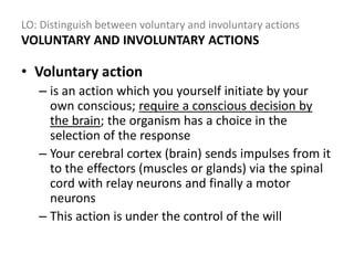
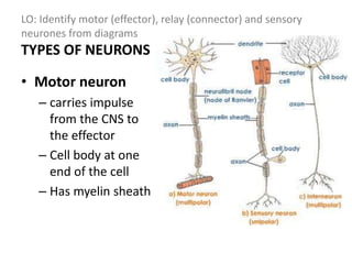
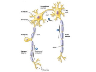
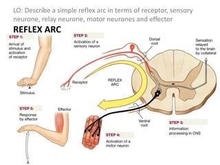
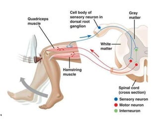
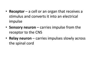
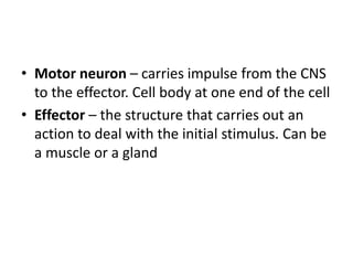
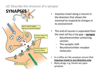
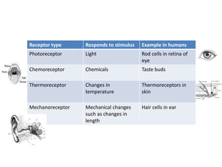
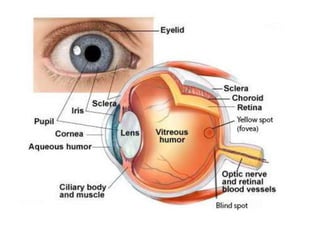
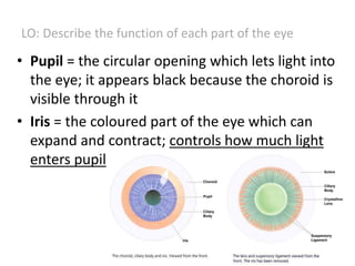
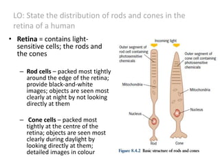
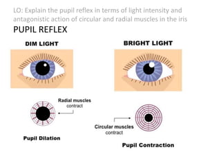
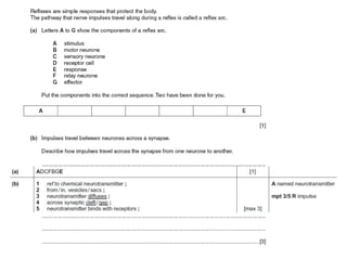
NERVOUS
CONTROL IN
HUMANS
LO:
• Describe a nerve impulse
• Describe the human nervous system in terms
of:
– the central nervous system
– the peripheral nervous system
– coordination and regulation of body functions
• Distinguish between voluntary and
involuntary actions
• Identify motor (effector), relay (connector) and
sensory neurones from diagrams
• Describe a simple reflex arc in terms of receptor,
sensory neurone, relay neurone, motor neurones
and effector
• Describe a reflex action
• Describe the structure of a synapse
• Describe how an impulse triggers the release of a
neurotransmitter and how the neurotransmitter
diffuses across to bind with receptor molecules,
causing the impulse to continue
LO: Describe the human nervous system
NERVOUS SYSTEM
FUNCTION: Coordination and control of all the
work of cells and organs in an organism;
coordination and regulation of body functions
• Works closely with the endocrine system
• Consists of:
– CNS – central nervous system – brain and spinal
cord: areas of coordination
– PNS – peripheral nervous system – nerves made
of neurons: serve to coordinate and regulate body
functions
LO: Describe a nerve impulse
NERVE IMPULSE
• Is an electrical signal that passes along nerve
cells called neurones
LO: Distinguish between voluntary and involuntary actions
VOLUNTARY AND INVOLUNTARY ACTIONS
• Voluntary action
– is an action which you yourself initiate by your
own conscious; require a conscious decision by
the brain; the organism has a choice in the
selection of the response
– Your cerebral cortex (brain) sends impulses from it
to the effectors (muscles or glands) via the spinal
cord with relay neurons and finally a motor
neurons
– This action is under the control of the will
LO: Distinguish between voluntary and involuntary actions
VOLUNTARY AND INVOLUNTARY ACTIONS
• Involuntary actions
– Are opposites of voluntary actions
– Reflex action is not under the control of the will
– Spinal cord takes total control, without your own
conscious
– Spinal reflexes = reflex actions controlled by the spinal
cord: example scratching
– Cranial reflexes = reflexes that are controlled by the
brain but not by the cerebral cortex: example blinking
– Blushing, sneezing and salivation are also reflex
actions however, salivation is also known an
conditioned reflex action
LO: Distinguish between voluntary and involuntary actions
LO: Identify motor (effector), relay (connector) and sensory
neurones from diagrams
Nerve cells - NEURONS
• Cell body – controls the
metabolism of the nerve
cell
• Dendrites – collect
information from other cells
• Axon – is a long fiber which
carries information away
from the cell body,
sometimes over long
distances
LO: Identify motor (effector), relay (connector) and sensory
neurones from diagrams
Nerve cells - NEURONS
• Nodes of Ranvier – allow
rapid conduction of
impulses by forcing them to
jump from one node to the
next
• Fatty sheath made of myelin
– gives electrical insulation
between neighboring cells
and makes impulses travel
faster
• End plate – synapses – with
another nerve cell, a muscle
or a gland
LO: Identify motor (effector), relay (connector) and sensory
neurones from diagrams
TYPES OF NEURONS
• Motor neuron
– carries impulse
from the CNS to
the effector
– Cell body at one
end of the cell
– Has myelin sheath
LO: Identify motor (effector), relay (connector) and sensory
neurones from diagrams
TYPES OF NEURONS
• Sensory neuron
– carries impulse
from the receptor
to the CNS
– Has myelin sheath
LO: Identify motor (effector), relay (connector) and sensory
neurones from diagrams
TYPES OF NEURONS
• Relay neuron
– carries impulses
slowly across the
spinal cord
– No myelin sheath:
this gives time for
the action to be
modified by
impulses carried
down the spinal
cord from the brain
LO: Identify motor (effector), relay (connector) and sensory
neurones from diagrams
LO: Describe a simple reflex arc in terms of receptor, sensory
neurone, relay neurone, motor neurones and effector
REFLEX ARC
LO: Describe a reflex action
REFLEX ACTION
Reflex action = the simlest type of response; it is rapid
automatic response to a stimulus (coughing, pupil reflex,
knee jerk, swallowing)
• a very quick response, one that does not need
the involvement of the brain
1. receptor detects a stimulus - change in the
environment
2. sensory neurone sends signal to relay neurone
3. motor neurone sends signal to effector
4. effector produces a response
• Receptor – a cell or an organ that receives a
stimulus and converts it into an electrical
impulse
• Sensory neuron – carries impulse from the
receptor to the CNS
• Relay neuron – carries impulses slowly across
the spinal cord
• Motor neuron – carries impulse from the CNS
to the effector. Cell body at one end of the cell
• Effector – the structure that carries out an
action to deal with the initial stimulus. Can be
a muscle or a gland
LO: Describe a reflex action
REFLEX ACTION
• REFLEX ARC
• IRIS REACTING TO LIGHT
LO: Describe the structure of a synapse
SYNAPSES • Impulses travel along a neuron in
the direction that allows the
mammal to respond to changes in
its environment
• The end of neuron is separated from
the next cell by a tiny gap – synapse
- Neurotransmitter containing
vesicles
- The synaptic cleft
- Neurotransmitter receptor
molecules
• In a reflex arc the synapses ensure that
impulses travel in one direction only
• Many drugs, e.g. heroin act upon
synapses
LO: Define sense organs
SENSE ORGANS
= groups of receptor cells responding to specific
stimuli: light, sound, touch, temperature and
chemicals
• Sense = ability to be aware of different
aspects of the environment
• Stimulus = a change in the environment that
affects an organism
- Receptor = part of the nervous system that is
adapted to recieve stimuli; receptor are
transducers = convert one form of energy into
another
- Internal receptors = inside the body; detect
blood temperature, pH (important for
homeostasis)
Receptor type Responds to stimulus Example in humans
Photoreceptor Light Rod cells in retina of
eye
Chemoreceptor Chemicals Taste buds
Thermoreceptor Changes in
temperature
Thermoreceptors in
skin
Mechanoreceptor Mechanical changes
such as changes in
length
Hair cells in ear
LO: Identify the structures of the eye, Describe the
function of each part of the eye
THE EYE
• Receptors: the rod and cone cells on the retina
• Systems of making the most of the light stimulus;
including lens and the iris
• Its own blood supply and physical protection; the
choroid and the sclera
LO: Describe the function of each part of the eye
• Cornea = a transparent
layer responsible for most
of the refraction (bending)
of light rays that enter the
eye
• Aqueous humour = watery
fluid which supports the
cornea and the front
chamber of the eye
• Pupil = the circular opening which lets light into
the eye; it appears black because the choroid is
visible through it
• Iris = the coloured part of the eye which can
expand and contract; controls how much light
enters pupil
LO: Describe the function of each part of the eye
• Ciliary muscle; suspensory ligament and Lens
= together control light focusing on the retina
• Vitreous humour = a jelly-like substance
which helps to keep the shape of the eyeball,
supports the lens and keeps the retina in place
at the back of the eye
LO: Describe the function of each part of the eye
• Sclera = the tough outer coat which protects
hte eye against damage; muscles attached
• Choroid = a darkly coloured layer which
reduces reflection inside the eye and contains
blood vessels which help to nouorish the cells
of retina
LO: Describe the function of each part of the eye
• Retina = contains light-
sensitive cells; the rods and
the cones
– Rod cells – packed most tightly
around the edge of the retina;
provide black-and-white
images; objects are seen most
clearly at night by not looking
directly at them
– Cone cells – packed most
tightly at the centre of the
retina; objects are seen most
clearly during daylight by
looking directly at them;
detailed images in colour
LO: State the distribution of rods and cones in the
retina of a human
LO: Identify the position of the fovea
The fovea is responsible for sharp
central vision
LO: Explain the pupil reflex in terms of light intensity and
antagonistic action of circular and radial muscles in the iris
PUPIL REFLEX
ACCOMODATION
• Explain accommodation to view near and
distant objects in terms of the contraction and
relaxation of the ciliary muscles, tension in the
suspensory ligaments, shape of the lens and
refraction of light
Nervous system
Nervous system
Nervous system

Nervous system

  • 1.
  • 2.
    LO: • Describe anerve impulse • Describe the human nervous system in terms of: – the central nervous system – the peripheral nervous system – coordination and regulation of body functions • Distinguish between voluntary and involuntary actions
  • 3.
    • Identify motor(effector), relay (connector) and sensory neurones from diagrams • Describe a simple reflex arc in terms of receptor, sensory neurone, relay neurone, motor neurones and effector • Describe a reflex action • Describe the structure of a synapse • Describe how an impulse triggers the release of a neurotransmitter and how the neurotransmitter diffuses across to bind with receptor molecules, causing the impulse to continue
  • 5.
    LO: Describe thehuman nervous system NERVOUS SYSTEM FUNCTION: Coordination and control of all the work of cells and organs in an organism; coordination and regulation of body functions • Works closely with the endocrine system • Consists of: – CNS – central nervous system – brain and spinal cord: areas of coordination – PNS – peripheral nervous system – nerves made of neurons: serve to coordinate and regulate body functions
  • 7.
    LO: Describe anerve impulse NERVE IMPULSE • Is an electrical signal that passes along nerve cells called neurones
  • 8.
    LO: Distinguish betweenvoluntary and involuntary actions VOLUNTARY AND INVOLUNTARY ACTIONS • Voluntary action – is an action which you yourself initiate by your own conscious; require a conscious decision by the brain; the organism has a choice in the selection of the response – Your cerebral cortex (brain) sends impulses from it to the effectors (muscles or glands) via the spinal cord with relay neurons and finally a motor neurons – This action is under the control of the will
  • 9.
    LO: Distinguish betweenvoluntary and involuntary actions VOLUNTARY AND INVOLUNTARY ACTIONS • Involuntary actions – Are opposites of voluntary actions – Reflex action is not under the control of the will – Spinal cord takes total control, without your own conscious – Spinal reflexes = reflex actions controlled by the spinal cord: example scratching – Cranial reflexes = reflexes that are controlled by the brain but not by the cerebral cortex: example blinking – Blushing, sneezing and salivation are also reflex actions however, salivation is also known an conditioned reflex action
  • 10.
    LO: Distinguish betweenvoluntary and involuntary actions
  • 11.
    LO: Identify motor(effector), relay (connector) and sensory neurones from diagrams Nerve cells - NEURONS • Cell body – controls the metabolism of the nerve cell • Dendrites – collect information from other cells • Axon – is a long fiber which carries information away from the cell body, sometimes over long distances
  • 12.
    LO: Identify motor(effector), relay (connector) and sensory neurones from diagrams Nerve cells - NEURONS • Nodes of Ranvier – allow rapid conduction of impulses by forcing them to jump from one node to the next • Fatty sheath made of myelin – gives electrical insulation between neighboring cells and makes impulses travel faster • End plate – synapses – with another nerve cell, a muscle or a gland
  • 13.
    LO: Identify motor(effector), relay (connector) and sensory neurones from diagrams TYPES OF NEURONS • Motor neuron – carries impulse from the CNS to the effector – Cell body at one end of the cell – Has myelin sheath
  • 15.
    LO: Identify motor(effector), relay (connector) and sensory neurones from diagrams TYPES OF NEURONS • Sensory neuron – carries impulse from the receptor to the CNS – Has myelin sheath
  • 17.
    LO: Identify motor(effector), relay (connector) and sensory neurones from diagrams TYPES OF NEURONS • Relay neuron – carries impulses slowly across the spinal cord – No myelin sheath: this gives time for the action to be modified by impulses carried down the spinal cord from the brain
  • 18.
    LO: Identify motor(effector), relay (connector) and sensory neurones from diagrams
  • 21.
    LO: Describe asimple reflex arc in terms of receptor, sensory neurone, relay neurone, motor neurones and effector REFLEX ARC
  • 22.
    LO: Describe areflex action REFLEX ACTION Reflex action = the simlest type of response; it is rapid automatic response to a stimulus (coughing, pupil reflex, knee jerk, swallowing) • a very quick response, one that does not need the involvement of the brain 1. receptor detects a stimulus - change in the environment 2. sensory neurone sends signal to relay neurone 3. motor neurone sends signal to effector 4. effector produces a response
  • 24.
    • Receptor –a cell or an organ that receives a stimulus and converts it into an electrical impulse • Sensory neuron – carries impulse from the receptor to the CNS • Relay neuron – carries impulses slowly across the spinal cord
  • 25.
    • Motor neuron– carries impulse from the CNS to the effector. Cell body at one end of the cell • Effector – the structure that carries out an action to deal with the initial stimulus. Can be a muscle or a gland
  • 26.
    LO: Describe areflex action REFLEX ACTION • REFLEX ARC • IRIS REACTING TO LIGHT
  • 27.
    LO: Describe thestructure of a synapse SYNAPSES • Impulses travel along a neuron in the direction that allows the mammal to respond to changes in its environment • The end of neuron is separated from the next cell by a tiny gap – synapse - Neurotransmitter containing vesicles - The synaptic cleft - Neurotransmitter receptor molecules • In a reflex arc the synapses ensure that impulses travel in one direction only • Many drugs, e.g. heroin act upon synapses
  • 30.
    LO: Define senseorgans SENSE ORGANS = groups of receptor cells responding to specific stimuli: light, sound, touch, temperature and chemicals • Sense = ability to be aware of different aspects of the environment • Stimulus = a change in the environment that affects an organism
  • 31.
    - Receptor =part of the nervous system that is adapted to recieve stimuli; receptor are transducers = convert one form of energy into another - Internal receptors = inside the body; detect blood temperature, pH (important for homeostasis)
  • 32.
    Receptor type Respondsto stimulus Example in humans Photoreceptor Light Rod cells in retina of eye Chemoreceptor Chemicals Taste buds Thermoreceptor Changes in temperature Thermoreceptors in skin Mechanoreceptor Mechanical changes such as changes in length Hair cells in ear
  • 33.
    LO: Identify thestructures of the eye, Describe the function of each part of the eye THE EYE • Receptors: the rod and cone cells on the retina • Systems of making the most of the light stimulus; including lens and the iris • Its own blood supply and physical protection; the choroid and the sclera
  • 35.
    LO: Describe thefunction of each part of the eye • Cornea = a transparent layer responsible for most of the refraction (bending) of light rays that enter the eye • Aqueous humour = watery fluid which supports the cornea and the front chamber of the eye
  • 36.
    • Pupil =the circular opening which lets light into the eye; it appears black because the choroid is visible through it • Iris = the coloured part of the eye which can expand and contract; controls how much light enters pupil LO: Describe the function of each part of the eye
  • 37.
    • Ciliary muscle;suspensory ligament and Lens = together control light focusing on the retina • Vitreous humour = a jelly-like substance which helps to keep the shape of the eyeball, supports the lens and keeps the retina in place at the back of the eye LO: Describe the function of each part of the eye
  • 38.
    • Sclera =the tough outer coat which protects hte eye against damage; muscles attached • Choroid = a darkly coloured layer which reduces reflection inside the eye and contains blood vessels which help to nouorish the cells of retina LO: Describe the function of each part of the eye
  • 39.
    • Retina =contains light- sensitive cells; the rods and the cones – Rod cells – packed most tightly around the edge of the retina; provide black-and-white images; objects are seen most clearly at night by not looking directly at them – Cone cells – packed most tightly at the centre of the retina; objects are seen most clearly during daylight by looking directly at them; detailed images in colour LO: State the distribution of rods and cones in the retina of a human
  • 40.
    LO: Identify theposition of the fovea The fovea is responsible for sharp central vision
  • 42.
    LO: Explain thepupil reflex in terms of light intensity and antagonistic action of circular and radial muscles in the iris PUPIL REFLEX
  • 43.
  • 45.
    • Explain accommodationto view near and distant objects in terms of the contraction and relaxation of the ciliary muscles, tension in the suspensory ligaments, shape of the lens and refraction of light