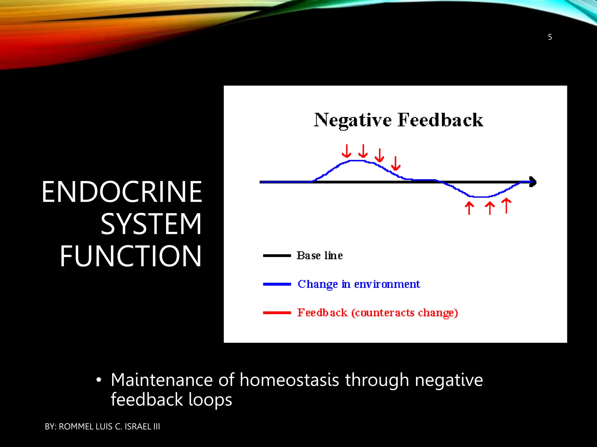
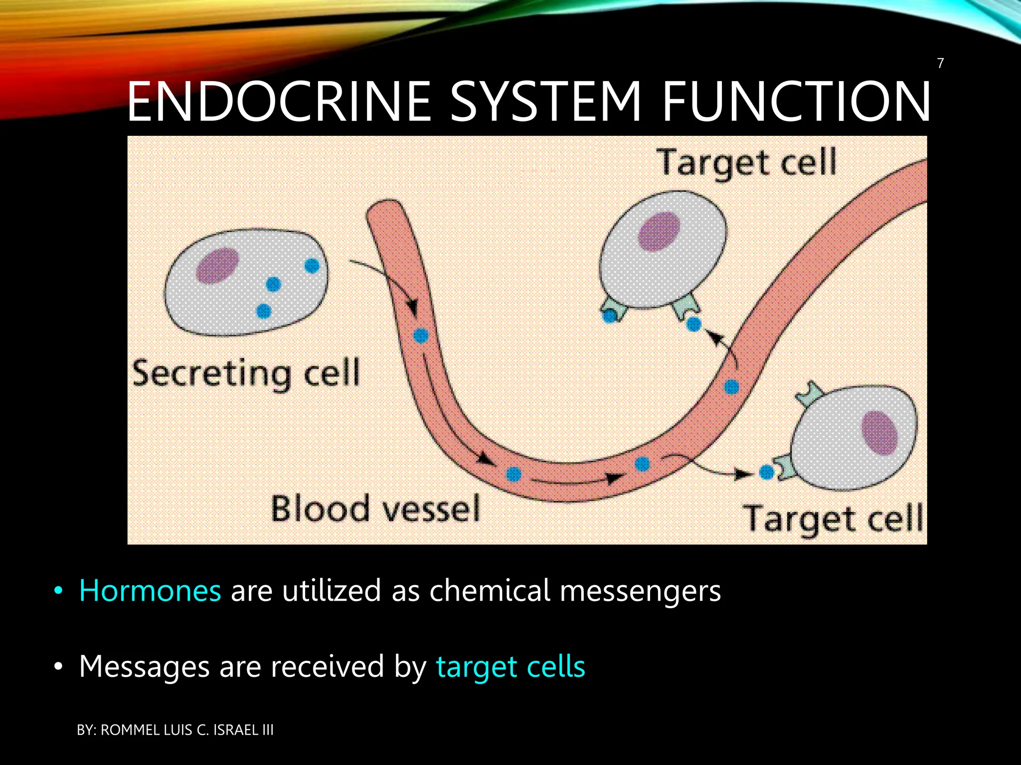
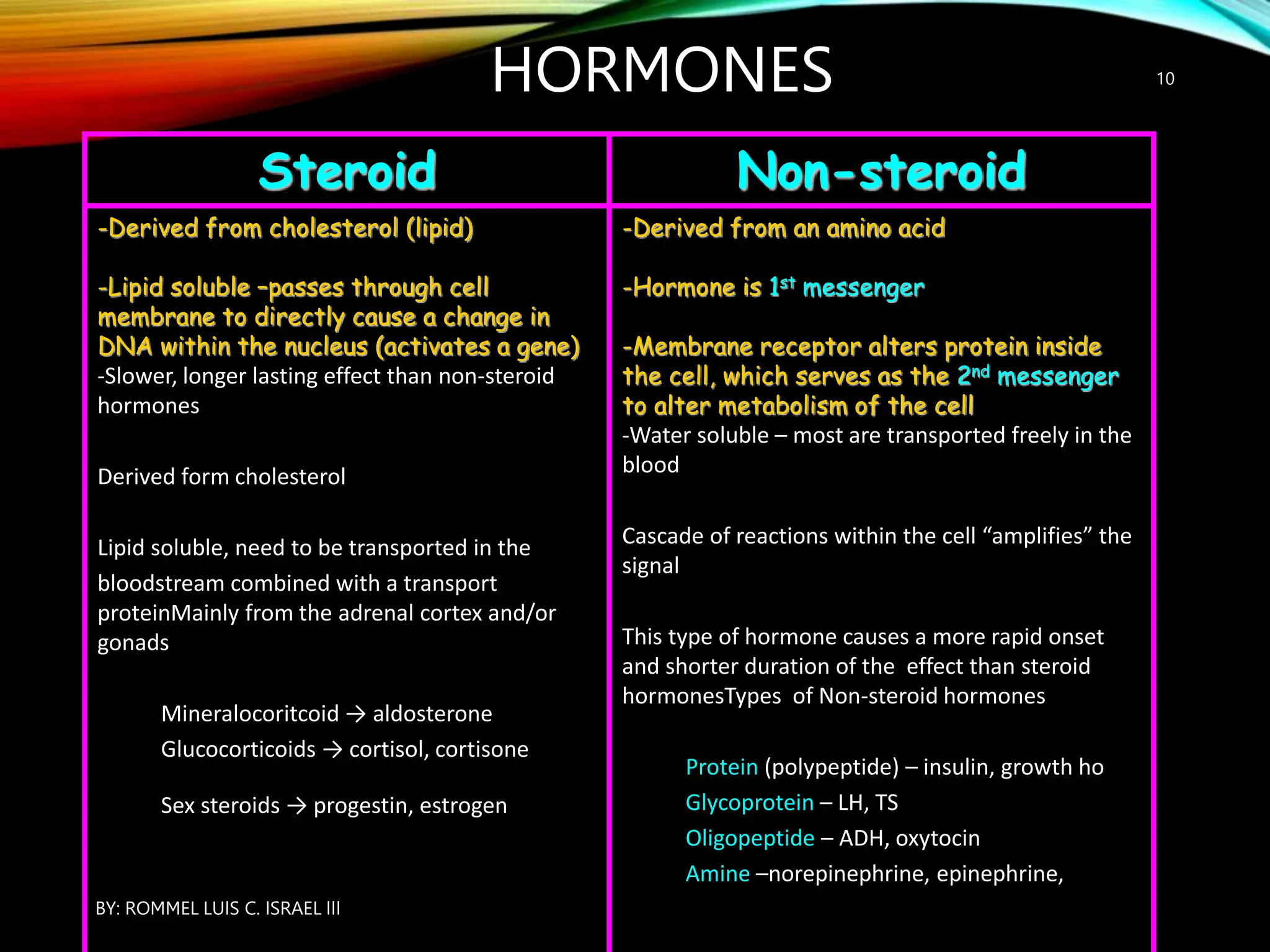
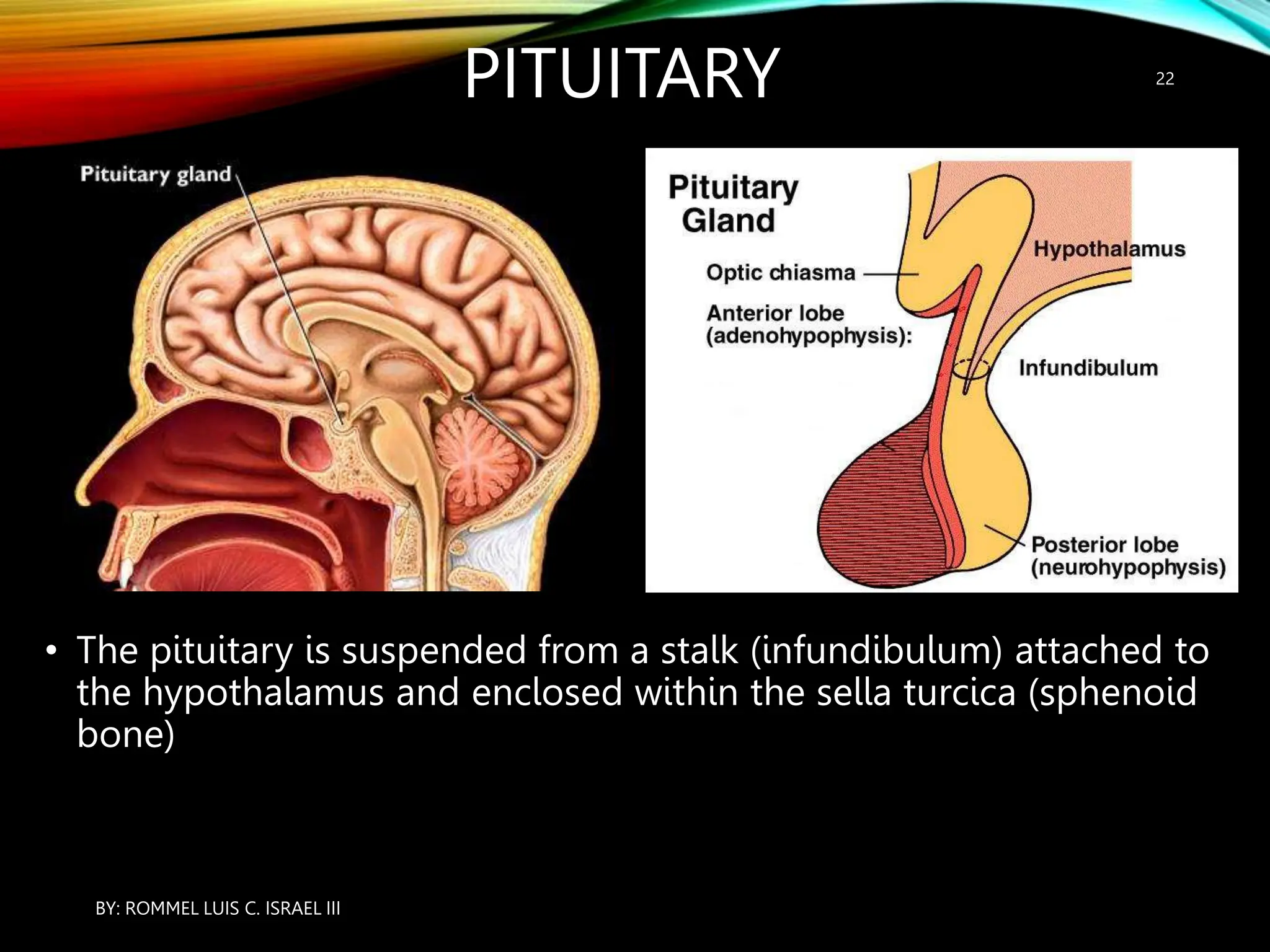
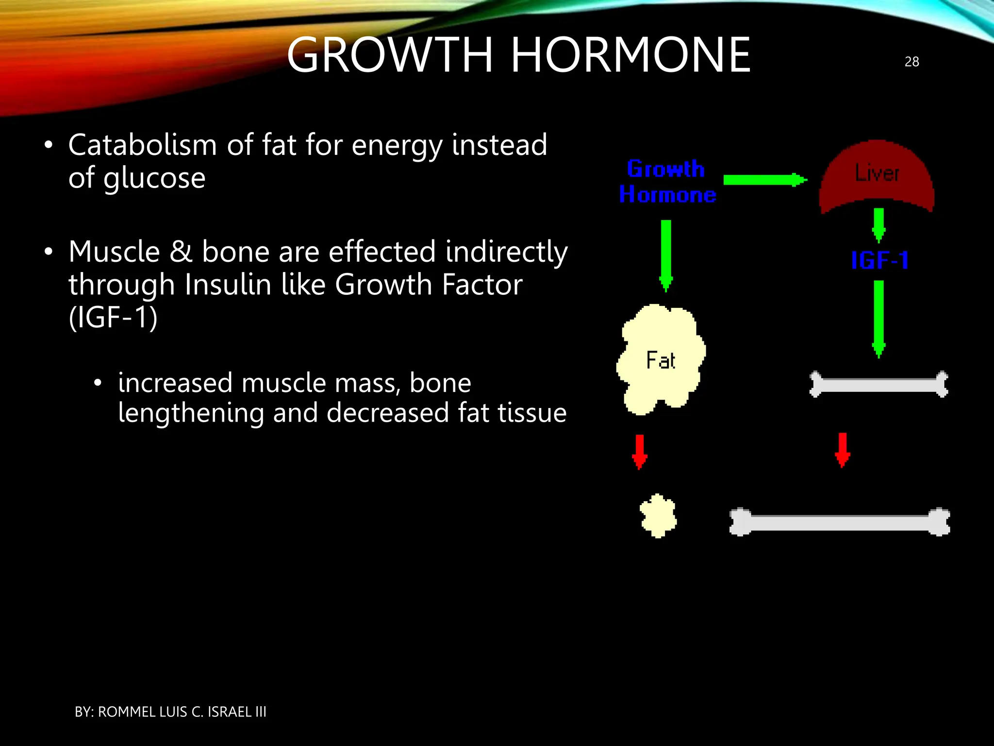
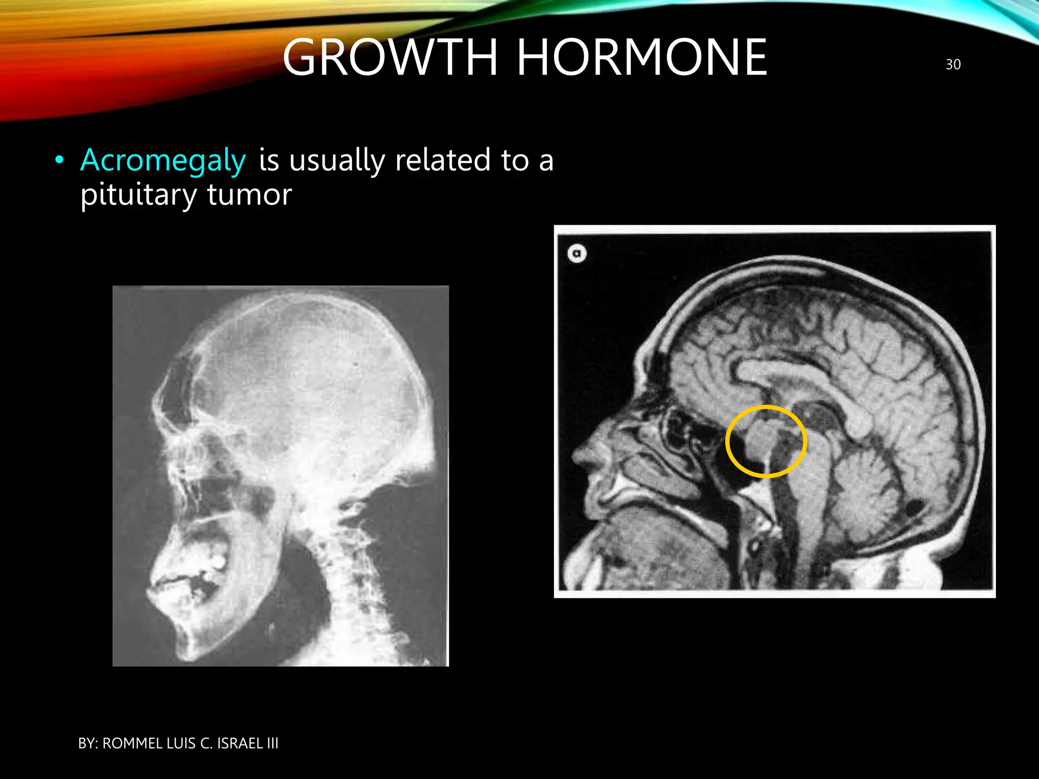
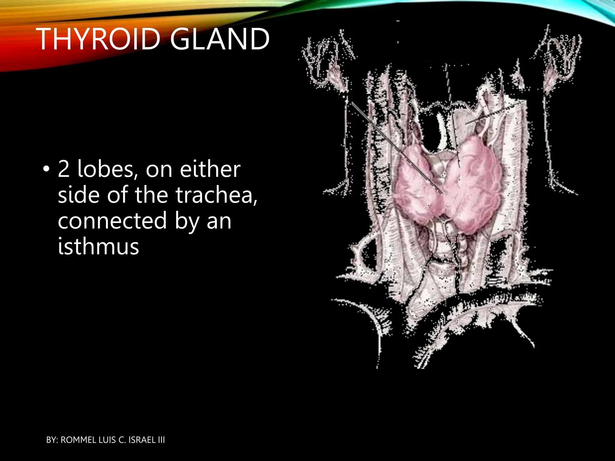
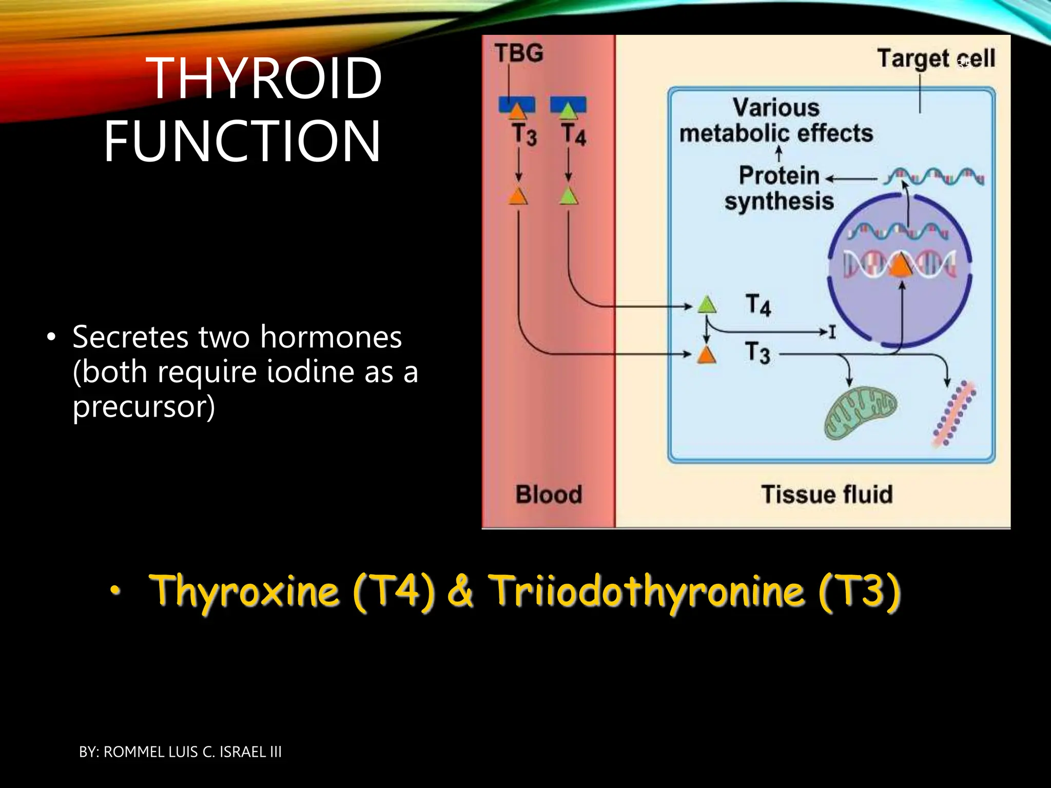
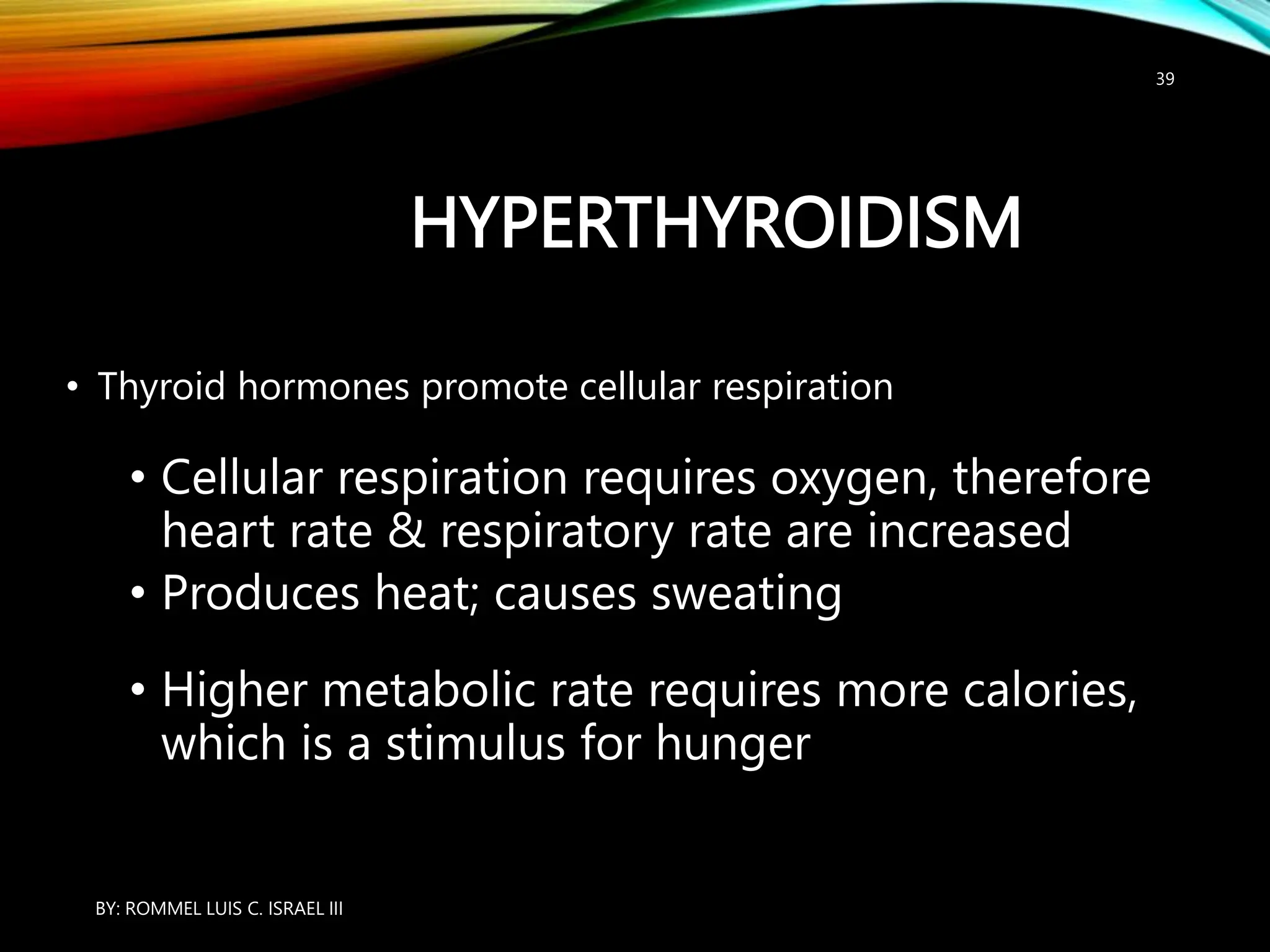
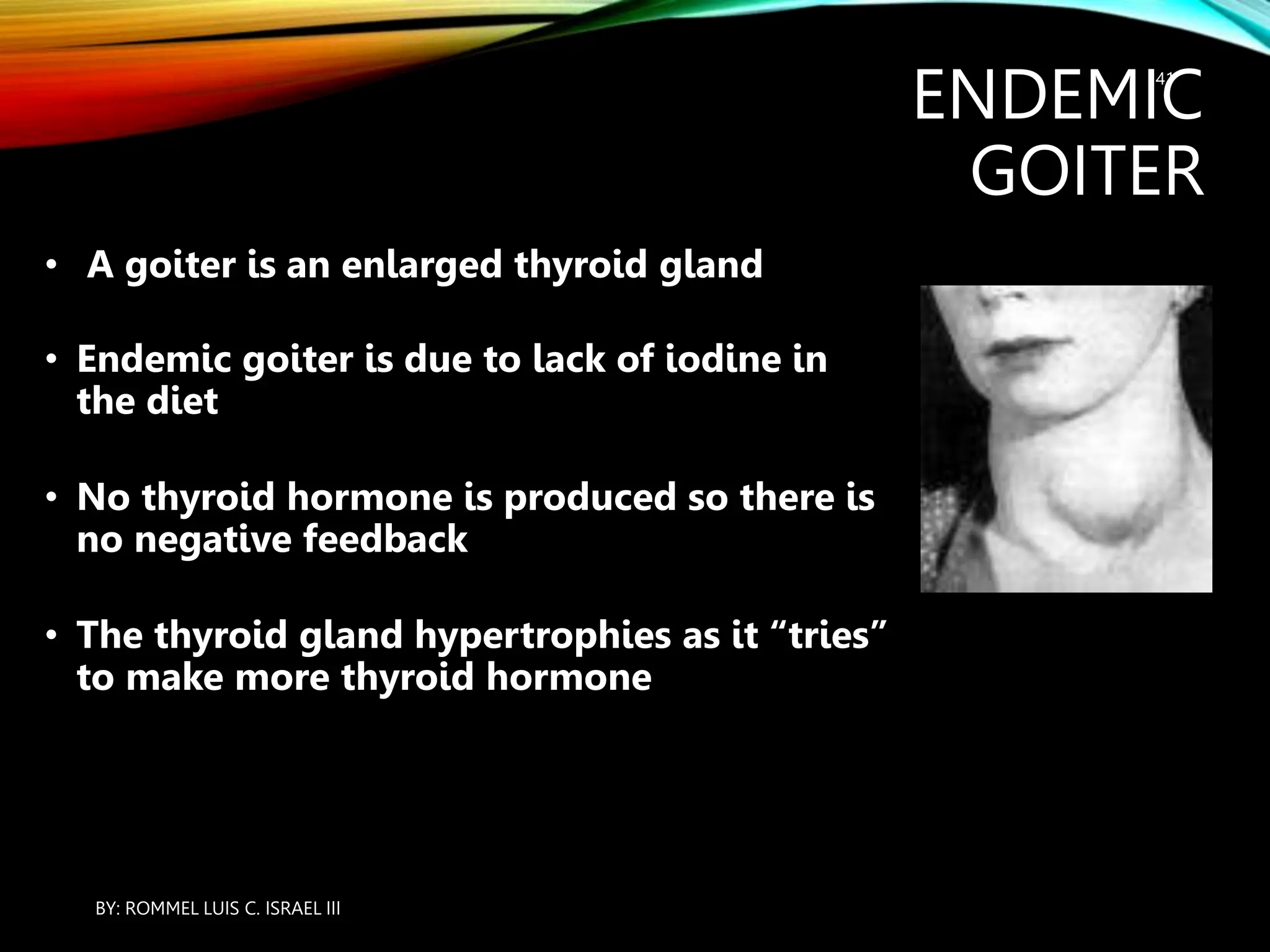
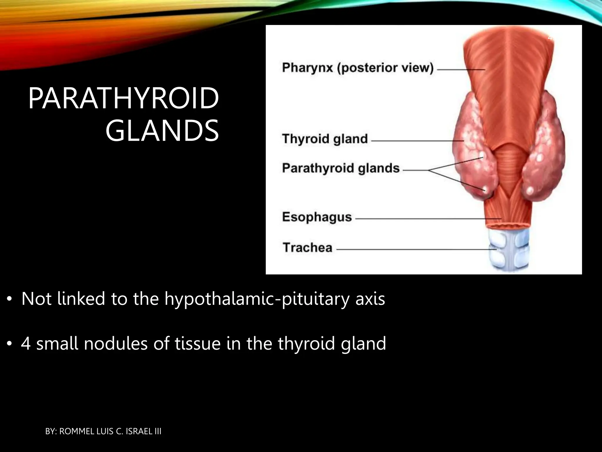
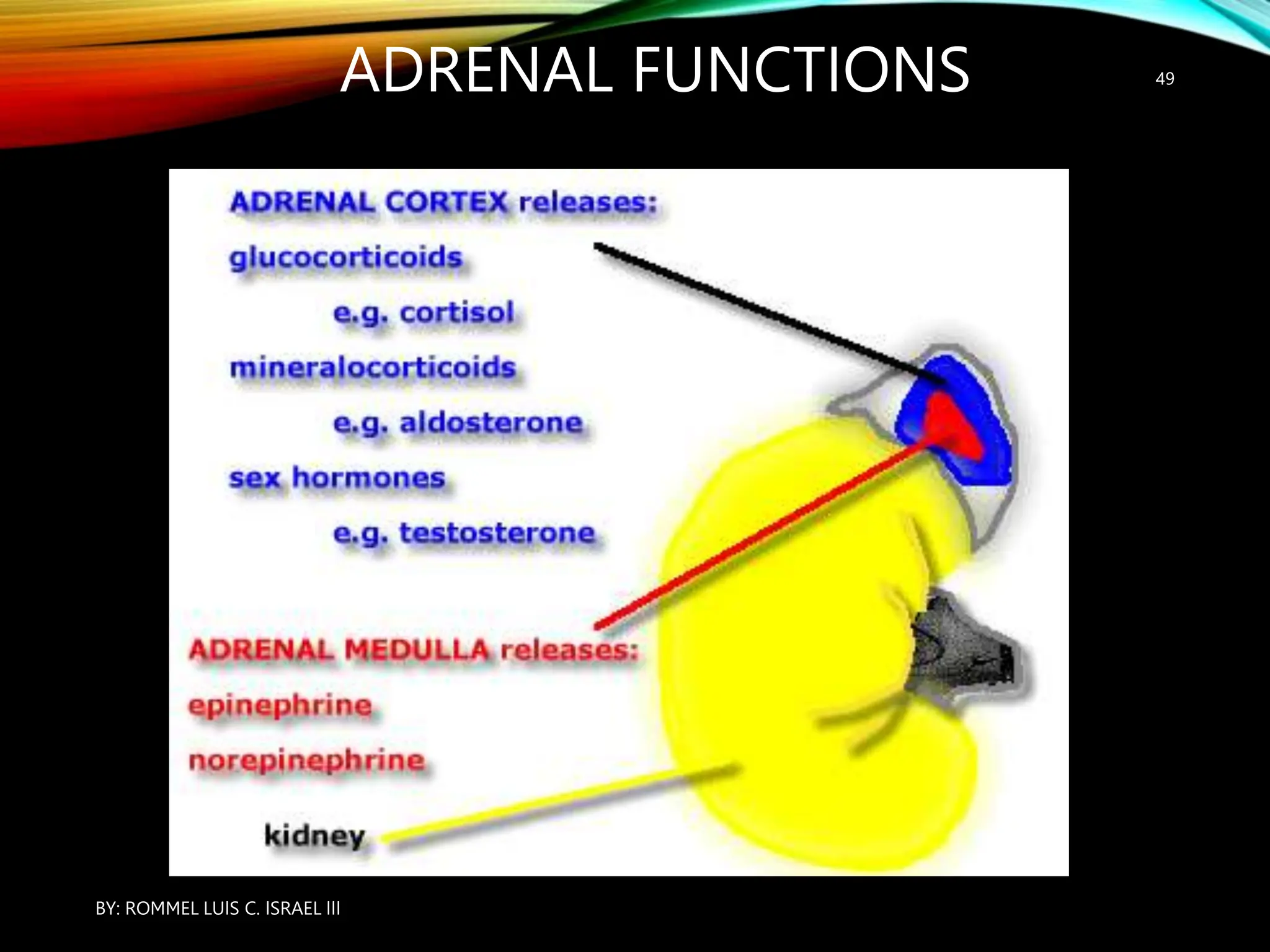
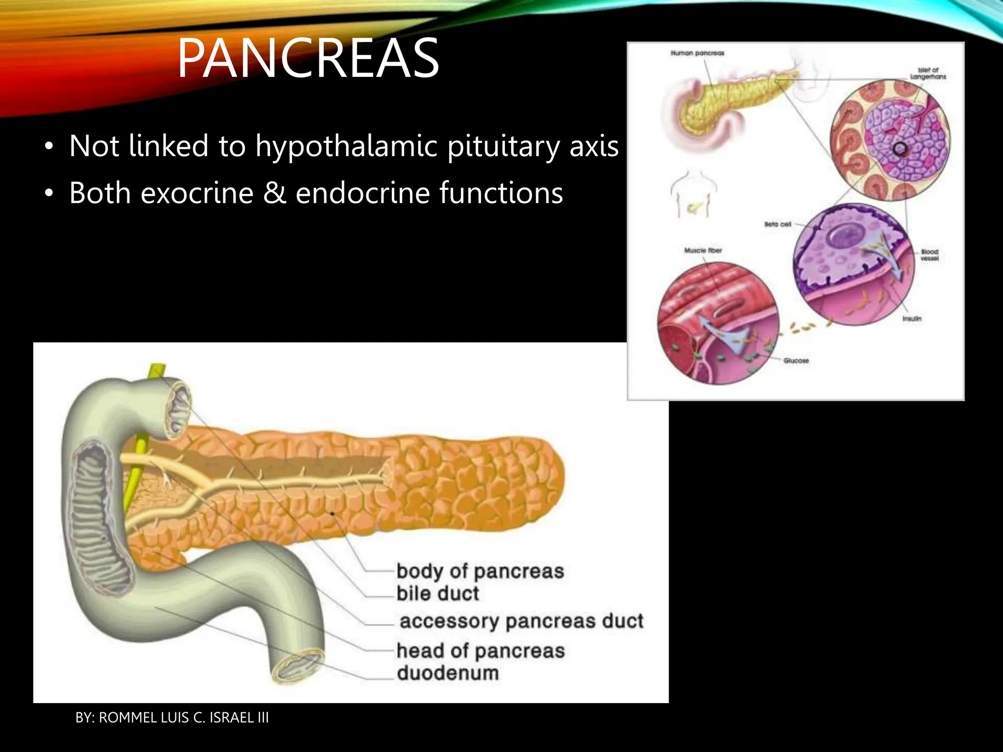
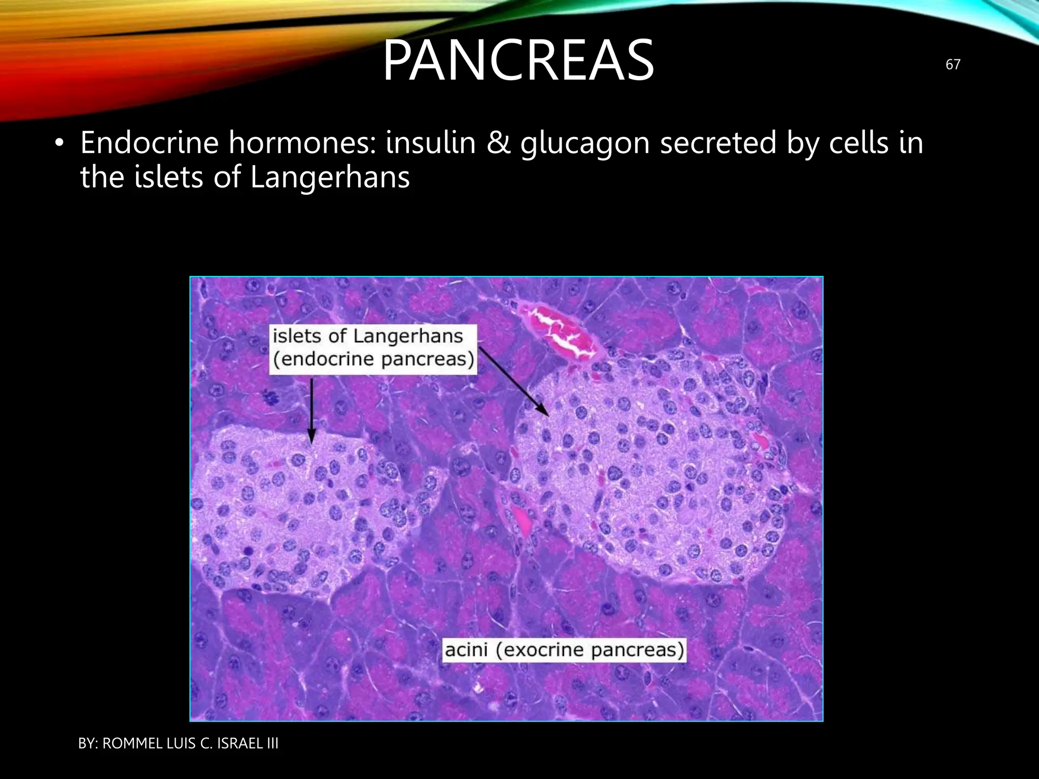
ENDOCRINE SYSTEM; A
REVIEW OF ANATOMY
AND PHYSIOLOGY
BY: ROMMEL LUIS C. ISRAEL III
THE CLASSICAL ENDOCRINE SYSTEM
• Pineal gland
• Hypothalamus / Pituitary
• Thyroid
• Parathyroid
• Thymus
• Adrenal glands
• Pancreas
• Gonads
BY: ROMMEL LUIS C. ISRAEL III
3
OTHER ENDOCRINE ORGANS
• Stomach - releases gastrin from G-cells
• Duodenum – CCK, secretin
• Heart – ANP
• Kidney – renin, erythropoietin
• Placenta - hCG
BY: ROMMEL LUIS C. ISRAEL III
4
ENDOCRINE
SYSTEM
FUNCTION
• Maintenance of homeostasis through negative
feedback loops
BY: ROMMEL LUIS C. ISRAEL III
5
ENDOCRINE SYSTEM FUNCTION
• Communication between organ systems within
the body (like nervous system)
• Differs from the nervous system:
• changes take longer to occur
• and changes persist longer as well.
BY: ROMMEL LUIS C. ISRAEL III
6
ENDOCRINE SYSTEM FUNCTION
• Hormones are utilized as chemical messengers
• Messages are received by target cells
BY: ROMMEL LUIS C. ISRAEL III
7
HORMONES
• Recall the distinction between exocrine
and endocrine glands
• Exocrine: through a duct
• Endocrine: into the blood
BY: ROMMEL LUIS C. ISRAEL III
8
HORMONES
• Endocrine hormones are transmitted in the
bloodstream to all parts of the body
• Hormones only have an effect on those cells that
have a hormone receptor embedded in the cell
membrane (target cells)
• 2 types of endocrine hormones:
• steroid & non-steroid
BY: ROMMEL LUIS C. ISRAEL III
9
HORMONES
Steroid Non-steroid
-Derived from cholesterol (lipid)
-Lipid soluble –passes through cell
membrane to directly cause a change in
DNA within the nucleus (activates a gene)
-Slower, longer lasting effect than non-steroid
hormones
Derived form cholesterol
Lipid soluble, need to be transported in the
bloodstream combined with a transport
proteinMainly from the adrenal cortex and/or
gonads
Mineralocoritcoid → aldosterone
Glucocorticoids → cortisol, cortisone
Sex steroids → progestin, estrogen
-Derived from an amino acid
-Hormone is 1st messenger
-Membrane receptor alters protein inside
the cell, which serves as the 2nd messenger
to alter metabolism of the cell
-Water soluble – most are transported freely in the
blood
Cascade of reactions within the cell “amplifies” the
signal
This type of hormone causes a more rapid onset
and shorter duration of the effect than steroid
hormonesTypes of Non-steroid hormones
Protein (polypeptide) – insulin, growth ho
Glycoprotein – LH, TS
Oligopeptide – ADH, oxytocin
Amine –norepinephrine, epinephrine,
BY: ROMMEL LUIS C. ISRAEL III
10
MECHANISM OF HORMONE
ACTION
• Hormones produce their effects on target
tissues by binding to specific proteins called
hormone receptors located in the target tissues
only.
• Hormone receptors present on the cell
membrane of the target cells are called
membrane-bound receptors and the receptors
present inside the target cell are called
intracellular receptors, mostly nuclear receptors
(present in the nucleus).
BY: ROMMEL LUIS C. ISRAEL III
11
MECHANISM OF HORMONE
ACTION
• Binding of a hormone to its receptor leads to
the formation of a hormone-receptor complex
• Each receptor is specific to one hormone only
and hence receptors are specific.
BY: ROMMEL LUIS C. ISRAEL III
12
MECHANISM OF HORMONE
ACTION
• Hormone-Receptor complex formation leads to
certain biochemical changes in the target tissue.
• Target tissue metabolism and hence
physiological functions are regulated by
hormones.
BY: ROMMEL LUIS C. ISRAEL III
13
HORMONES
BY: ROMMEL LUIS C. ISRAEL III
14
SECOND MESSENGER
• ACTION OF MEMBRANE BOUND RECEPTORS
Hormones which interact with membrane-bound
receptors normally do not enter the target cell, but
generate second messengers (e.g., cyclic AMP, IP3,
Ca++ etc) which in turn regulate cellular metabolism .
• ACTION OF NUCLEAR RECEPTORS Hormones which
interact with intracellular receptors (e.g., steroid
hormones, iodothyronines, etc.) mostly regulate gene
expression orchromosome function by the interaction
of hormone-receptor complex with the genome.
Cumulative biochemical actions result in physiological
and developmental effects .
•
BY: ROMMEL LUIS C. ISRAEL III
15
HYPOTHALAMUS
• the hypothalamus is the basal part of diencephalon,
forebrain.
• These hormones regulate the synthesis and secretion of
pituitary hormones.
• However, the hormones produced by hypothalamus
are of two types, the releasing hormones (which
stimulate secretion of pituitary hormones) and the
inhibiting hormones (which inhibit secretions of
pituitary hormones).
BY: ROMMEL LUIS C. ISRAEL III
16
HYPOTHALAMUS
For example
• hypothalamic hormone called Gonadotrophin releasing hormone (GnRH)
• stimulates the pituitary synthesis and release of gonadotrophins.
• On the other hand, somatostatin from the hypothalamus inhibits the
release of
• growth hormone from the pituitary.
• These hormones originating in the hypothlamic neurons, pass through
axons and are released from their
• nerve endings.
• These hormones reach the pituitary gland through a portal circulatory
system and regulate the functions of the anterior pituitary.
• The posterior pituitary is under the direct neural regulation of the
hypothalamus
BY: ROMMEL LUIS C. ISRAEL III
17
ANTERIOR
PITUITARY
• The releasing hormone is secreted into a portal system
• Cells in the anterior pituitary are stimulated to secrete another
hormone
BY: ROMMEL LUIS C. ISRAEL III
18
POSTERIOR PITUITARY
• Neurons from the
hypothalamus enter
the posterior
pituitary and
stimulate cells to
secrete hormones
BY: ROMMEL LUIS C. ISRAEL III
19
PITUITARY
• Interacts with many (not all) endocrine glands as part of a
feedback loop between the hypothalamus and the respective
gland.
BY: ROMMEL LUIS C. ISRAEL III
20
PITUITARY
• The pituitary serves as a link between the CNS
(hypothalamus) and the rest of the endocrine system
BY: ROMMEL LUIS C. ISRAEL III
21
PITUITARY
• The pituitary is suspended from a stalk (infundibulum) attached to
the hypothalamus and enclosed within the sella turcica (sphenoid
bone)
BY: ROMMEL LUIS C. ISRAEL III
22
PITUITARY
• Actually 2 separate glands: anterior pituitary & posterior
pituitary
• No blood brain barrier in the hypothalamus
BY: ROMMEL LUIS C. ISRAEL III
23
ANTERIOR PITUITARY
hypothalamus
(releasing
hormones)
GHRH PRF TRH CRH GnRH
pituitary
(tropic
hormones)
GH PRL TSH ACTH FSH/LH
end organ muscle,bone
adipose
mammary
gl.
thyroid adrenal
cortex
ovary &
testes
Note:
1. GH is also called somatotropin
2. There are also inhibitory factors from the hypothalamus
for GH and PRL
BY: ROMMEL LUIS C. ISRAEL III
24
POSTERIOR PITUITARY
ADH anti-diuretic hormone, causes kidney to retain
water
Oxytocin contraction of smooth muscle
(uterus/childbirth & mammary gland ducts)
* failure of the posterior pituitary to secrete ADH
results in Diabetes Insipidus (DI)……if no ADH is
produced the result is production of LARGE volumes of
dilute urine
BY: ROMMEL LUIS C. ISRAEL III
25
GROWTH HORMONE
• GHRH/somatostatin  GH or somatotropin  cells related
to body growth
• Produced in the ant. Pituitary
• Secretion declines gradually with age
• Many more GH producing cells in ant. pituitary than any
other type
BY: ROMMEL LUIS C. ISRAEL III
26
GROWTH HORMONE
• Increased cell division (mitosis) & increased
cell growth & differentiation
• The main effects are on cartilage, muscle,
bone, fat
• Promotes protein synthesis
(transcription/translation)
• Anti-insulin effect - elevates blood sugar
BY: ROMMEL LUIS C. ISRAEL III
27
GROWTH HORMONE
• Catabolism of fat for energy instead
of glucose
• Muscle & bone are effected indirectly
through Insulin like Growth Factor
(IGF-1)
• increased muscle mass, bone
lengthening and decreased fat tissue
BY: ROMMEL LUIS C. ISRAEL III
28
GROWTH HORMONE
• Continued secretion of high levels after epiphysial plate closure
is called acromegaly
• Bones continue to thicken even after epiphysial plate closure
BY: ROMMEL LUIS C. ISRAEL III
29
GROWTH HORMONE
• Acromegaly is usually related to a
pituitary tumor
BY: ROMMEL LUIS C. ISRAEL III
30
PROLACTIN
• In women
• increased levels during pregnancy
• after delivery causes mammary glands
to produce milk
• In men
• increases sensitivity to LH and indirectly
enhances testosterone production
BY: ROMMEL LUIS C. ISRAEL III
31
THYROID
GLAND
• TRH (hypothalamus) → TSH (pituitary) → Thyroid hormone
(Thyroid gland)
BY: ROMMEL LUIS C. ISRAEL III
32
THYROID GLAND
• 2 lobes, on either
side of the trachea,
connected by an
isthmus
BY: ROMMEL LUIS C. ISRAEL III
33
THYROID GLAND
Microscopic anatomy
• Thyroid follicles, lined by
secretory epithelium
(follicular cells)
• Filled with thyroglobulin
(transport protein)
• Parafollicular cells between
the follicles
BY: ROMMEL LUIS C. ISRAEL III
34
THYROID
FUNCTION
• Secretes two hormones
(both require iodine as a
precursor)
• Thyroxine (T4) & Triiodothyronine (T3)
BY: ROMMEL LUIS C. ISRAEL III
35
THYROID FUNCTION
• (T4): 90% of all Thyroid
hormone
• Less active
• Converted to T3 in the cell
• Most remains in the
bloodstream as reservoir of
thyroid hormone
BY: ROMMEL LUIS C. ISRAEL III
36
THYROID FUNCTION
• (T3): 5 times more
metabolically active,
but only 10% of total
hormone that is
produced
BY: ROMMEL LUIS C. ISRAEL III
37
THYROID
FUNCTION
1.Mitochondria: increased cellular respiration
2.Ribosomes: increased protein synthesis
3.Chromatin: increased transcription DNA to mRNA
• Binds to three
sites in the cell
BY: ROMMEL LUIS C. ISRAEL III
38
HYPERTHYROIDISM
• Thyroid hormones promote cellular respiration
• Cellular respiration requires oxygen, therefore
heart rate & respiratory rate are increased
• Produces heat; causes sweating
• Higher metabolic rate requires more calories,
which is a stimulus for hunger
BY: ROMMEL LUIS C. ISRAEL III
39
GRAVES DISEASE
• The most common form of
hyperthyroidism (autoimmune)
• Signs and symptoms may
include heat intolerance, 
appetite, weight loss, warm
moist skin, nervousness, tremor,
 BP, tachycardia, goiter,
exophthalmos
Exophthalmos may be due
to an autoimmune,
inflammatory reaction in
the soft tissue that is
confined within the boney
orbit
BY: ROMMEL LUIS C. ISRAEL III
40
ENDEMIC
GOITER
• A goiter is an enlarged thyroid gland
• Endemic goiter is due to lack of iodine in
the diet
• No thyroid hormone is produced so there is
no negative feedback
• The thyroid gland hypertrophies as it “tries”
to make more thyroid hormone
BY: ROMMEL LUIS C. ISRAEL III
41
HYPOTHYROIDISM
• In children (cretinism)
• permanent mental retardation due to
inadequate nervous system development
• In adults
• common cause of fatigue
• other symptoms are related to low BMR
• cold intolerance, weight gain,  CNS function
(mentation), BP, dry skin
BY: ROMMEL LUIS C. ISRAEL III
42
PARATHYROID
GLANDS
• Not linked to the hypothalamic-pituitary axis
• 4 small nodules of tissue in the thyroid gland
BY: ROMMEL LUIS C. ISRAEL III
43
PARATHYROID GLANDS
• PTH is secreted in response to low serum Ca++
(hypocalcemia)
1. Promotes synthesis of calcitrol (active
metabolite of vitamin D), which ↑ GI absorption
of Ca++
2. Limits Ca++ secretion by the kidney
3. Stimulates osteoclasts to reabsorb bone
BY: ROMMEL LUIS C. ISRAEL III
44
PARATHYROIDS
• In humans, four parathyroid glands are present
on the back side of the thyroid gland, one pair
each in the two lobes of the thyroid gland
• The parathyroid glands secrete a peptide
hormone called parathyroid hormone (PTH).
The secretion of PTH is regulated by the
circulating levels of calcium ions.
BY: ROMMEL LUIS C. ISRAEL III
45
PARATHYROIDS
• Parathyroid hormone (PTH) increases the Ca2+ levels in
the blood. PTH acts on bones and stimulates the
process of bone resorption
(dissolution/demineralisation).
• PTH also stimulates reabsorption of Ca2+ by the renal
tubules and increases Ca2+ absorption from the
digested food. It is, thus clear that PTH is a
hypercalcemic hormone, i.e., it increases the blood
Ca2+ levels.
• Along with TCT(THYROID CALCITONIN) it plays a
significant role in calcium balance in the body.
BY: ROMMEL LUIS C. ISRAEL III
46
• PTH is opposed by the action of
calcitonin
• Secreted in response to elevated
Ca++ (hypercalcemia) by
parafollicular (C-cells) of the
thyroid gland
• Little effect in adults
• Stimulates mineral deposition in
bone by osteoblasts
Parathyroid
Glands
BY: ROMMEL LUIS C. ISRAEL III
47
ADRENAL
GLANDS
• Each gland is
actually 2 separate
glands: the cortex
& medulla
BY: ROMMEL LUIS C. ISRAEL III
48
ADRENAL FUNCTIONS
BY: ROMMEL LUIS C. ISRAEL III
49
ADRENAL MEDULLA
• Secretes Catecholamines
• (Epinephrine & Norepinephrine)
• Derived from SNS neurons (Chromaffin cells) that lack dendrites
& axons
BY: ROMMEL LUIS C. ISRAEL III
50
CATECHOLAMINES
• Adrenaline and noradrenaline are called catecholamines
which are rapidly secreted in response to stress of any
kind and during emergency situations and are called
emergency hormones or hormones of Fight or Flight.
• These hormones increase alertness, pupilary dilation,
piloerection (raising of hairs), sweating etc. Both the
hormones increase the heart beat, the strength of heart
contraction and the rate of respiration.
BY: ROMMEL LUIS C. ISRAEL III
51
CATECHOLAMINES
• Catecholamines also stimulate the breakdown
of glycogen resulting in an increased
concentration of glucose in blood. In addition,
they also stimulate the breakdown of lipids and
proteins.
BY: ROMMEL LUIS C. ISRAEL III
52
ADRENAL MEDULLA
• Innervated by Symphatetic Nervous System
• Secretes SNS neurotransmitters mostly
Epinephrine (Adrenalin)
• The presence of these neurotransmitters in the
circulation lowers the threshold for transmission of
an impulse in the SNS for about 30 min.
BY: ROMMEL LUIS C. ISRAEL III
53
EPINEPHRINE
• Elevates blood sugar – glucose sparing effects to
preserve glucose for the CNS
• glycogenolysis
• gluconeogenesis
BY: ROMMEL LUIS C. ISRAEL III
54
ADRENAL CORTEX
3 layers
BY: ROMMEL LUIS C. ISRAEL III
55
Zona
Glomerulosa
mineralocorticoids Aldosterone:
renal Na+ & water
reabsorption
Zona
Fasciculata
gluccorticoids Cortisol
Zona
Reticularis
sex steroids Androgen,
Estrogen
ADRENAL CORTEX
BY: ROMMEL LUIS C. ISRAEL III
56
GLUCOCORTICIODS
• Glucocorticoids stimulate, gluconeogenesis, lipolysis
and proteolysis; and inhibit cellular uptake and
utilisation of amino acids.
• Cortisol is also involved in maintaining the cardio-
vascular system as well as the kidney functions.
• Glucocorticoids, particularly cortisol, produces anti-
inflamatory reactions and suppresses the immune
response.
• Cortisol stimulates the RBC production.
BY: ROMMEL LUIS C. ISRAEL III
57
GLUCOCORTICIODS
• ldosterone acts mainly at the renal tubules and
stimulates the reabsorption of Na+ and water and
excretion of K+ and phosphate ions. Thus, aldosterone
helps in the maintenance of electrolytes, body fluid
volume, osmotic pressure and blood pressure.
• Small amounts of androgenic steroids are also secreted
by the adrenal cortex which play a role in the growth of
axial hair, pubic hair and facial hair during puberty
BY: ROMMEL LUIS C. ISRAEL III
58
• Aldosterone – Na+ retention/ K+ secretion
• Water follows Sodium, so fluid volume and
BP increase too
• Recall that aldosterone secretion can be
promoted by:
1. ACTH
2. Angiotensin II
3. Low Na+
4. High K+
Adrenal Cortex: Z. Glomerulosa
BY: ROMMEL LUIS C. ISRAEL III
59
Secretes Cortisol (hydrocortisone)
• Corticosterone and cortisone are two other
similar hormones that are secreted
• Steroid medication can suppress adrenal
function
Adrenal Cortex: Z. Fasciculata
BY: ROMMEL LUIS C. ISRAEL III
60
Two basic effects of Cortisol:
1. Glucose sparing effects: increased glucose
synthesis & protein/lipid catabolism
(gluconeogenesis)
2. Anti-inflammatory effects: inhibits WBC
function, decreased phagocytosis, decreased
chemotaxis, decreased mast cell degranulation
Adrenal Cortex: Z. Fasciculata
BY: ROMMEL LUIS C. ISRAEL III
61
ADRENAL CORTEX: Z. RETICULARIS
• Sex steroids – small amount of estrogens & weak
androgens (DHEA), which is a precursor of
testosterone
• Testosterone is required in men & women – pubic
& axillary hair, libido, apocrine sweat glands
• most testosterone is produced in testes in men
• 50% produced by the adrenal gland in women
BY: ROMMEL LUIS C. ISRAEL III
62
CUSHING’S
DISEASE
• Hyperfunction of the Adrenal glands
• Disrupts normal carbohydrate & protein
metabolism (Cortisol)
• characteristic lipid deposits in the face
• Potential electrolyte imbalance
(Aldosterone)
• Mood changes (Testosterone)
BY: ROMMEL LUIS C. ISRAEL III
63
GONADS
Ovaries
• Secrete estrogen &
progesterone during the
menstrual cycle
• FSH from the pituitary
causes maturation of the
follicle & egg
• LH causes ovulation (rupture
of the follicle – release of
the egg)
Testes
• FSH promotes spermatogenesis
• LH causes interstitial cells to
secrete testosterone
• Testosterone from adrenal gland
is also present in women, but at
much lower levels
• Produces secondary sexual
characteristics in men
BY: ROMMEL LUIS C. ISRAEL III
64
• PTH is opposed by the action of
calcitonin
• Secreted in response to elevated
Ca++ (hypercalcemia) by
parafollicular (C-cells) of the
thyroid gland
• Little effect in adults
• Stimulates mineral deposition in
bone by osteoblasts
Parathyroid
Glands
BY: ROMMEL LUIS C. ISRAEL III
65
PANCREAS
• Not linked to hypothalamic pituitary axis
• Both exocrine & endocrine functions
BY: ROMMEL LUIS C. ISRAEL III
66
PANCREAS
• Endocrine hormones: insulin & glucagon secreted by cells in
the islets of Langerhans
BY: ROMMEL LUIS C. ISRAEL III
67
GLUCAGON
Alpha cells secrete glucagon
• Glucagon is secreted in response to low blood
sugar, causing glycogen to be converted to
glucose (glycogenolysis)
BY: ROMMEL LUIS C. ISRAEL III
68
GLUCAGON
• Glucagon is a peptide hormone, and plays an
important role in maintaining the normal blood
glucose levels.
• Glucagon acts mainly on the liver cells
(hepatocytes) and stimulates glycogenolysis
resulting in an increased blood sugar
(hyperglycemia).
BY: ROMMEL LUIS C. ISRAEL III
69
GLUCAGON
• In addition, this hormone stimulates the
process of gluconeogenesis which also
contributes to hyperglycemia.
• Glucagon reduces the cellular glucose uptake
and utilisation. Thus, glucagon is a
hyperglycemic hormone.
BY: ROMMEL LUIS C. ISRAEL III
70
INSULIN
• Beta cells secrete insulin
• insulin is secreted in response to ↑ blood sugar, causes ↑
permeability of cell membranes throughout the body to glucose
• Except
CNS...does not
require insulin
to take up
glucose
BY: ROMMEL LUIS C. ISRAEL III
71
INSULIN
• Insulin is a peptide hormone, which plays a major role
in the regulation of glucose homeostasis
• Insulin acts mainly on hepatocytes and adipocytes (cells
of adipose tissue), and enhances cellular glucose uptake
and utilisation.
• As a result, there is a rapid movement of glucose from
blood to hepatocytes and adipocytes resulting in
decreased blood glucose levels (hypoglycemia).
BY: ROMMEL LUIS C. ISRAEL III
72
INSULIN
• Insulin also stimulates conversion of glucose to
glycogen (glycogenesis) in the target cells.
• The glucose homeostasis in blood is thus
maintained jointly by the two – insulin and
glucagons
BY: ROMMEL LUIS C. ISRAEL III
73
DIABETES MELLITUS
• Elevated blood sugar
• Normal fasting blood sugar is 70 – 100 mg/dL
• 100 – 125 mg/dL is “prediabetic”
• Higher than 125 mg/dL is either Type I or Type II
diabetes
BY: ROMMEL LUIS C. ISRAEL III
74
TYPE I DIABETES MELLITUS
• A failure of beta cells to produce insulin
• Also called IDDM (Insulin Dependent DM)
• complete loss of insulin means that replacement is required
• Usually onset is in childhood (juvenile onset)
BY: ROMMEL LUIS C. ISRAEL III
75
TYPE I DIABETES MELLITUS
• Abrupt onset of symptoms
• DKA (diabetic ketoacidosis)
• No glucose is available to cells
• They utilize lipids instead
• This produces ketoacids (lower the pH)
• Polydipsia (thirst)
• Polyphagia (hunger)
• Polyuria (osmotic diuresis)
BY: ROMMEL LUIS C. ISRAEL III
76
• The renal threshold for glucose resorption is exceeded
resulting in glycosuria
• Poorly controlled DM is a disease of small blood vessels
• diabetic nephropathy
• diabetic retinopathy
• diabetic neuropathy
• changes in coronary & peripheral vessels increase the
risk of vascular disease
Type I Diabetes Mellitus
BY: ROMMEL LUIS C. ISRAEL III
77
DIABETES MELLITUS TYPE II
• An insensitivity of an insulin receptor in cell
membranes to insulin
• NIDDM (Non Insulin Dependent DM)
• usually does not require insulin (oral
hypoglycemics)
• Usually onset is as an adult
• Onset is insidious
• Usually no ketoacidosis
BY: ROMMEL LUIS C. ISRAEL III
78
DIABETES MELLITUS
• Long term monitoring of blood sugar levels
(months)
• Hgb A1C (glycosylated hemoglobin)
• A type of hgb that incorporates a sugar molecule
• More Hgb A1C is made when blood sugar levels
are high
BY: ROMMEL LUIS C. ISRAEL III
79
PINEAL GLAND
• The main hormone is
melatonin
• The precursor is a CNS
neurotransmitter,
serotonin
BY: ROMMEL LUIS C. ISRAEL III
80
PINEAL
GLAND
• Production and secretion of melatonin is stimulated by
darkness
• Information about light levels is provided through the retina in
mammals
BY: ROMMEL LUIS C. ISRAEL III
81
PINEAL GLAND
• The pineal gland is located on the dorsal side of
forebrain.
• Pineal secretes a hormone called melatonin.
• Melatonin plays a very important role in the
regulationof a 24-hour (diurnal) rhythm of our
body.
• For example, it helps in maintaining the normal
rhythms of sleep-wake cycle, body temperature.
• In addition, melatonin also influences metabolism,
pigmentation,the menstrual cycle as well as our
defense capability.
BY: ROMMEL LUIS C. ISRAEL III
82
PINEAL GLAND
• Circadian rhythm Regulates sleep / wake cycle
• Undergoes involution during childhood, which may bring
about the onset of puberty
• Near the skin in non-mammal vertebrates
• Light levels are perceived directly
• Regulates seasonal behavior in other animals
• Migratory patterns in birds
• Breeding cycles in animals with seasonal reproductive patterns
BY: ROMMEL LUIS C. ISRAEL III
83
THYMUS
• Posterior to the
sternum
• Larger in adolescence
• Regresses at puberty
• Secretes thymosin
BY: ROMMEL LUIS C. ISRAEL III
84
THYMUS
• The thymus gland is a lobular structure located
on the dorsal side of the heart and the aorta.
• The thymus plays a major role in the
development of the immune system.
• This gland secretes the peptide hormones
called thymosins.
BY: ROMMEL LUIS C. ISRAEL III
85
THYMUS
• Thymosins play a major role in the differentiation of T-
lymphocytes, which provide cell-mediated
immunity.
• thymosins also promote production of antibodies to
provide humoral immunity.
• Thymus is degenerated in old individuals resulting in a
decreased production of thymosins. As a result, the
immune responses of old persons become weak.
BY: ROMMEL LUIS C. ISRAEL III
86
THYMUS
• Thymosin causes undifferentiated lymphocytes to become T cells
• Blood – thymus barrier in the cortex
• Only T cells that are “self tolerant” are released to the medulla and
into the rest of the body
cortex
medulla
BY: ROMMEL LUIS C. ISRAEL III
87
TESTIS
• The Leydig cells or interstitial cells, which are present in
the intertubular spacesproduce a group of hormones
called androgens mainly testosterone.
• Androgens regulate the development, maturation and
functions of the male accessory sex organs like
epididymis, vas deferens, seminal vesicles, prostate
gland, urethra etc.
BY: ROMMEL LUIS C. ISRAEL III
88
TESTIS
• These hormones stimulate muscular growth, growth of facial and
axillary hair, aggressiveness, low pitch of voice etc.
•
• Androgens play a major stimulatory role in the process of
spermatogenesis (formation of spermatozoa).
• Androgens act on the central neural system and influence the male
sexual behaviour (libido).
•
• These hormones produce anabolic (synthetic) effects on protein
and carbohydrate metabolism.
BY: ROMMEL LUIS C. ISRAEL III
89
OVARY
• Females have a pair of ovaries which produce two
groups of steroid hormones called estrogen and
progesterone.
• Ovary is composed of ovarian follicles and stromal
tissues. The estrogen is synthesised and secreted mainly
by the growing ovarian follicles.
• After ovulation, the ruptured follicle is converted to a
structure called corpus luteum, which secretes mainly
progesterone.
BY: ROMMEL LUIS C. ISRAEL III
90
OVARY
• Estrogens produce wide ranging actions such as
stimulation of growth and activities of female
secondary sex organs, development of growing ovarian
follicles, appearance of female secondary sex characters
(e.g., high pitch of voice, etc.), mammary gland
development. Estrogens also regulate female sexual
behaviour.
• Progesterone supports pregnancy. Progesterone also
acts on the mammary glands and stimulates the
formation of alveoli (sac-like structures which store
milk) and milk secretion.
BY: ROMMEL LUIS C. ISRAEL III
91
ANF (ATRIAL NATRIURETIC
FACTOR)
• hormones are also secreted by some tissues which
are not endocrine glands.
For example
• the atrial wall of our heart secretes a very
important peptide hormone called atrial natriuretic
factor (ANF),which decreases blood pressure.
• When blood pressure is increased, ANF is secreted
which causes dilation of the blood vessels. This
reduces the blood pressure.
BY: ROMMEL LUIS C. ISRAEL III
92
OTHER HORMONES
• The juxtaglomerular cells of kidney produce a peptide hormone
called erythropoietin which stimulates erythropoiesis (formation of
RBC).
• Endocrine cells present in different parts of the gastro-intestinal
tract secrete four major peptide hormones, namely gastrin,
secretin,
• cholecystokinin (CCK) and gastric inhibitory peptide (GIP).
• Gastrin acts on the gastric glands and stimulates the secretion of
hydrochloric acid and pepsinogen.
• Secretin acts on the exocrine pancreas and stimulates secretion of
water and bicarbonate ions.
• CCK acts on both pancreas and gall bladder and stimulates the
secretion of pancreatic enzymes and bile juice, respectively.
• GIP inhibits gastric secretion and motility
BY: ROMMEL LUIS C. ISRAEL III
93
TYPES OF HORMONES
On the basis of their chemical nature, hormones can be
divided into groups :
• (i) peptide, polypeptide, protein hormones (e.g., insulin,
glucagon,pituitary hormones, hypothalamic hormones, etc.)
• (ii) steroids (e.g., cortisol, testosterone, estradiol and
progesterone)
• (iii) iodothyronines (thyroid hormones)
• (iv) amino-acid derivatives (e.g., epinephrine).
BY: ROMMEL LUIS C. ISRAEL III
94
TYPES OF HORMONES
On the basis of their chemical nature, hormones can be divided into
groups :
• (i) peptide, polypeptide, protein hormones (e.g., insulin,
glucagon,pituitary hormones, hypothalamic hormones, etc.)
• (ii) steroids (e.g., cortisol, testosterone, estradiol and progesterone)
• (iii) iodothyronines (thyroid hormones)
• (iv) amino-acid derivatives (e.g., epinephrine).
BY: ROMMEL LUIS C. ISRAEL III
95
STRESS
• A stressor is a stimulus to promote a response to a threatening
situation
physical stress - an actual
physical change
psychological stress
 post surgical
 hot/cold
 trauma
 malnutrition
 hemorrhage
 originates in the
cerebral cortex
 abstraction about a
potentially harmful
situation
 anger, grief,
depression, anxiety, guilt
BY: ROMMEL LUIS C. ISRAEL III
96
STRESS
3 phases of the stress response
1. The alarm phase
2. Resistance phase: occurs as glycogen is
consumed
3. Exhaustion phase: chronic stress occurs over
a period of weeks
BY: ROMMEL LUIS C. ISRAEL III
97
• An immediate response
• Increased sympathetic output (fight or flight)
• The adrenal medulla secretes catecholamines like
adrenalin (SNS neurotransmitter)
Stress: The Alarm Phase
BY: ROMMEL LUIS C. ISRAEL III
98
Sympathetic Nervous System input to the kidney
initiates the Right Artery Aneurysm cascade which
leads to :
• Increased BP to supply large skeletal muscles
• Increased fluid retention to compensate for
potential fluid loss through sweat or
hemorrhage
• Glycogen stores are consumed in a few hours
Stress: The Alarm Phase
BY: ROMMEL LUIS C. ISRAEL III
99
Occurs as glycogen is consumed:
• Hypothalamus → CRH →
ACTH → Cortisol
Stress:
The Resistance
Phase
BY: ROMMEL LUIS C. ISRAEL III
100
• Cortisol decreases glucose use peripherally
(glucose sparing for the CNS)
• Promotes the breakdown of protein & fatty
acids, which are converted to glucose in the liver
(gluconeogenesis)
Stress: The Resistance Phase
BY: ROMMEL LUIS C. ISRAEL III
101
• Both Adrenaline (alarm phase) and Cortisol (resistance
phase) elevate blood sugar
Stress and Blood Sugar
BY: ROMMEL LUIS C. ISRAEL III
102
STRESS: THE EXHAUSTION PHASE
Chronic stress occurs over a period of weeks:
• Less protein is available for immune system function
(gluconeogenesis)
• ↓ ability to make antibodies (protein)
• ↑ susceptibility to infections
• ↓ protein available for wound healing
BY: ROMMEL LUIS C. ISRAEL III
103
PARACRINE SECRETIONS
• Paracrine hormones exert a
local effect
• Eicosanoids are paracrine
secretions
• They are produced by the
Arachidonic acid pathway
BY: ROMMEL LUIS C. ISRAEL III
104
• Arachidonic acid (a fatty
acid) is produced from
phospholipids in the cell
membrane
• This reaction is catalyzed
by phospholipase A2
Arachidonic Acid Pathway
BY: ROMMEL LUIS C. ISRAEL III
105
Arachidonic acid is then
subjected to either of two
metabolic pathways
1. lipoxygenase – leads to
the production of
leukotrienes, chemical
mediators of
inflammation
Arachidonic Acid Pathway
BY: ROMMEL LUIS C. ISRAEL III
106
2. cyclooxygenase –
leads to the
production of
prostaglandins,
thromboxane, and
prostacyclin
Arachidonic Acid Pathway
BY: ROMMEL LUIS C. ISRAEL III
107
PARACRINE SECRETIONS
Thromboxane is secreted by platelets to enhance
platelet aggregation
Prostaglandins have many effects depending upon
the specific metabolite
• fever
• pain
• etc.
BY: ROMMEL LUIS C. ISRAEL III
108
PARACRINE SECRETIONS
• Steroid medication (e.g.
cortisol/prednisone) blocks
the production of
arachidonic acid
(phosphlipase)
• *Non-steroidal anti-inflammatories (NSAID’s)
like aspirin and Ibuprofen block the
cyclooxygenase pathway to reduce
inflammation & fever
BY: ROMMEL LUIS C. ISRAEL III
109

ENDOCRINE SYSTEM - ANATOMY AND PHYSIOLOGY

  • 1.
    ENDOCRINE SYSTEM; A REVIEWOF ANATOMY AND PHYSIOLOGY BY: ROMMEL LUIS C. ISRAEL III
  • 3.
    THE CLASSICAL ENDOCRINESYSTEM • Pineal gland • Hypothalamus / Pituitary • Thyroid • Parathyroid • Thymus • Adrenal glands • Pancreas • Gonads BY: ROMMEL LUIS C. ISRAEL III 3
  • 4.
    OTHER ENDOCRINE ORGANS •Stomach - releases gastrin from G-cells • Duodenum – CCK, secretin • Heart – ANP • Kidney – renin, erythropoietin • Placenta - hCG BY: ROMMEL LUIS C. ISRAEL III 4
  • 5.
    ENDOCRINE SYSTEM FUNCTION • Maintenance ofhomeostasis through negative feedback loops BY: ROMMEL LUIS C. ISRAEL III 5
  • 6.
    ENDOCRINE SYSTEM FUNCTION •Communication between organ systems within the body (like nervous system) • Differs from the nervous system: • changes take longer to occur • and changes persist longer as well. BY: ROMMEL LUIS C. ISRAEL III 6
  • 7.
    ENDOCRINE SYSTEM FUNCTION •Hormones are utilized as chemical messengers • Messages are received by target cells BY: ROMMEL LUIS C. ISRAEL III 7
  • 8.
    HORMONES • Recall thedistinction between exocrine and endocrine glands • Exocrine: through a duct • Endocrine: into the blood BY: ROMMEL LUIS C. ISRAEL III 8
  • 9.
    HORMONES • Endocrine hormonesare transmitted in the bloodstream to all parts of the body • Hormones only have an effect on those cells that have a hormone receptor embedded in the cell membrane (target cells) • 2 types of endocrine hormones: • steroid & non-steroid BY: ROMMEL LUIS C. ISRAEL III 9
  • 10.
    HORMONES Steroid Non-steroid -Derived fromcholesterol (lipid) -Lipid soluble –passes through cell membrane to directly cause a change in DNA within the nucleus (activates a gene) -Slower, longer lasting effect than non-steroid hormones Derived form cholesterol Lipid soluble, need to be transported in the bloodstream combined with a transport proteinMainly from the adrenal cortex and/or gonads Mineralocoritcoid → aldosterone Glucocorticoids → cortisol, cortisone Sex steroids → progestin, estrogen -Derived from an amino acid -Hormone is 1st messenger -Membrane receptor alters protein inside the cell, which serves as the 2nd messenger to alter metabolism of the cell -Water soluble – most are transported freely in the blood Cascade of reactions within the cell “amplifies” the signal This type of hormone causes a more rapid onset and shorter duration of the effect than steroid hormonesTypes of Non-steroid hormones Protein (polypeptide) – insulin, growth ho Glycoprotein – LH, TS Oligopeptide – ADH, oxytocin Amine –norepinephrine, epinephrine, BY: ROMMEL LUIS C. ISRAEL III 10
  • 11.
    MECHANISM OF HORMONE ACTION •Hormones produce their effects on target tissues by binding to specific proteins called hormone receptors located in the target tissues only. • Hormone receptors present on the cell membrane of the target cells are called membrane-bound receptors and the receptors present inside the target cell are called intracellular receptors, mostly nuclear receptors (present in the nucleus). BY: ROMMEL LUIS C. ISRAEL III 11
  • 12.
    MECHANISM OF HORMONE ACTION •Binding of a hormone to its receptor leads to the formation of a hormone-receptor complex • Each receptor is specific to one hormone only and hence receptors are specific. BY: ROMMEL LUIS C. ISRAEL III 12
  • 13.
    MECHANISM OF HORMONE ACTION •Hormone-Receptor complex formation leads to certain biochemical changes in the target tissue. • Target tissue metabolism and hence physiological functions are regulated by hormones. BY: ROMMEL LUIS C. ISRAEL III 13
  • 14.
    HORMONES BY: ROMMEL LUISC. ISRAEL III 14
  • 15.
    SECOND MESSENGER • ACTIONOF MEMBRANE BOUND RECEPTORS Hormones which interact with membrane-bound receptors normally do not enter the target cell, but generate second messengers (e.g., cyclic AMP, IP3, Ca++ etc) which in turn regulate cellular metabolism . • ACTION OF NUCLEAR RECEPTORS Hormones which interact with intracellular receptors (e.g., steroid hormones, iodothyronines, etc.) mostly regulate gene expression orchromosome function by the interaction of hormone-receptor complex with the genome. Cumulative biochemical actions result in physiological and developmental effects . • BY: ROMMEL LUIS C. ISRAEL III 15
  • 16.
    HYPOTHALAMUS • the hypothalamusis the basal part of diencephalon, forebrain. • These hormones regulate the synthesis and secretion of pituitary hormones. • However, the hormones produced by hypothalamus are of two types, the releasing hormones (which stimulate secretion of pituitary hormones) and the inhibiting hormones (which inhibit secretions of pituitary hormones). BY: ROMMEL LUIS C. ISRAEL III 16
  • 17.
    HYPOTHALAMUS For example • hypothalamichormone called Gonadotrophin releasing hormone (GnRH) • stimulates the pituitary synthesis and release of gonadotrophins. • On the other hand, somatostatin from the hypothalamus inhibits the release of • growth hormone from the pituitary. • These hormones originating in the hypothlamic neurons, pass through axons and are released from their • nerve endings. • These hormones reach the pituitary gland through a portal circulatory system and regulate the functions of the anterior pituitary. • The posterior pituitary is under the direct neural regulation of the hypothalamus BY: ROMMEL LUIS C. ISRAEL III 17
  • 18.
    ANTERIOR PITUITARY • The releasinghormone is secreted into a portal system • Cells in the anterior pituitary are stimulated to secrete another hormone BY: ROMMEL LUIS C. ISRAEL III 18
  • 19.
    POSTERIOR PITUITARY • Neuronsfrom the hypothalamus enter the posterior pituitary and stimulate cells to secrete hormones BY: ROMMEL LUIS C. ISRAEL III 19
  • 20.
    PITUITARY • Interacts withmany (not all) endocrine glands as part of a feedback loop between the hypothalamus and the respective gland. BY: ROMMEL LUIS C. ISRAEL III 20
  • 21.
    PITUITARY • The pituitaryserves as a link between the CNS (hypothalamus) and the rest of the endocrine system BY: ROMMEL LUIS C. ISRAEL III 21
  • 22.
    PITUITARY • The pituitaryis suspended from a stalk (infundibulum) attached to the hypothalamus and enclosed within the sella turcica (sphenoid bone) BY: ROMMEL LUIS C. ISRAEL III 22
  • 23.
    PITUITARY • Actually 2separate glands: anterior pituitary & posterior pituitary • No blood brain barrier in the hypothalamus BY: ROMMEL LUIS C. ISRAEL III 23
  • 24.
    ANTERIOR PITUITARY hypothalamus (releasing hormones) GHRH PRFTRH CRH GnRH pituitary (tropic hormones) GH PRL TSH ACTH FSH/LH end organ muscle,bone adipose mammary gl. thyroid adrenal cortex ovary & testes Note: 1. GH is also called somatotropin 2. There are also inhibitory factors from the hypothalamus for GH and PRL BY: ROMMEL LUIS C. ISRAEL III 24
  • 25.
    POSTERIOR PITUITARY ADH anti-diuretichormone, causes kidney to retain water Oxytocin contraction of smooth muscle (uterus/childbirth & mammary gland ducts) * failure of the posterior pituitary to secrete ADH results in Diabetes Insipidus (DI)……if no ADH is produced the result is production of LARGE volumes of dilute urine BY: ROMMEL LUIS C. ISRAEL III 25
  • 26.
    GROWTH HORMONE • GHRH/somatostatin GH or somatotropin  cells related to body growth • Produced in the ant. Pituitary • Secretion declines gradually with age • Many more GH producing cells in ant. pituitary than any other type BY: ROMMEL LUIS C. ISRAEL III 26
  • 27.
    GROWTH HORMONE • Increasedcell division (mitosis) & increased cell growth & differentiation • The main effects are on cartilage, muscle, bone, fat • Promotes protein synthesis (transcription/translation) • Anti-insulin effect - elevates blood sugar BY: ROMMEL LUIS C. ISRAEL III 27
  • 28.
    GROWTH HORMONE • Catabolismof fat for energy instead of glucose • Muscle & bone are effected indirectly through Insulin like Growth Factor (IGF-1) • increased muscle mass, bone lengthening and decreased fat tissue BY: ROMMEL LUIS C. ISRAEL III 28
  • 29.
    GROWTH HORMONE • Continuedsecretion of high levels after epiphysial plate closure is called acromegaly • Bones continue to thicken even after epiphysial plate closure BY: ROMMEL LUIS C. ISRAEL III 29
  • 30.
    GROWTH HORMONE • Acromegalyis usually related to a pituitary tumor BY: ROMMEL LUIS C. ISRAEL III 30
  • 31.
    PROLACTIN • In women •increased levels during pregnancy • after delivery causes mammary glands to produce milk • In men • increases sensitivity to LH and indirectly enhances testosterone production BY: ROMMEL LUIS C. ISRAEL III 31
  • 32.
    THYROID GLAND • TRH (hypothalamus)→ TSH (pituitary) → Thyroid hormone (Thyroid gland) BY: ROMMEL LUIS C. ISRAEL III 32
  • 33.
    THYROID GLAND • 2lobes, on either side of the trachea, connected by an isthmus BY: ROMMEL LUIS C. ISRAEL III 33
  • 34.
    THYROID GLAND Microscopic anatomy •Thyroid follicles, lined by secretory epithelium (follicular cells) • Filled with thyroglobulin (transport protein) • Parafollicular cells between the follicles BY: ROMMEL LUIS C. ISRAEL III 34
  • 35.
    THYROID FUNCTION • Secretes twohormones (both require iodine as a precursor) • Thyroxine (T4) & Triiodothyronine (T3) BY: ROMMEL LUIS C. ISRAEL III 35
  • 36.
    THYROID FUNCTION • (T4):90% of all Thyroid hormone • Less active • Converted to T3 in the cell • Most remains in the bloodstream as reservoir of thyroid hormone BY: ROMMEL LUIS C. ISRAEL III 36
  • 37.
    THYROID FUNCTION • (T3):5 times more metabolically active, but only 10% of total hormone that is produced BY: ROMMEL LUIS C. ISRAEL III 37
  • 38.
    THYROID FUNCTION 1.Mitochondria: increased cellularrespiration 2.Ribosomes: increased protein synthesis 3.Chromatin: increased transcription DNA to mRNA • Binds to three sites in the cell BY: ROMMEL LUIS C. ISRAEL III 38
  • 39.
    HYPERTHYROIDISM • Thyroid hormonespromote cellular respiration • Cellular respiration requires oxygen, therefore heart rate & respiratory rate are increased • Produces heat; causes sweating • Higher metabolic rate requires more calories, which is a stimulus for hunger BY: ROMMEL LUIS C. ISRAEL III 39
  • 40.
    GRAVES DISEASE • Themost common form of hyperthyroidism (autoimmune) • Signs and symptoms may include heat intolerance,  appetite, weight loss, warm moist skin, nervousness, tremor,  BP, tachycardia, goiter, exophthalmos Exophthalmos may be due to an autoimmune, inflammatory reaction in the soft tissue that is confined within the boney orbit BY: ROMMEL LUIS C. ISRAEL III 40
  • 41.
    ENDEMIC GOITER • A goiteris an enlarged thyroid gland • Endemic goiter is due to lack of iodine in the diet • No thyroid hormone is produced so there is no negative feedback • The thyroid gland hypertrophies as it “tries” to make more thyroid hormone BY: ROMMEL LUIS C. ISRAEL III 41
  • 42.
    HYPOTHYROIDISM • In children(cretinism) • permanent mental retardation due to inadequate nervous system development • In adults • common cause of fatigue • other symptoms are related to low BMR • cold intolerance, weight gain,  CNS function (mentation), BP, dry skin BY: ROMMEL LUIS C. ISRAEL III 42
  • 43.
    PARATHYROID GLANDS • Not linkedto the hypothalamic-pituitary axis • 4 small nodules of tissue in the thyroid gland BY: ROMMEL LUIS C. ISRAEL III 43
  • 44.
    PARATHYROID GLANDS • PTHis secreted in response to low serum Ca++ (hypocalcemia) 1. Promotes synthesis of calcitrol (active metabolite of vitamin D), which ↑ GI absorption of Ca++ 2. Limits Ca++ secretion by the kidney 3. Stimulates osteoclasts to reabsorb bone BY: ROMMEL LUIS C. ISRAEL III 44
  • 45.
    PARATHYROIDS • In humans,four parathyroid glands are present on the back side of the thyroid gland, one pair each in the two lobes of the thyroid gland • The parathyroid glands secrete a peptide hormone called parathyroid hormone (PTH). The secretion of PTH is regulated by the circulating levels of calcium ions. BY: ROMMEL LUIS C. ISRAEL III 45
  • 46.
    PARATHYROIDS • Parathyroid hormone(PTH) increases the Ca2+ levels in the blood. PTH acts on bones and stimulates the process of bone resorption (dissolution/demineralisation). • PTH also stimulates reabsorption of Ca2+ by the renal tubules and increases Ca2+ absorption from the digested food. It is, thus clear that PTH is a hypercalcemic hormone, i.e., it increases the blood Ca2+ levels. • Along with TCT(THYROID CALCITONIN) it plays a significant role in calcium balance in the body. BY: ROMMEL LUIS C. ISRAEL III 46
  • 47.
    • PTH isopposed by the action of calcitonin • Secreted in response to elevated Ca++ (hypercalcemia) by parafollicular (C-cells) of the thyroid gland • Little effect in adults • Stimulates mineral deposition in bone by osteoblasts Parathyroid Glands BY: ROMMEL LUIS C. ISRAEL III 47
  • 48.
    ADRENAL GLANDS • Each glandis actually 2 separate glands: the cortex & medulla BY: ROMMEL LUIS C. ISRAEL III 48
  • 49.
    ADRENAL FUNCTIONS BY: ROMMELLUIS C. ISRAEL III 49
  • 50.
    ADRENAL MEDULLA • SecretesCatecholamines • (Epinephrine & Norepinephrine) • Derived from SNS neurons (Chromaffin cells) that lack dendrites & axons BY: ROMMEL LUIS C. ISRAEL III 50
  • 51.
    CATECHOLAMINES • Adrenaline andnoradrenaline are called catecholamines which are rapidly secreted in response to stress of any kind and during emergency situations and are called emergency hormones or hormones of Fight or Flight. • These hormones increase alertness, pupilary dilation, piloerection (raising of hairs), sweating etc. Both the hormones increase the heart beat, the strength of heart contraction and the rate of respiration. BY: ROMMEL LUIS C. ISRAEL III 51
  • 52.
    CATECHOLAMINES • Catecholamines alsostimulate the breakdown of glycogen resulting in an increased concentration of glucose in blood. In addition, they also stimulate the breakdown of lipids and proteins. BY: ROMMEL LUIS C. ISRAEL III 52
  • 53.
    ADRENAL MEDULLA • Innervatedby Symphatetic Nervous System • Secretes SNS neurotransmitters mostly Epinephrine (Adrenalin) • The presence of these neurotransmitters in the circulation lowers the threshold for transmission of an impulse in the SNS for about 30 min. BY: ROMMEL LUIS C. ISRAEL III 53
  • 54.
    EPINEPHRINE • Elevates bloodsugar – glucose sparing effects to preserve glucose for the CNS • glycogenolysis • gluconeogenesis BY: ROMMEL LUIS C. ISRAEL III 54
  • 55.
    ADRENAL CORTEX 3 layers BY:ROMMEL LUIS C. ISRAEL III 55
  • 56.
    Zona Glomerulosa mineralocorticoids Aldosterone: renal Na+& water reabsorption Zona Fasciculata gluccorticoids Cortisol Zona Reticularis sex steroids Androgen, Estrogen ADRENAL CORTEX BY: ROMMEL LUIS C. ISRAEL III 56
  • 57.
    GLUCOCORTICIODS • Glucocorticoids stimulate,gluconeogenesis, lipolysis and proteolysis; and inhibit cellular uptake and utilisation of amino acids. • Cortisol is also involved in maintaining the cardio- vascular system as well as the kidney functions. • Glucocorticoids, particularly cortisol, produces anti- inflamatory reactions and suppresses the immune response. • Cortisol stimulates the RBC production. BY: ROMMEL LUIS C. ISRAEL III 57
  • 58.
    GLUCOCORTICIODS • ldosterone actsmainly at the renal tubules and stimulates the reabsorption of Na+ and water and excretion of K+ and phosphate ions. Thus, aldosterone helps in the maintenance of electrolytes, body fluid volume, osmotic pressure and blood pressure. • Small amounts of androgenic steroids are also secreted by the adrenal cortex which play a role in the growth of axial hair, pubic hair and facial hair during puberty BY: ROMMEL LUIS C. ISRAEL III 58
  • 59.
    • Aldosterone –Na+ retention/ K+ secretion • Water follows Sodium, so fluid volume and BP increase too • Recall that aldosterone secretion can be promoted by: 1. ACTH 2. Angiotensin II 3. Low Na+ 4. High K+ Adrenal Cortex: Z. Glomerulosa BY: ROMMEL LUIS C. ISRAEL III 59
  • 60.
    Secretes Cortisol (hydrocortisone) •Corticosterone and cortisone are two other similar hormones that are secreted • Steroid medication can suppress adrenal function Adrenal Cortex: Z. Fasciculata BY: ROMMEL LUIS C. ISRAEL III 60
  • 61.
    Two basic effectsof Cortisol: 1. Glucose sparing effects: increased glucose synthesis & protein/lipid catabolism (gluconeogenesis) 2. Anti-inflammatory effects: inhibits WBC function, decreased phagocytosis, decreased chemotaxis, decreased mast cell degranulation Adrenal Cortex: Z. Fasciculata BY: ROMMEL LUIS C. ISRAEL III 61
  • 62.
    ADRENAL CORTEX: Z.RETICULARIS • Sex steroids – small amount of estrogens & weak androgens (DHEA), which is a precursor of testosterone • Testosterone is required in men & women – pubic & axillary hair, libido, apocrine sweat glands • most testosterone is produced in testes in men • 50% produced by the adrenal gland in women BY: ROMMEL LUIS C. ISRAEL III 62
  • 63.
    CUSHING’S DISEASE • Hyperfunction ofthe Adrenal glands • Disrupts normal carbohydrate & protein metabolism (Cortisol) • characteristic lipid deposits in the face • Potential electrolyte imbalance (Aldosterone) • Mood changes (Testosterone) BY: ROMMEL LUIS C. ISRAEL III 63
  • 64.
    GONADS Ovaries • Secrete estrogen& progesterone during the menstrual cycle • FSH from the pituitary causes maturation of the follicle & egg • LH causes ovulation (rupture of the follicle – release of the egg) Testes • FSH promotes spermatogenesis • LH causes interstitial cells to secrete testosterone • Testosterone from adrenal gland is also present in women, but at much lower levels • Produces secondary sexual characteristics in men BY: ROMMEL LUIS C. ISRAEL III 64
  • 65.
    • PTH isopposed by the action of calcitonin • Secreted in response to elevated Ca++ (hypercalcemia) by parafollicular (C-cells) of the thyroid gland • Little effect in adults • Stimulates mineral deposition in bone by osteoblasts Parathyroid Glands BY: ROMMEL LUIS C. ISRAEL III 65
  • 66.
    PANCREAS • Not linkedto hypothalamic pituitary axis • Both exocrine & endocrine functions BY: ROMMEL LUIS C. ISRAEL III 66
  • 67.
    PANCREAS • Endocrine hormones:insulin & glucagon secreted by cells in the islets of Langerhans BY: ROMMEL LUIS C. ISRAEL III 67
  • 68.
    GLUCAGON Alpha cells secreteglucagon • Glucagon is secreted in response to low blood sugar, causing glycogen to be converted to glucose (glycogenolysis) BY: ROMMEL LUIS C. ISRAEL III 68
  • 69.
    GLUCAGON • Glucagon isa peptide hormone, and plays an important role in maintaining the normal blood glucose levels. • Glucagon acts mainly on the liver cells (hepatocytes) and stimulates glycogenolysis resulting in an increased blood sugar (hyperglycemia). BY: ROMMEL LUIS C. ISRAEL III 69
  • 70.
    GLUCAGON • In addition,this hormone stimulates the process of gluconeogenesis which also contributes to hyperglycemia. • Glucagon reduces the cellular glucose uptake and utilisation. Thus, glucagon is a hyperglycemic hormone. BY: ROMMEL LUIS C. ISRAEL III 70
  • 71.
    INSULIN • Beta cellssecrete insulin • insulin is secreted in response to ↑ blood sugar, causes ↑ permeability of cell membranes throughout the body to glucose • Except CNS...does not require insulin to take up glucose BY: ROMMEL LUIS C. ISRAEL III 71
  • 72.
    INSULIN • Insulin isa peptide hormone, which plays a major role in the regulation of glucose homeostasis • Insulin acts mainly on hepatocytes and adipocytes (cells of adipose tissue), and enhances cellular glucose uptake and utilisation. • As a result, there is a rapid movement of glucose from blood to hepatocytes and adipocytes resulting in decreased blood glucose levels (hypoglycemia). BY: ROMMEL LUIS C. ISRAEL III 72
  • 73.
    INSULIN • Insulin alsostimulates conversion of glucose to glycogen (glycogenesis) in the target cells. • The glucose homeostasis in blood is thus maintained jointly by the two – insulin and glucagons BY: ROMMEL LUIS C. ISRAEL III 73
  • 74.
    DIABETES MELLITUS • Elevatedblood sugar • Normal fasting blood sugar is 70 – 100 mg/dL • 100 – 125 mg/dL is “prediabetic” • Higher than 125 mg/dL is either Type I or Type II diabetes BY: ROMMEL LUIS C. ISRAEL III 74
  • 75.
    TYPE I DIABETESMELLITUS • A failure of beta cells to produce insulin • Also called IDDM (Insulin Dependent DM) • complete loss of insulin means that replacement is required • Usually onset is in childhood (juvenile onset) BY: ROMMEL LUIS C. ISRAEL III 75
  • 76.
    TYPE I DIABETESMELLITUS • Abrupt onset of symptoms • DKA (diabetic ketoacidosis) • No glucose is available to cells • They utilize lipids instead • This produces ketoacids (lower the pH) • Polydipsia (thirst) • Polyphagia (hunger) • Polyuria (osmotic diuresis) BY: ROMMEL LUIS C. ISRAEL III 76
  • 77.
    • The renalthreshold for glucose resorption is exceeded resulting in glycosuria • Poorly controlled DM is a disease of small blood vessels • diabetic nephropathy • diabetic retinopathy • diabetic neuropathy • changes in coronary & peripheral vessels increase the risk of vascular disease Type I Diabetes Mellitus BY: ROMMEL LUIS C. ISRAEL III 77
  • 78.
    DIABETES MELLITUS TYPEII • An insensitivity of an insulin receptor in cell membranes to insulin • NIDDM (Non Insulin Dependent DM) • usually does not require insulin (oral hypoglycemics) • Usually onset is as an adult • Onset is insidious • Usually no ketoacidosis BY: ROMMEL LUIS C. ISRAEL III 78
  • 79.
    DIABETES MELLITUS • Longterm monitoring of blood sugar levels (months) • Hgb A1C (glycosylated hemoglobin) • A type of hgb that incorporates a sugar molecule • More Hgb A1C is made when blood sugar levels are high BY: ROMMEL LUIS C. ISRAEL III 79
  • 80.
    PINEAL GLAND • Themain hormone is melatonin • The precursor is a CNS neurotransmitter, serotonin BY: ROMMEL LUIS C. ISRAEL III 80
  • 81.
    PINEAL GLAND • Production andsecretion of melatonin is stimulated by darkness • Information about light levels is provided through the retina in mammals BY: ROMMEL LUIS C. ISRAEL III 81
  • 82.
    PINEAL GLAND • Thepineal gland is located on the dorsal side of forebrain. • Pineal secretes a hormone called melatonin. • Melatonin plays a very important role in the regulationof a 24-hour (diurnal) rhythm of our body. • For example, it helps in maintaining the normal rhythms of sleep-wake cycle, body temperature. • In addition, melatonin also influences metabolism, pigmentation,the menstrual cycle as well as our defense capability. BY: ROMMEL LUIS C. ISRAEL III 82
  • 83.
    PINEAL GLAND • Circadianrhythm Regulates sleep / wake cycle • Undergoes involution during childhood, which may bring about the onset of puberty • Near the skin in non-mammal vertebrates • Light levels are perceived directly • Regulates seasonal behavior in other animals • Migratory patterns in birds • Breeding cycles in animals with seasonal reproductive patterns BY: ROMMEL LUIS C. ISRAEL III 83
  • 84.
    THYMUS • Posterior tothe sternum • Larger in adolescence • Regresses at puberty • Secretes thymosin BY: ROMMEL LUIS C. ISRAEL III 84
  • 85.
    THYMUS • The thymusgland is a lobular structure located on the dorsal side of the heart and the aorta. • The thymus plays a major role in the development of the immune system. • This gland secretes the peptide hormones called thymosins. BY: ROMMEL LUIS C. ISRAEL III 85
  • 86.
    THYMUS • Thymosins playa major role in the differentiation of T- lymphocytes, which provide cell-mediated immunity. • thymosins also promote production of antibodies to provide humoral immunity. • Thymus is degenerated in old individuals resulting in a decreased production of thymosins. As a result, the immune responses of old persons become weak. BY: ROMMEL LUIS C. ISRAEL III 86
  • 87.
    THYMUS • Thymosin causesundifferentiated lymphocytes to become T cells • Blood – thymus barrier in the cortex • Only T cells that are “self tolerant” are released to the medulla and into the rest of the body cortex medulla BY: ROMMEL LUIS C. ISRAEL III 87
  • 88.
    TESTIS • The Leydigcells or interstitial cells, which are present in the intertubular spacesproduce a group of hormones called androgens mainly testosterone. • Androgens regulate the development, maturation and functions of the male accessory sex organs like epididymis, vas deferens, seminal vesicles, prostate gland, urethra etc. BY: ROMMEL LUIS C. ISRAEL III 88
  • 89.
    TESTIS • These hormonesstimulate muscular growth, growth of facial and axillary hair, aggressiveness, low pitch of voice etc. • • Androgens play a major stimulatory role in the process of spermatogenesis (formation of spermatozoa). • Androgens act on the central neural system and influence the male sexual behaviour (libido). • • These hormones produce anabolic (synthetic) effects on protein and carbohydrate metabolism. BY: ROMMEL LUIS C. ISRAEL III 89
  • 90.
    OVARY • Females havea pair of ovaries which produce two groups of steroid hormones called estrogen and progesterone. • Ovary is composed of ovarian follicles and stromal tissues. The estrogen is synthesised and secreted mainly by the growing ovarian follicles. • After ovulation, the ruptured follicle is converted to a structure called corpus luteum, which secretes mainly progesterone. BY: ROMMEL LUIS C. ISRAEL III 90
  • 91.
    OVARY • Estrogens producewide ranging actions such as stimulation of growth and activities of female secondary sex organs, development of growing ovarian follicles, appearance of female secondary sex characters (e.g., high pitch of voice, etc.), mammary gland development. Estrogens also regulate female sexual behaviour. • Progesterone supports pregnancy. Progesterone also acts on the mammary glands and stimulates the formation of alveoli (sac-like structures which store milk) and milk secretion. BY: ROMMEL LUIS C. ISRAEL III 91
  • 92.
    ANF (ATRIAL NATRIURETIC FACTOR) •hormones are also secreted by some tissues which are not endocrine glands. For example • the atrial wall of our heart secretes a very important peptide hormone called atrial natriuretic factor (ANF),which decreases blood pressure. • When blood pressure is increased, ANF is secreted which causes dilation of the blood vessels. This reduces the blood pressure. BY: ROMMEL LUIS C. ISRAEL III 92
  • 93.
    OTHER HORMONES • Thejuxtaglomerular cells of kidney produce a peptide hormone called erythropoietin which stimulates erythropoiesis (formation of RBC). • Endocrine cells present in different parts of the gastro-intestinal tract secrete four major peptide hormones, namely gastrin, secretin, • cholecystokinin (CCK) and gastric inhibitory peptide (GIP). • Gastrin acts on the gastric glands and stimulates the secretion of hydrochloric acid and pepsinogen. • Secretin acts on the exocrine pancreas and stimulates secretion of water and bicarbonate ions. • CCK acts on both pancreas and gall bladder and stimulates the secretion of pancreatic enzymes and bile juice, respectively. • GIP inhibits gastric secretion and motility BY: ROMMEL LUIS C. ISRAEL III 93
  • 94.
    TYPES OF HORMONES Onthe basis of their chemical nature, hormones can be divided into groups : • (i) peptide, polypeptide, protein hormones (e.g., insulin, glucagon,pituitary hormones, hypothalamic hormones, etc.) • (ii) steroids (e.g., cortisol, testosterone, estradiol and progesterone) • (iii) iodothyronines (thyroid hormones) • (iv) amino-acid derivatives (e.g., epinephrine). BY: ROMMEL LUIS C. ISRAEL III 94
  • 95.
    TYPES OF HORMONES Onthe basis of their chemical nature, hormones can be divided into groups : • (i) peptide, polypeptide, protein hormones (e.g., insulin, glucagon,pituitary hormones, hypothalamic hormones, etc.) • (ii) steroids (e.g., cortisol, testosterone, estradiol and progesterone) • (iii) iodothyronines (thyroid hormones) • (iv) amino-acid derivatives (e.g., epinephrine). BY: ROMMEL LUIS C. ISRAEL III 95
  • 96.
    STRESS • A stressoris a stimulus to promote a response to a threatening situation physical stress - an actual physical change psychological stress  post surgical  hot/cold  trauma  malnutrition  hemorrhage  originates in the cerebral cortex  abstraction about a potentially harmful situation  anger, grief, depression, anxiety, guilt BY: ROMMEL LUIS C. ISRAEL III 96
  • 97.
    STRESS 3 phases ofthe stress response 1. The alarm phase 2. Resistance phase: occurs as glycogen is consumed 3. Exhaustion phase: chronic stress occurs over a period of weeks BY: ROMMEL LUIS C. ISRAEL III 97
  • 98.
    • An immediateresponse • Increased sympathetic output (fight or flight) • The adrenal medulla secretes catecholamines like adrenalin (SNS neurotransmitter) Stress: The Alarm Phase BY: ROMMEL LUIS C. ISRAEL III 98
  • 99.
    Sympathetic Nervous Systeminput to the kidney initiates the Right Artery Aneurysm cascade which leads to : • Increased BP to supply large skeletal muscles • Increased fluid retention to compensate for potential fluid loss through sweat or hemorrhage • Glycogen stores are consumed in a few hours Stress: The Alarm Phase BY: ROMMEL LUIS C. ISRAEL III 99
  • 100.
    Occurs as glycogenis consumed: • Hypothalamus → CRH → ACTH → Cortisol Stress: The Resistance Phase BY: ROMMEL LUIS C. ISRAEL III 100
  • 101.
    • Cortisol decreasesglucose use peripherally (glucose sparing for the CNS) • Promotes the breakdown of protein & fatty acids, which are converted to glucose in the liver (gluconeogenesis) Stress: The Resistance Phase BY: ROMMEL LUIS C. ISRAEL III 101
  • 102.
    • Both Adrenaline(alarm phase) and Cortisol (resistance phase) elevate blood sugar Stress and Blood Sugar BY: ROMMEL LUIS C. ISRAEL III 102
  • 103.
    STRESS: THE EXHAUSTIONPHASE Chronic stress occurs over a period of weeks: • Less protein is available for immune system function (gluconeogenesis) • ↓ ability to make antibodies (protein) • ↑ susceptibility to infections • ↓ protein available for wound healing BY: ROMMEL LUIS C. ISRAEL III 103
  • 104.
    PARACRINE SECRETIONS • Paracrinehormones exert a local effect • Eicosanoids are paracrine secretions • They are produced by the Arachidonic acid pathway BY: ROMMEL LUIS C. ISRAEL III 104
  • 105.
    • Arachidonic acid(a fatty acid) is produced from phospholipids in the cell membrane • This reaction is catalyzed by phospholipase A2 Arachidonic Acid Pathway BY: ROMMEL LUIS C. ISRAEL III 105
  • 106.
    Arachidonic acid isthen subjected to either of two metabolic pathways 1. lipoxygenase – leads to the production of leukotrienes, chemical mediators of inflammation Arachidonic Acid Pathway BY: ROMMEL LUIS C. ISRAEL III 106
  • 107.
    2. cyclooxygenase – leadsto the production of prostaglandins, thromboxane, and prostacyclin Arachidonic Acid Pathway BY: ROMMEL LUIS C. ISRAEL III 107
  • 108.
    PARACRINE SECRETIONS Thromboxane issecreted by platelets to enhance platelet aggregation Prostaglandins have many effects depending upon the specific metabolite • fever • pain • etc. BY: ROMMEL LUIS C. ISRAEL III 108
  • 109.
    PARACRINE SECRETIONS • Steroidmedication (e.g. cortisol/prednisone) blocks the production of arachidonic acid (phosphlipase) • *Non-steroidal anti-inflammatories (NSAID’s) like aspirin and Ibuprofen block the cyclooxygenase pathway to reduce inflammation & fever BY: ROMMEL LUIS C. ISRAEL III 109