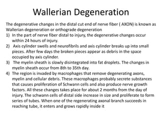
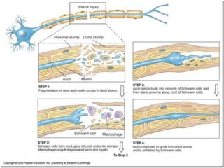
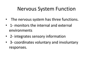
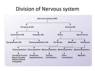
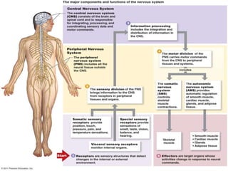
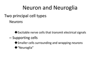
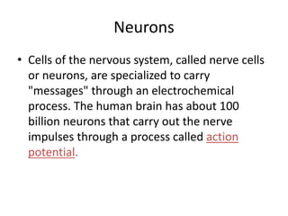
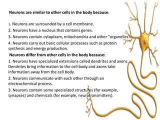
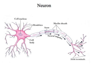
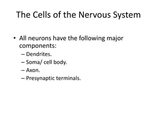
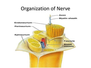
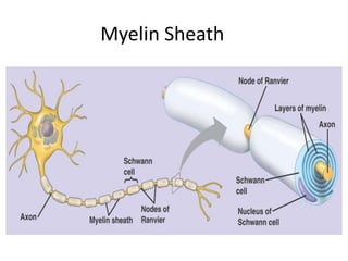
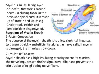
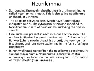
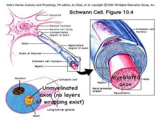
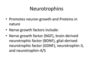
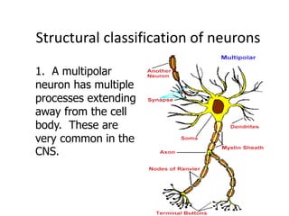
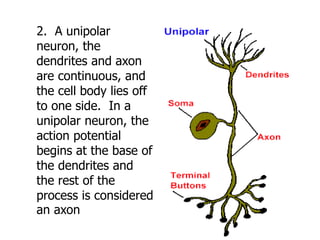
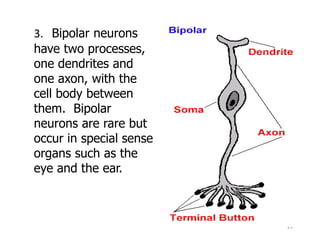
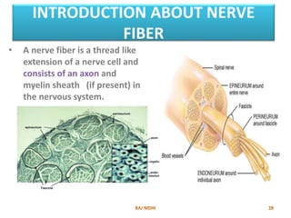
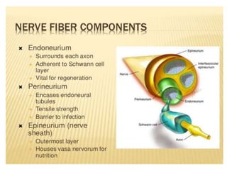
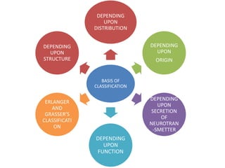
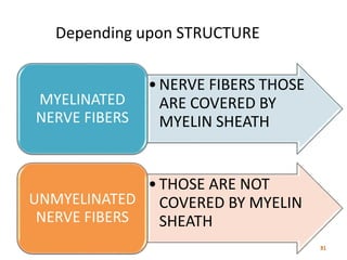
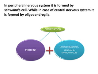
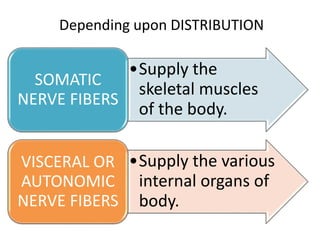
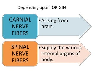
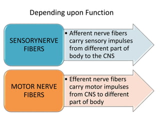
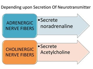
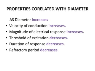
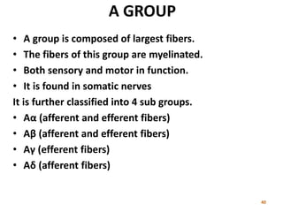
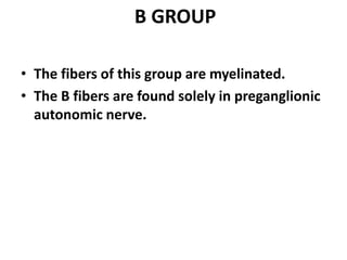
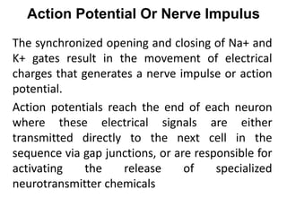
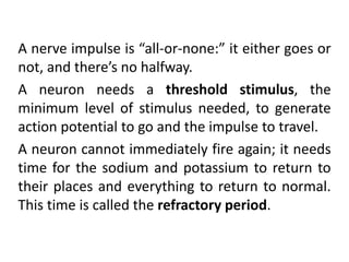
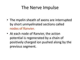
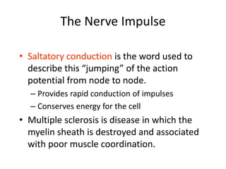
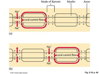
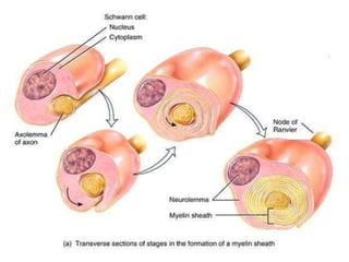
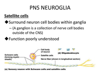
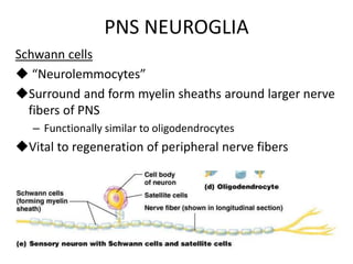
The document provides an overview of the nervous system, including its functions and major components. It discusses the two principal cell types - neurons and neuroglia. Neurons are specialized to transmit electrical signals through a process called an action potential. All neurons have dendrites, a cell body, an axon, and presynaptic terminals. The document also describes the basic structure and function of a nerve, including the myelin sheath that surrounds many axons. Nerve fibers are classified in different ways, including based on their structure, function, neurotransmitter secretion, origin and distribution.
Classification of nerve fibers, Nervous System PhysiologyShaista Jabeen
https://www.youtube.com/channel/UCrrAABI7QDRCJ1yMrQCip_w/videos
https://www.facebook.com/ShaistaJabeeen/
https://www.facebook.com/Human-Physiology-Lectures-100702741804409/
Classification of nerve fibers
Nervous System Physiology
BASIS OF CLASSIFICATION
DEPENDING UPON STRUCTURE
DEPENDING UPON DISTRIBUTION
DEPENDING UPON ORIGIN
DEPENDING UPON FUNCTION
DEPENDING UPON SECRETION OF NEUROTRANSMITTER
DEPENDING UPON DIAMETER AND CONDUCTION OF IMPULSE
Short Notes
pdf ppt
Axoplasmic flow in Axons - Mechanisms and Applications in Clinical NeurologyRahul Kumar
Discussion about the historical aspects of axoplasmic flow, the mechanisms, microtubule motors, and applications in neurological diseases and therapeutics.
Lecture notes and diagrams to help high school anatomy and physiology students learn the general functions of the nervous system and types of glial support nerve cells, types of neurons and anatomy of typical neurons.
NERVE CELLS FINAL( NEURON AND GLIAL CELLS.pptx FOR NURSING STUDENTSWINCY THIRUMURUGAN
THE NERVOUS SYSTEM CONTAINS TWO MAIN TYPES OF CELLS.
A neuron is a nerve cell that is the basic building block of the
nervous system.
Neurons are the structural and functional unit of the nervous
system.
Neurons are specialized to transmit information throughout the
body.
They constitute the communication network of the nervous system and transfer electrical impulses between the central nervous system and sensory organs such as eye,ear.nose,tongue and skin.
There are Approximately 86-100 billion neurons in the brain.
DENDRITES
Dendrites are the tree-like branched structures that arise from the nerve cell body.
Apart from the main dendrite branches, dendrites may contain additional protrusions
known as dendrite spines.
The axon hillock is a specialized region from which the
axon extends.
The axon is a single elongated tubal structure that extends from the Axon Hillock.
Each neuron has a single axon that extends and branches at its end.
The inner most Plasma membrane around the axon is Axolemma.
Neurilemma is the plasma membrane of schwann cells .
The spaces/gaps between the Schwann cells are known as the nodes of Ranvier and they serve to propagate electrical signals along the axon.
The branched end of the axon is known as the axon terminal[arborization] and
branches at the middle of the axon is axon collaterals .
This is the distal part of the axon that comes in contact with other cells. Also called as terminal boutons.
This part of the axon is largely involved in the release of the neurotransmitter.The cell body, also called the soma, is the spherical part of the neuron that contains the nucleus ,cytoplasm and organelles.
The cell body connects to the dendrites, and send information to the
axon depending on the strength of the signal.
The neuronal cytoplasm have the following
The Nucleus,
Nucleolus,
Endoplasmic Reticulum,
Golgi Apparatus,
Mitochondria,
Ribosomes,
Lysosomes,
Endosomes,
And Peroxisomes. A bipolar neuron is a type of neuron which has two extensions (one axon and one dendrite).
A multipolar neuron is a type of neuron that possesses a single axon and many dendrites (and dendritic branches), allowing for the integration of a great deal of information from other neurons.
TYPES OF NEURON:
A unipolar neuron is a type of neuron in which only one process called a neurite extends from the cell body. A pseudounipolar neuron is a type of neuron which has one extension from its cell body. This type of neuron contains an axon that has split into two branches; one branch travels to the PNS and the other to the CNS.They are three types of neurons based on the function as follows Sensory Neuron
Inter-Neuron
Motor Neuron
Interneurons are the central nodes of neural circuits, enabling communication between sensory or motor neurons and the (CNS).
Glial cells (named from the Greek word for "glue") are non- neuronal cells that
provide support and nutrition,
maintain homeostasis,
form myelin,
and participate in signal transmission.
Structures of Axon Terminals and Presynaptic Membrane
Presynaptic axon terminal has a definite intact membrane known as presynaptic
membrane.
Axon terminal has two important structures:
i. Mitochondria, which help in the synthesis of neurotransmitter substance
ii. Synaptic vesicles, which store neurotransmitter substanceMain function of the synapse is to transmit the
impulses, i.e. action potential from one neuron to
Another
1. Excitatory synapses
2. Inhibitory synapses,
A brief information about the SCOP protein database used in bioinformatics.
The Structural Classification of Proteins (SCOP) database is a comprehensive and authoritative resource for the structural and evolutionary relationships of proteins. It provides a detailed and curated classification of protein structures, grouping them into families, superfamilies, and folds based on their structural and sequence similarities.
(May 29th, 2024) Advancements in Intravital Microscopy- Insights for Preclini...Scintica Instrumentation
Intravital microscopy (IVM) is a powerful tool utilized to study cellular behavior over time and space in vivo. Much of our understanding of cell biology has been accomplished using various in vitro and ex vivo methods; however, these studies do not necessarily reflect the natural dynamics of biological processes. Unlike traditional cell culture or fixed tissue imaging, IVM allows for the ultra-fast high-resolution imaging of cellular processes over time and space and were studied in its natural environment. Real-time visualization of biological processes in the context of an intact organism helps maintain physiological relevance and provide insights into the progression of disease, response to treatments or developmental processes.
In this webinar we give an overview of advanced applications of the IVM system in preclinical research. IVIM technology is a provider of all-in-one intravital microscopy systems and solutions optimized for in vivo imaging of live animal models at sub-micron resolution. The system’s unique features and user-friendly software enables researchers to probe fast dynamic biological processes such as immune cell tracking, cell-cell interaction as well as vascularization and tumor metastasis with exceptional detail. This webinar will also give an overview of IVM being utilized in drug development, offering a view into the intricate interaction between drugs/nanoparticles and tissues in vivo and allows for the evaluation of therapeutic intervention in a variety of tissues and organs. This interdisciplinary collaboration continues to drive the advancements of novel therapeutic strategies.
Seminar of U.V. Spectroscopy by SAMIR PANDASAMIR PANDA
Spectroscopy is a branch of science dealing the study of interaction of electromagnetic radiation with matter.
Ultraviolet-visible spectroscopy refers to absorption spectroscopy or reflect spectroscopy in the UV-VIS spectral region.
Ultraviolet-visible spectroscopy is an analytical method that can measure the amount of light received by the analyte.
Earliest Galaxies in the JADES Origins Field: Luminosity Function and Cosmic ...Sérgio Sacani
We characterize the earliest galaxy population in the JADES Origins Field (JOF), the deepest
imaging field observed with JWST. We make use of the ancillary Hubble optical images (5 filters
spanning 0.4−0.9µm) and novel JWST images with 14 filters spanning 0.8−5µm, including 7 mediumband filters, and reaching total exposure times of up to 46 hours per filter. We combine all our data
at > 2.3µm to construct an ultradeep image, reaching as deep as ≈ 31.4 AB mag in the stack and
30.3-31.0 AB mag (5σ, r = 0.1” circular aperture) in individual filters. We measure photometric
redshifts and use robust selection criteria to identify a sample of eight galaxy candidates at redshifts
z = 11.5 − 15. These objects show compact half-light radii of R1/2 ∼ 50 − 200pc, stellar masses of
M⋆ ∼ 107−108M⊙, and star-formation rates of SFR ∼ 0.1−1 M⊙ yr−1
. Our search finds no candidates
at 15 < z < 20, placing upper limits at these redshifts. We develop a forward modeling approach to
infer the properties of the evolving luminosity function without binning in redshift or luminosity that
marginalizes over the photometric redshift uncertainty of our candidate galaxies and incorporates the
impact of non-detections. We find a z = 12 luminosity function in good agreement with prior results,
and that the luminosity function normalization and UV luminosity density decline by a factor of ∼ 2.5
from z = 12 to z = 14. We discuss the possible implications of our results in the context of theoretical
models for evolution of the dark matter halo mass function.
Introduction:
RNA interference (RNAi) or Post-Transcriptional Gene Silencing (PTGS) is an important biological process for modulating eukaryotic gene expression.
It is highly conserved process of posttranscriptional gene silencing by which double stranded RNA (dsRNA) causes sequence-specific degradation of mRNA sequences.
dsRNA-induced gene silencing (RNAi) is reported in a wide range of eukaryotes ranging from worms, insects, mammals and plants.
This process mediates resistance to both endogenous parasitic and exogenous pathogenic nucleic acids, and regulates the expression of protein-coding genes.
What are small ncRNAs?
micro RNA (miRNA)
short interfering RNA (siRNA)
Properties of small non-coding RNA:
Involved in silencing mRNA transcripts.
Called “small” because they are usually only about 21-24 nucleotides long.
Synthesized by first cutting up longer precursor sequences (like the 61nt one that Lee discovered).
Silence an mRNA by base pairing with some sequence on the mRNA.
Discovery of siRNA?
The first small RNA:
In 1993 Rosalind Lee (Victor Ambros lab) was studying a non- coding gene in C. elegans, lin-4, that was involved in silencing of another gene, lin-14, at the appropriate time in the
development of the worm C. elegans.
Two small transcripts of lin-4 (22nt and 61nt) were found to be complementary to a sequence in the 3' UTR of lin-14.
Because lin-4 encoded no protein, she deduced that it must be these transcripts that are causing the silencing by RNA-RNA interactions.
Types of RNAi ( non coding RNA)
MiRNA
Length (23-25 nt)
Trans acting
Binds with target MRNA in mismatch
Translation inhibition
Si RNA
Length 21 nt.
Cis acting
Bind with target Mrna in perfect complementary sequence
Piwi-RNA
Length ; 25 to 36 nt.
Expressed in Germ Cells
Regulates trnasposomes activity
MECHANISM OF RNAI:
First the double-stranded RNA teams up with a protein complex named Dicer, which cuts the long RNA into short pieces.
Then another protein complex called RISC (RNA-induced silencing complex) discards one of the two RNA strands.
The RISC-docked, single-stranded RNA then pairs with the homologous mRNA and destroys it.
THE RISC COMPLEX:
RISC is large(>500kD) RNA multi- protein Binding complex which triggers MRNA degradation in response to MRNA
Unwinding of double stranded Si RNA by ATP independent Helicase
Active component of RISC is Ago proteins( ENDONUCLEASE) which cleave target MRNA.
DICER: endonuclease (RNase Family III)
Argonaute: Central Component of the RNA-Induced Silencing Complex (RISC)
One strand of the dsRNA produced by Dicer is retained in the RISC complex in association with Argonaute
ARGONAUTE PROTEIN :
1.PAZ(PIWI/Argonaute/ Zwille)- Recognition of target MRNA
2.PIWI (p-element induced wimpy Testis)- breaks Phosphodiester bond of mRNA.)RNAse H activity.
MiRNA:
The Double-stranded RNAs are naturally produced in eukaryotic cells during development, and they have a key role in regulating gene expression .
Nutraceutical market, scope and growth: Herbal drug technologyLokesh Patil
As consumer awareness of health and wellness rises, the nutraceutical market—which includes goods like functional meals, drinks, and dietary supplements that provide health advantages beyond basic nutrition—is growing significantly. As healthcare expenses rise, the population ages, and people want natural and preventative health solutions more and more, this industry is increasing quickly. Further driving market expansion are product formulation innovations and the use of cutting-edge technology for customized nutrition. With its worldwide reach, the nutraceutical industry is expected to keep growing and provide significant chances for research and investment in a number of categories, including vitamins, minerals, probiotics, and herbal supplements.
Multi-source connectivity as the driver of solar wind variability in the heli...Sérgio Sacani
The ambient solar wind that flls the heliosphere originates from multiple
sources in the solar corona and is highly structured. It is often described
as high-speed, relatively homogeneous, plasma streams from coronal
holes and slow-speed, highly variable, streams whose source regions are
under debate. A key goal of ESA/NASA’s Solar Orbiter mission is to identify
solar wind sources and understand what drives the complexity seen in the
heliosphere. By combining magnetic feld modelling and spectroscopic
techniques with high-resolution observations and measurements, we show
that the solar wind variability detected in situ by Solar Orbiter in March
2022 is driven by spatio-temporal changes in the magnetic connectivity to
multiple sources in the solar atmosphere. The magnetic feld footpoints
connected to the spacecraft moved from the boundaries of a coronal hole
to one active region (12961) and then across to another region (12957). This
is refected in the in situ measurements, which show the transition from fast
to highly Alfvénic then to slow solar wind that is disrupted by the arrival of
a coronal mass ejection. Our results describe solar wind variability at 0.5 au
but are applicable to near-Earth observatories.
2. Nervous System Function
• The nervous system has three functions.
• 1- monitors the internal and external
environments
• 2- integrates sensory information
• 3- coordinates voluntary and involuntary
responses.
5. Neuron and Neuroglia
Two principal cell types
Neurons
Excitable nerve cells that transmit electrical signals
– Supporting cells
Smaller cells surrounding and wrapping neurons
“Neuroglia”
6. Neurons
• Cells of the nervous system, called nerve cells
or neurons, are specialized to carry
"messages" through an electrochemical
process. The human brain has about 100
billion neurons that carry out the nerve
impulses through a process called action
potential.
7. Neurons are similar to other cells in the body because:
1. Neurons are surrounded by a cell membrane.
2. Neurons have a nucleus that contains genes.
3. Neurons contain cytoplasm, mitochondria and other "organelles".
4. Neurons carry out basic cellular processes such as protein
synthesis and energy production.
Neurons differ from other cells in the body because:
1. Neurons have specialized extensions called dendrites and axons.
Dendrites bring information to the cell body and axons take
information away from the cell body.
2. Neurons communicate with each other through an
electrochemical process.
3. Neurons contain some specialized structures (for example,
synapses) and chemicals (for example, neurotransmitters).
8.
9. The Cells of the Nervous System
• All neurons have the following major
components:
– Dendrites.
– Soma/ cell body.
– Axon.
– Presynaptic terminals.
10. The Cells of the Nervous System
• Dendrites- branching fibers with a surface
lined with synaptic receptors responsible for
bringing in information from other neurons.
• Some dendrites also contain dendritic spines
that further branch out and increase the
surface area of the dendrite.
12. The Cells of the Nervous System
• Soma - contains the nucleus, mitochondria,
ribosomes, and other structures found in
other cells.
– Also responsible for the metabolic work of the
neuron.
13. The Cells of the Nervous System
• Axon - thin fiber of a neuron responsible for
transmitting nerve impulses away to other
neurons, glands, or muscles.
• Some neurons are covered with an insulating
material called the myelin sheath with
interruptions in the sheath known as nodes of
Ranvier.
14. The Cells of the Nervous System
• Presynaptic terminals refer to the end points
of an axon responsible for releasing chemicals
to communicate with other neurons.
15. Axons
• Take information away
from the cell body
• Smooth Surface
• Generally only 1 axon per
cell
• No ribosomes
• Can have myelin
• Branch further from the cell
body
Dendrites
• Bring information to the
cell body
• Rough Surface (dendritic
spines)
• Usually many dendrites
per cell
• Have ribosomes
• No myelin insulation
• Branch near the cell body
There are several differences between
axons and dendrites
17. Nerve
• Each nerve is a cable-like structure that contains
many axons that are sometimes referred to as
"fibers. " Within a nerve, each axon is surrounded
by a layer of connective tissue called the
endoneurium. The axons are bundled together
into groups called fascicles. Each fascicle is
wrapped in a layer of connective tissue called the
perineurium. Finally, the entire nerve is wrapped
in a layer of connective tissue called the
epineurium
19. Myelin is an insulating layer,
or sheath, that forms around
nerves, including those in the
brain and spinal cord. It is made
up of protein and Lipids e.g
Cholesterol, lecithin and
cerebroside (spingomyelin).
Functions of Myelin Sheath
1)Faster Conduction
The purpose of the myelin sheath is to allow electrical impulses
to transmit quickly and efficiently along the nerve cells. If myelin
is damaged, the impulses slow down.
2)Insulating Capacity
Myelin sheath has a high insulating capacity means its restricts
the nerve impulses within the signal never fiber and prevents the
stimulation of neighboring nerve fibers.
20. Neurilemma
• Surrounding the myelin sheath, there is a thin membrane
called neurilemmal sheath. This is also called neurilemma
or sheath of Schwann.
• This contains Schwann cells, which have flattened and
elongated nuclei. The cytoplasm is thin and modified to
form the thin sheath of neurilemma enclosing the myelin
sheath.
• One nucleus is present in each internode of the axon. The
nucleus is situated between myelin sheath . At the node of
Ranvier (where myelin sheath is absent), the neurilemma
invaginates and runs up to axolemma in the form of a finger
like process.
• In nonmyelinated nerve fiber, the neurilemma continuously
surrounds axolemma. Neurilemma is absent in central
nervous system. Neurilemma is necessary for the formation
of myelin sheath (myelinogeneis).
23. 23
Structural classification of neurons
1. A multipolar
neuron has multiple
processes extending
away from the cell
body. These are
very common in the
CNS.
24. 24
2. A unipolar
neuron, the
dendrites and axon
are continuous, and
the cell body lies off
to one side. In a
unipolar neuron, the
action potential
begins at the base of
the dendrites and
the rest of the
process is considered
an axon
25. 25
3. Bipolar neurons
have two processes,
one dendrites and
one axon, with the
cell body between
them. Bipolar
neurons are rare but
occur in special sense
organs such as the
eye and the ear.
26. Neurons can also be classified by the direction
that they send information:
• Sensory (or afferent) neurons: send information
from sensory receptors (e.g., in skin, eyes, nose,
tongue, ears) TOWARD the central nervous
system.
• Motor (or efferent) neurons: send information
AWAY from the central nervous system to
muscles or glands.
• Interneurons: send information between
sensory neurons and motor neurons. Most
interneurons are located in the central nervous
system.
28. INTRODUCTION ABOUT NERVE
FIBER
• A nerve fiber is a thread like
extension of a nerve cell and
consists of an axon and
myelin sheath (if present) in
the nervous system.
31. •NERVE FIBERS THOSE
ARE COVERED BY
MYELIN SHEATH
MYELINATED
NERVE FIBERS
•THOSE ARE NOT
COVERED BY MYELIN
SHEATH
UNMYELINATED
NERVE FIBERS
Depending upon STRUCTURE
32. MYELINSHEATH
In peripheral nervous system it is formed by
schwann’s cell. While in case of central nervous system it
is formed by oligodendroglia.
COMPOSITION
PROTEINS
LIPIDS(CHOLESTEROL,
LECITHIN &
SPHINGOMYELIN)
33. •Supply the
skeletal muscles
of the body.
SOMATIC
NERVE FIBERS
•Supply the various
internal organs of
body.
VISCERAL OR
AUTONOMIC
NERVE FIBERS
Depending upon DISTRIBUTION
35. • Afferent nerve fibers
carry sensory impulses
from different part of
body to the CNS
SENSORYNERVE
FIBERS
• Efferent nerve fibers
carry motor impulses
from CNS to different
part of body
MOTOR NERVE
FIBERS
Depending upon Function
37. ERLANGER AND GRASSER’S CLASSIFICATION
• Erlanger and Grasser studied the
action potential of mixed nerve trunk
by means of cathode ray oscilloscope
and they obtained the compounded
spike. So they divided nerve fibers into
3 groups. They observed that the main
cause of difference in nerve fibers is
diameter
38. • A GROUP
• B GROUP
• C GROUP
GROUPS OF NERVE FIBERS
Depending of Diameter
39. PROPERTIES CORELATED WITH DIAMETER
AS Diameter increases
• Velocity of conduction increases.
• Magnitude of electrical response increases.
• Threshold of excitation decreases.
• Duration of response decreases.
• Refractory period decreases.
40. A GROUP
• A group is composed of largest fibers.
• The fibers of this group are myelinated.
• Both sensory and motor in function.
• It is found in somatic nerves
It is further classified into 4 sub groups.
• Aα (afferent and efferent fibers)
• Aβ (afferent and efferent fibers)
• Aγ (efferent fibers)
• Aδ (afferent fibers)
41. B GROUP
• The fibers of this group are myelinated.
• The B fibers are found solely in preganglionic
autonomic nerve.
42. C GROUP
• It is composed of smallest fibers.
• All the fibers of this group are unmyelinated.
• Mostly found in visceral and cutaneous nerve.
• They have high threshold i.e. 30 folds that of A group.
• Generally they are found in postganglionic
sympathetic nerve.
45. Properties of Nerve Fibers
1) Excitability:
It is the ability of generating electrochemical
impulse (action potentials) at the cell
membrane in response to any stimulus.
Stimulus*
The stimulus is defined as an external agent
that produce excitabilty in tissues.
46. Types of Stimulus
-Chemical transmitters
- Hormones.
- Drugs.
-Ions (Na+, K+, .... etc).
- Gases (O2 and CO2).
-Thermal.
e.g. cooling
or warming.
- Mechanical.
e.g. stretch, touch,
pressure and injury.
- Electromagnetic.
e.g. light rays
Chemical Physical1 2 Electrical3
- Galvanic Current:
Low intensity
Long Duration
- Faradic Current:
High intensity
Short duration
47. Resting membrane potential
A voltage exists across the plasma membrane
– Due to separation of oppositely charged ions
Potential difference in a resting membrane is
termed its “resting membrane potential”
~ -70 mV in a resting
neuron
Membrane is “polarized”
48. Action Potential Or Nerve Impulus
The synchronized opening and closing of Na+ and
K+ gates result in the movement of electrical
charges that generates a nerve impulse or action
potential.
Action potentials reach the end of each neuron
where these electrical signals are either
transmitted directly to the next cell in the
sequence via gap junctions, or are responsible for
activating the release of specialized
neurotransmitter chemicals
49. A nerve impulse is “all-or-none:” it either goes or
not, and there’s no halfway.
A neuron needs a threshold stimulus, the
minimum level of stimulus needed, to generate
action potential to go and the impulse to travel.
A neuron cannot immediately fire again; it needs
time for the sodium and potassium to return to
their places and everything to return to normal.
This time is called the refractory period.
50.
51.
52. Conductivity
• Conductivity:
• It is the ability to propagate action potential
from the point of generation to the rest of the
membrane.
53. The Nerve Impulse
• The myelin sheath of axons are interrupted
by short unmyelinated sections called
nodes of Ranvier.
• At each node of Ranvier, the action
potential is regenerated by a chain of
positively charged ion pushed along by the
previous segment.
54. The Nerve Impulse
• Saltatory conduction is the word used to
describe this “jumping” of the action
potential from node to node.
– Provides rapid conduction of impulses
– Conserves energy for the cell
• Multiple sclerosis is disease in which the
myelin sheath is destroyed and associated
with poor muscle coordination.
57. DEGENERATION OF NERVE FIBERS
When a peripheral nerve is injured, the
degenerative changes occurs in the nerve cell
body and in the nerve fibres.
Degenerative Changes are Classified in to Three
Types.
Wallarian Degeneration
Retrograde Degeneration
Transneural Degeneration
58. Wallerian Degeneration
The degenerative changes in the distal cut end of nerve fiber ( AXON) is known as
Wallerian degeneration or orthograde degeneration
1) In the part of nerve fiber distal to injury, the degenerative changes occur
within 24 hours of injury.
2) Axis cylinder swells and neurofibrils and axis cylinder breaks up into small
pieces. After few days the broken pieces appear as debris in the space
occupied by axis cylinder.
3) The myelin sheath is slowly disintegrated into fat droplets. The changes in
myelin sheath occur from 8th to 35th day.
4) The region is invaded by macrophages that remove degenerating axons,
myelin and cellular debris. These macrophages probably secrete substances
that causes proliferation of Schwann cells and also produce nerve growth
factors. All these changes takes place for about 2 months from the day of
injury. The schwann cells of distal side increase in size and proliferate to form
series of tubes. When one of the regenerating axonal branch succeeds in
reaching tube, it enters and grows rapidly inside it
59.
60.
61. Retrograde Degeneration
The Retrograde degeneration changes in
the nerve cell body and part of axon
attached to nerve cell body, axon proximal
to the cut are together known as
RETROGRADE DEGENERATION .
63. Regeneration Of Nerve Fiber
The injured and degenerated nerve fiber can
regenerated, but regeneration is possible only
If degenerated nerve fiber meets with following
criteria
1)The between the cut ends of the nerve should
not exceed 3mm
2) The neurilemma should be present
3) The nucleus must be intact
4) The cut ends should remain in same line
64. NEUROGLIA
“Nerve glue”
Six types of small cells associated with neurons
– 4 in CNS
– 2 in PNS
Most have central cell body and branching
processes
Several functions
– e.g., Supportive scaffolding for neurons
– e.g., Electrical isolation of neurons
– e.g., Neuron health and growth
66. CNS NEUROGLIA
Astrocytes
Most abundant and versatile glial cells
Numerous processes support branching
neurons
– Anchor neurons to capillary blood supply
Guide migration of young neurons
Facilitate nutrient delivery to neurons
– (blood glial cell neuron)
Control chemical environment
around neurons
– Uptake of K+, neurotransmitters
Communicate with astrocytes
& neurons
– Gap junctions
67. CNS NEUROGLIA
Microglia
Small ovoid cells
Relatively long “thorny”
processes
– Processes touch nearby neurons
Migrate toward injured neurons
Transform into macrophage
– Phagocytize microorganisms, debris
– (Cells of immune system cannot enter the CNS)
68. CNS NEUROGLIA
Ependymal Cells
Line central cavities of brain and spinal cord
– Form permeable barrier between cerebrospinal fluid inside
these cavities and tissue fluid of CNS tissue
Shapes range from squamous to columnar
Many are ciliated
– Beating helps circulate cerebrospinal fluid cushioning brain
and spinal cord
71. PNS NEUROGLIA
Satellite cells
Surround neuron cell bodies within ganglia
– (A ganglion is a collection of nerve cell bodies
outside of the CNS)
Function poorly understood
72. PNS NEUROGLIA
Schwann cells
“Neurolemmocytes”
Surround and form myelin sheaths around larger nerve
fibers of PNS
– Functionally similar to oligodendrocytes
Vital to regeneration of peripheral nerve fibers