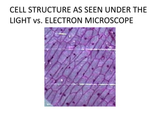
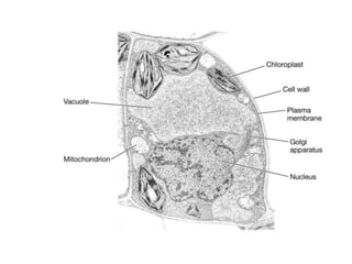
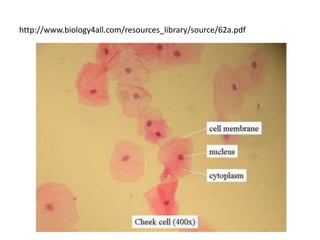
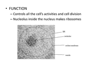
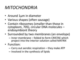
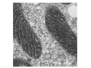
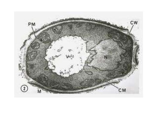
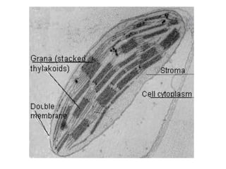
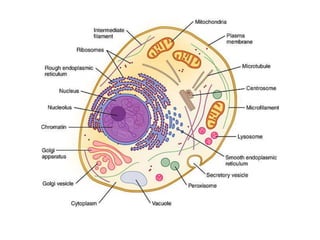
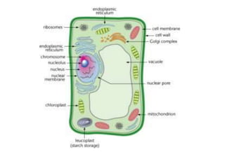
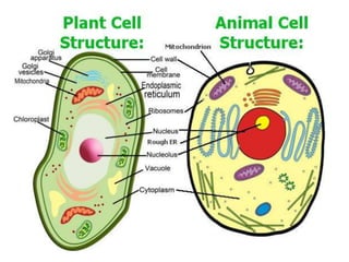
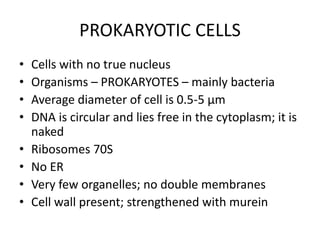
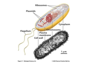
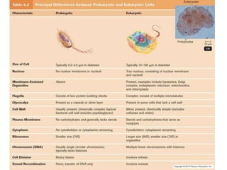
This document provides information on cell structure and organization. It describes typical animal and plant cell structures as seen under light and electron microscopes. Key structures discussed include the nucleus, endoplasmic reticulum, Golgi apparatus, mitochondria, chloroplasts, cell wall, and vacuoles. It also covers tissues, organs, organ systems, and the differences between prokaryotic and eukaryotic cells.
The cell membrane (also known as the plasma membrane or cytoplasmic membrane) is a biological membrane that separates the interior of all cells from the outside environment. The cell membrane is selectively permeable to ions and organic molecules and controls the movement of substances in and out of cells.The basic function of the cell membrane is to protect the cell from its surroundings. It consists of the phospholipid bilayer with embedded proteins. Cell membranes are involved in a variety of cellular processes such as cell adhesion, ion conductivity and cell signalling and serve as the attachment surface for several extracellular structures, including the cell wall, glycocalyx, and intracellular cytoskeleton.
This is a PowerPoint presentation for Topic 1 in the Edexcel Biology B A Level course that starts in 2015.
This is a free sample, the full PowerPoint presentation is available to purchase here: https://sellfy.com/MrExham
This is the first PowerPoint in the mrexham IGCSE Biology series. It is also available on iBooks.
It covers the Cells section from life processes of the Edexcel IGCSE Biology course
The cell membrane is selectively permeable, allowing only a limited number of materials to diffuse through its lipid bilayer. During passive transport, materials move by simple diffusion or by facilitated diffusion through the membrane, down their concentration gradient.
Instructions for Submissions thorugh G- Classroom.pptxJheel Barad
This presentation provides a briefing on how to upload submissions and documents in Google Classroom. It was prepared as part of an orientation for new Sainik School in-service teacher trainees. As a training officer, my goal is to ensure that you are comfortable and proficient with this essential tool for managing assignments and fostering student engagement.
Read| The latest issue of The Challenger is here! We are thrilled to announce that our school paper has qualified for the NATIONAL SCHOOLS PRESS CONFERENCE (NSPC) 2024. Thank you for your unwavering support and trust. Dive into the stories that made us stand out!
How to Make a Field invisible in Odoo 17Celine George
It is possible to hide or invisible some fields in odoo. Commonly using “invisible” attribute in the field definition to invisible the fields. This slide will show how to make a field invisible in odoo 17.
Biological screening of herbal drugs: Introduction and Need for
Phyto-Pharmacological Screening, New Strategies for evaluating
Natural Products, In vitro evaluation techniques for Antioxidants, Antimicrobial and Anticancer drugs. In vivo evaluation techniques
for Anti-inflammatory, Antiulcer, Anticancer, Wound healing, Antidiabetic, Hepatoprotective, Cardio protective, Diuretics and
Antifertility, Toxicity studies as per OECD guidelines
Model Attribute Check Company Auto PropertyCeline George
In Odoo, the multi-company feature allows you to manage multiple companies within a single Odoo database instance. Each company can have its own configurations while still sharing common resources such as products, customers, and suppliers.
The French Revolution, which began in 1789, was a period of radical social and political upheaval in France. It marked the decline of absolute monarchies, the rise of secular and democratic republics, and the eventual rise of Napoleon Bonaparte. This revolutionary period is crucial in understanding the transition from feudalism to modernity in Europe.
For more information, visit-www.vavaclasses.com
Welcome to TechSoup New Member Orientation and Q&A (May 2024).pdfTechSoup
In this webinar you will learn how your organization can access TechSoup's wide variety of product discount and donation programs. From hardware to software, we'll give you a tour of the tools available to help your nonprofit with productivity, collaboration, financial management, donor tracking, security, and more.
2. LO
• Draw and label low power plan diagrams of
tissues and organs (including a transverse
section of stems, roots and leaves)
• Describe and interpret drawings and
photographs of typical animal and plant cells
as seen using the light microscope
3. • Describe and interpret drawings and photographs of
typical animal and plant cells as seen using the electron
microscope, recognizing:
– rough and smooth endoplasmic reticulum,
– Golgi apparatus,
– mitochondria,
– ribosomes,
– lysosomes,
– cell surface membrane,
– centrioles,
– nucleus and microvilli,
– chloroplasts, cell wall,
– vacuole,
– tonoplast and plasmodesmata
4. Organization of cells
• Cell
• Tissue (e.g. palisade tissue, blood, …)
• Organ (e.g. leaf, heart, …)
• Organ system (e.g. circulatory system)
• Organism (e.g. bufo bufo – common toad)
5. LOW POWER PLAN DRAWINGS
• Shows distribution of tissues but not details of
individual cells
• How to draw a low power plan diagram?
• https://www.youtube.com/watch?v=t3FuoBo
E9XE
6. Rules for biological drawings
• Always use a pencil, not a pen!
• Don’t use shading
• Use clear, continuous lines
• Use accurate proportions and observation –
not a text book version
• Label all tissues and relevant structures
• Identify parts correctly
• Use a ruler for label lines
7. Low-power drawings
• Don’t draw individual cells
• Draw all tissues completely enclosed by lines
• Draw a correct interpretation of the
distribution of tissues
• A representative portion may be drawn
8. High-power drawing
• Draw only a few representative cells
• Draw the cell wall of all plant cells
• Don’t draw the nucleus as a solid blob
12. CELL
• Robert Hook – English scientist, in 1665
examined cork, he called the pore-like
structures CELLS
13. • Cell is the fundamental unit of all living things
• CELL THEORY
– The basic unit of structure and function of all
living organisms is the cell (Schleiden - botanist
and Schwann – zoologist) 1838 and 1839
– All cells arise from pre-existing cells by cell division
(Virchow) 1855
14. CELL BIOLOGY AND MICROSCOPY
• The light microscope
– Uses light as a source of radiation
– Golden age in 19th century – new branch of
biology = CYTOLOGY
• The electron microscope
– Uses electrons and source of radiation
17. UNITS O MEASUREMENT IN CELL
STUDIES
• The basic unit of measurements is METRE (m)
18.
19. • Magnification
– The number of times larger an image is, compared
with the real size of the object
– Observed size – what you can measure with a
ruler
– Actual size – the real size, the size of a cell before
it’s magnified
20. • Resolution
– The ability to distinguish between two separate
points
– If two points cannot be resolved, they will be seen
as one point
– In practice – the amount of detail that can be seen
– the greater the resolution, the greater the detail
– Max for light microscope is 200 nm
21. THE ELECTROMAGNETIC SPECTRUM
• Light travels in waves with variable lengths
• Different lengths are in our brain translated
into different colors
• The whole range of different wave lengths is
called = electromagnetic spectrum
22.
23. • The limit of resolution is about one half the
wavelength of the radiation used to view the
specimen
• If an object is smaller than half the
wavelength of the radiation used to view it, it
cannot be seen separately from nearby
objects
(ribosomes can never be seen under the light
microscope as their diameter is 25 nm)
24. • If an object is transparent, it will allow waves
to pass through it and therefore needs to be
stained before it can be seen
25. THE ELECTRON MICROSCOPE
• Uses radiation of shorter wavelength than
light – electrons (negatively charged particles
which orbit the nucleus of an atom)
– Free electrons behave like electromagnetic
radiation
– They have very short wavelength
– Thanks to the negative charge they can be easily
focused using electromagnets
– Resolution of 0.5 nm can be obtained
26. • Transmission electron microscope (TEM)
– The beam of electrons is passed through the
specimen before being viewed
– Only those electrons that are transmitted are seen
– This allows to see thin sections and inside of cells
• Scanning electron microscope (SEM)
– The electron beam is used to scan the surface of
structures and only the reflected beam is
observed
– Three dimensional appearance is obtained
27. • Disadvantages of electron microscopes:
– Electron beam has to be projected onto a
florescent screen
– The resulting picture is like an X-ray photograph
– The electron beam and the specimen and the
screen must be in vacuum and therefore all
specimens must be dehydrated – only dead
material can be examined
28. ULTRASTRUCTURE OF AN ANIMAL
CELL
• Ultrastructure = detailed structure of a cell as
revealed by the electron microscope
31. NUCLEUS
• The largest cell organelle
• Surrounded by – nuclear envelope
• Two membranes
• Outer layer is continuous with the endoplasmic
reticulum
• Has small openings called NUCLEAR PORES that allow
and control exchange between the nucleus and
cytoplasm
(entering substances: proteins, ATP, hormones
Leaving substances: mRNA, ribosomes)
32. • Chromatin – loosely coiled chromosomes that
contain DNA organized into functional units =
genes
– Genes control the activities of the cell and
inheritance
33. • FUNCTION
– Controls all the cell’s activities and cell division
– Nucleolus inside the nucleus makes ribosomes
34. ENDOPLASMIC RETICULUM (ER)
• Extensive system of membranes running
through the cytoplasm
• It is continuous with the outer membrane of
the nuclear envelope
35. • Two types:
– Rough ER – it is covered with many tiny ribosomes
visible as black dots
• Forms and extensive system of flattened sacs spreading
in sheets throughout the cell
• Proteins produced by ribosomes enter the sacs and
move through them
• Ribosomes
– consist of two subunits (small and large); 80S
- they are the site of protein synthesis;
- can be found free in cytoplasm or on the rough ER
- very small – 25 nm in diameter
- made of RNA (ribonucleic acid) and protein
36. – Smooth ER
» Lacks ribosomes
» Makes LIPIDS and STEROIDS such as
cholesterol, oestrogen and testosterone
37.
38. GOLGI APPARATUS
• Stack of flattened sacs
• The stack is formed as the vesicles bud off
from the ER; are further broken down to form
Golgi vesicles
• Function:
– Collects, processes and sorts molecules (proteins)
– Golgi vesicles transport these either to another
part of the cell or out of the cell
– Golgi vesicles are used to make lysosomes
39.
40. LYSOSOMES
• Spherical sacs, surrounded by a single
membrane with no internal structure
• 0.1 – 0.5 μm in diameter
• Contain digestive (hydrolytic) enzymes
• Function:
– Responsible for the breakdown of unwanted
structures (old organelles, bacteria in
lymphocytes)
41.
42. MITOCHONDRIA
• Around 1μm in diameter
• Various shapes (often sausage)
• Contain ribosomes (smaller than those in
cytoplasm, 70S), circular DNA molecules –
endosymbiont theory
• Surrounded by two membranes (an envelope)
– Inner membrane – folded to form CRISTAE which
project into the interior solution called MATRIX
• Function:
– Carry out aerobic respiration – they make ATP
– Involved in the synthesis of lipids
43.
44. CELL SURFACE MEMBRANE
• Extremely thin (about 7nm)
• Has three layers
• Semipermeable = partially permeable
• Controls exchange between the cell and its
environment
45.
46. MICROVILLI
• Finger-like extensions of the cell surface
membrane
• Typical of epithelial cells
• Increase the surface area of the cell surface
membrane (absorption in guts)
47.
48. CENTRIOLES
• 2 centrioles just outside the nucleus
• Lye close together at right angles to each
other
• Not found in plant cells!
• = hollow cylinder about 0.4 μm long formed of
short microtubules
– Tiny tubes made of protein = tubulin
– Used as a starting point for growing the spindle
microtubules for nuclear division
49.
50.
51. ULTRASTRUCTURE OF A PLANT CELL
• He only structure commonly found in animal
cell which is absent from plant cell is the
centriole
• Plant cells posses cell wall, large permanent
vacuoles and chloroplasts
52.
53. CELL WALL
• Outside the cell surface membrane
• Rigid thanks to the fibres and cellulose
• Gives the cell a definite shape and prevents the
cell from bursting
• May be reinforced with extra cellulose or lignin
• Freely permeable
• Plasmodesmata – fine strands of cytoplasm
which pass through the walls of neighbouring
cells
54.
55. VACUOLES
• Large, permanent, central
• Surrounded by a membrane = tonoplast
– Controls exchange between the vacuole and the
cytoplasm
• Fluid inside a vacuole – solution of mineral salts,
sugars, oxygen, carbon dioxide, pigments,
enzymes including waste products
• Help to regulate the osmotic properties of cells;
pigments give color to the different flower parts
56.
57. CHLOROPLASTS
• Relatively large organelles; green thanks to
chlorophyll (green pigment)
• Granum – small structures inside chloroplast;
responsible for light absorption during
photosynthesis
– Consist of stacks of membrane-bound sacs called
thylakoids
58.
59.
60.
61.
62. PROKARYOTIC CELLS
• Cells with no true nucleus
• Organisms – PROKARYOTES – mainly bacteria
• Average diameter of cell is 0.5-5 μm
• DNA is circular and lies free in the cytoplasm; it is
naked
• Ribosomes 70S
• No ER
• Very few organelles; no double membranes
• Cell wall present; strengthened with murein
63.
64.
65. • Organisms made of eukaryotic cells =
EUKARYOTES
– Animals, plants, fungi and unicellular eukaryotes
called PROTOCTISTS
66. TISSUES AND ORGANS
• Unicellular organisms (e.g. bacteria)
• Multicellular organisms
– Greater independence from the environment
– Functions of the organisms are divided among
groups of cells
– Structurally and functionally specialized cells are
grouped into TISSUES
67. A TISSUE
• Is a collection of cells, together with any
intercellular secretion produced by them, that
is specialized to perform one or more
particular functions
• The cells may be of the same type (e.g.
parenchyma) or they may be of mixed type
(e.g. blood, bones,…)
• The study of tissues is called HISTOLOGY
68. • Messophyll
- Tissue made up of many similar cells, all with the same
function
• Epidermis
– A protective layer one cell thick
– May be covered with waxy cuticule which is
waterproof and protects the organ from drying
out and infection
– Pores called stomata
– Extensions called root hairs
69. • Parenchyma
– Thin-walled cells used as packing tissue
– The cells are very active and can be used for many
functions
– Forms cortex in roots and stems and the pith in
stems
– Palisade mesophyll and spongy mesophyll
• Endodermis
– Like the epidermis one cell thick
– Surrounds the vascular tissue in stem and roots
70. • Pericycle
– A layer of cells just inside the endodermis and
next to the vascular tissue
– In roots one cell thick and new roots can grow
from this layer
– In stems – is formed from a tissue called
sclerenchyma
• Xylem and phloem
71. AN ORGAN
• Is a part of the body which forms a structural
and functional unit and is composed of more
than one tissue
• Leaf, stem, root, brain, liver, kidney
72. A SYSTEM
• Is a collection of organs with a particular
function: secretory, excretory, reproductive, …
• E.g. vascular system