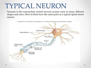
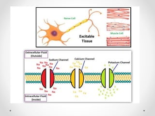
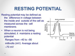
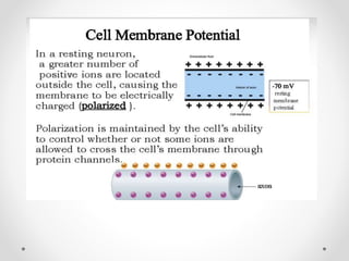
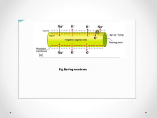
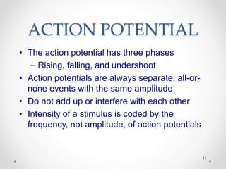
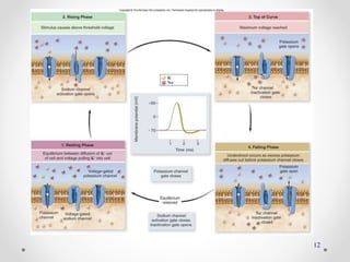
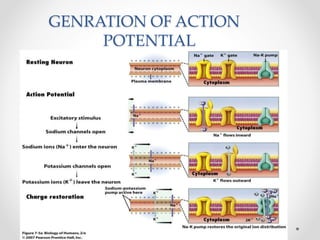
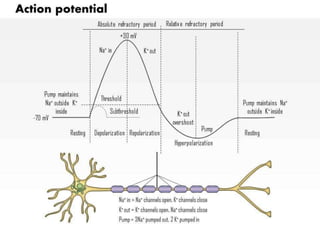
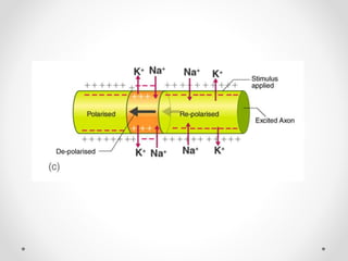
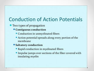
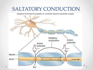
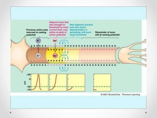
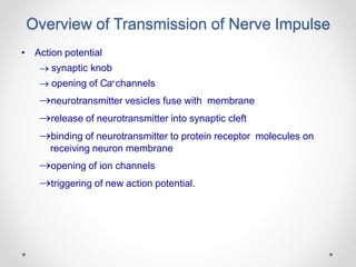
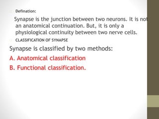
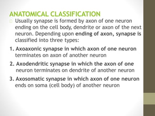
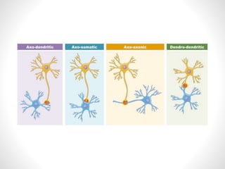
NERVE IMPULSE CONDUCTION&
SYNAPSES
MS.SHILPI DAMOR
ASSISTANT PROFESSOR,DEPTT. OF ZOOLOGY
PODDAR GROUP OF INSTITUTIONS,JAIPUR
CONTENTS
1. Structure of a nerve cell
2. Resting Potential
3. Action Potential
• Formation of an action potential
4. Propagation of Action Potentials as an Impulse
• Saltatory conduction
5. Neurotransmission: Jumping the Synaptic Cleft
TYPICAL NEURON
Neurons in the mammalian central nervous system come in many different
shapes and sizes. Most of them have the same parts as a typical spinal motor
neuron
Direction of impulse
The cell body (soma) contains the nucleus and is the metabolic center of the neuron.
Neurons have processes known as dendrites which extend outward from the cell body and
arborize extensively. Particularly in the cerebral and cerebellar cortex, the small knobby
projections over dendrites are called dendritic spines. The dendrites are the receptive part
of the neuron. A typical neuron has long fibrous axon that originates from thickened area of
the cell body, the axon of hillock. The first portion of the axon is called the initial segment.
The axon divides into presynaptic terminals, each ending in a number of synaptic knobs
which are also called terminal buttons or boutons. They contain granules or vesicles in
which the synaptic transmitters secreted by the nerves are stored. The axonal process is
responsible for transmission of propagated impulses to the nerve endings.
RESTING POTENTIAL
Resting potential may be defined as
the difference in voltage between
the inside and outside of the cell as
measured across the cell
membrane.
• When a neuron is not being
stimulated, it maintains a resting
potential
Ranges from –40 to –90
millivolts (mV) Average about
–70 mV
RESTING POTENTIAL
• Two major forces act on ions in establishing the
resting membrane potential
1. Electrical potential produced by unequal
distribution of charges
2. Concentration gradient produced by
unequal concentrations of molecules from
one side of the membrane to the other
• During this process,
potassium floods out
of the neuron cell.
• Depolarization results
because inward
diffusion of sodium is
much greater than a
outward diffusion of
potassium
DE
DEP
POLARIZAT
OL
DEPOLARIZATION
10
ACTION POTENTIAL
Action potential may be defined as the entire series of
changes which contribute towards the changes in membrane
potential.
ACTION POTENTIAL
• Voltage-gated Na+ channels
– Activation gate and inactivation gate
– At rest, activation gate closed, inactivation gate open
– Transient influx of Na+causes the membrane to
depolarize
• Voltage-gated K+channels
– Single activation gate that is closed in the resting state
– K+channel opens slowly
– Efflux of K+repolarizes the membrane
ACTION POTENTIAL
• The action potential has three phases
– Rising, falling, and undershoot
• Action potentials are always separate, all-or-
none events with the same amplitude
• Do not add up or interfere with each other
• Intensity of a stimulus is coded by the
frequency, not amplitude, of action potentials
11
12
GENRATION OF ACTION
POTENTIAL
PROPAGATION OF ACTION
POTENTIAL
• Propagation of action potentials
– Each action potential, in its rising phase,
reflects a reversal in membrane polarity
– Positive charges due to influx of Na+can
depolarize the adjacent region to threshold
– And so the next region produces its own
action potential
– Meanwhile, the previous region repolarizes back
to the resting membrane potential
• Signal does not go back toward cell
body
15
PROPAGATION OF ACTION
POTENTIAL
• Two ways to increase velocity of conduction
–Axon has a large diameter
• Less resistance to current flow
• Found primarily in invertebrates
–Axon is myelinated
• Action potential is only produced at the
nodes of Ranvier
• Impulse jumps from node to node
• Saltatory conduction
16
17
SALTATORY CONDUCTION
Overview of Transmission of Nerve Impulse
• Action potential
 synaptic knob
 opening of Ca+channels
neurotransmitter vesicles fuse with membrane
release of neurotransmitter into synaptic cleft
binding of neurotransmitter to protein receptor molecules on
receiving neuron membrane
opening of ion channels
triggering of new action potential.
Synapses
Defination:
Synapse is the junction between two neurons. It is not
an anatomical continuation. But, it is only a
physiological continuity between two nerve cells.
CLASSIFICATION OF SYNAPSE
Synapse is classified by two methods:
A. Anatomical classification
B. Functional classification.
ANATOMICAL CLASSIFICATION
Usually synapse is formed by axon of one neuron
ending on the cell body, dendrite or axon of the next
neuron. Depending upon ending of axon, synapse is
classified into three types:
1. Axoaxonic synapse in which axon of one neuron
terminates on axon of another neuron
2. Axodendritic synapse in which the axon of one
neuron terminates on dendrite of another neuron
3. Axosomatic synapse in which axon of one neuron
ends on soma (cell body) of another neuron
„FUNCTIONAL CLASSIFICATION
Functional classification of synapse is on the basis of
mode of impulse transmission
1. Electrical Synapse
Electrical synapse is the synapse in which the
physiological continuity between the presynaptic and
the postsynaptic neurons is provided by gap junction
between the two neurons.
2. Chemical synapse
Is the junction between a nerve fiber and a muscle
fiber or between two nerve fibers, through which the
signals are transmitted by the release of chemical
transmitter
On the basis of functions, synapses are divided into two types:
1. Excitatory synapses, which transmit the impulses (excitatory
function)
Excitatory Postsynaptic Potential
Excitatory postsynaptic potential (EPSP) is the non propagated
electrical potential that develops during the process of synaptic
transmission
2. Inhibitory synapses, which inhibit the transmission of
impulses (inhibitory function
Postsynaptic or Direct Inhibition
Postsynaptic inhibition (IPSP) is the type of synaptic
inhibition that occurs due to the release of an inhibitory
neurotransmitter from presynaptic terminal instead of an
excitatory neurotransmitter substance. It is also called direct
inhibition. Inhibitory neurotransmitters are
gammaaminobutyric acid (GABA), dopamine and glycine
PROPERTIES OF SYNAPSE
1. ONE WAY CONDUCTION – BELL-
MAGENDIE LAW
According to BellMagendie law, the
impulses are transmitted only in one
direction in synapse, i.e. from
presynaptic neuron to postsynaptic
neuron
2. SYNAPTIC DELAY
Synaptic delay is a short delay that occurs during the
transmission of impulses through the synapse. It is due to
the time taken for:
i. Release of neurotransmitter
ii. Passage of neurotransmitter from axon terminal to
postsynaptic membrane
iii. Action of the neurotransmitter to open the ionic channels in
postsynaptic membrane.
• Normal duration = is 0.3 to 0.5 millisecond.
3. Fatigue
Fatigue at synapse is due to the depletion of neurotransmitter
substance, acetylcholine.
Depletion of acetylcholine occurs because of two
factors:
Soon after the action, acetylcholine is destroyed by
acetylcholinesterase.
Due to continuous action, new acetylcholine is not
synthesized.
4. SUMMATION
Summation is the fusion of effects or progressive
increase in the excitatory postsynaptic potential in
post synaptic neuron when many presynaptic
excitatory terminals are stimulated simultaneously or
when single presynaptic terminal is stimulated
repeatedly.
i. Spatial Summation
Spatial summation occurs when many presynaptic
terminals are stimulated simultaneously
ii. Temporal Summation
Temporal summation occurs when one presynaptic
terminal is stimulated repeatedly.
THANKS
End of Presentation
That’s all
folks

Nerve Impulse Conduction & Synapses

  • 1.
    NERVE IMPULSE CONDUCTION& SYNAPSES MS.SHILPIDAMOR ASSISTANT PROFESSOR,DEPTT. OF ZOOLOGY PODDAR GROUP OF INSTITUTIONS,JAIPUR
  • 2.
    CONTENTS 1. Structure ofa nerve cell 2. Resting Potential 3. Action Potential • Formation of an action potential 4. Propagation of Action Potentials as an Impulse • Saltatory conduction 5. Neurotransmission: Jumping the Synaptic Cleft
  • 3.
    TYPICAL NEURON Neurons inthe mammalian central nervous system come in many different shapes and sizes. Most of them have the same parts as a typical spinal motor neuron
  • 4.
    Direction of impulse Thecell body (soma) contains the nucleus and is the metabolic center of the neuron. Neurons have processes known as dendrites which extend outward from the cell body and arborize extensively. Particularly in the cerebral and cerebellar cortex, the small knobby projections over dendrites are called dendritic spines. The dendrites are the receptive part of the neuron. A typical neuron has long fibrous axon that originates from thickened area of the cell body, the axon of hillock. The first portion of the axon is called the initial segment. The axon divides into presynaptic terminals, each ending in a number of synaptic knobs which are also called terminal buttons or boutons. They contain granules or vesicles in which the synaptic transmitters secreted by the nerves are stored. The axonal process is responsible for transmission of propagated impulses to the nerve endings.
  • 6.
    RESTING POTENTIAL Resting potentialmay be defined as the difference in voltage between the inside and outside of the cell as measured across the cell membrane. • When a neuron is not being stimulated, it maintains a resting potential Ranges from –40 to –90 millivolts (mV) Average about –70 mV
  • 9.
    RESTING POTENTIAL • Twomajor forces act on ions in establishing the resting membrane potential 1. Electrical potential produced by unequal distribution of charges 2. Concentration gradient produced by unequal concentrations of molecules from one side of the membrane to the other
  • 10.
    • During thisprocess, potassium floods out of the neuron cell. • Depolarization results because inward diffusion of sodium is much greater than a outward diffusion of potassium DE DEP POLARIZAT OL DEPOLARIZATION 10
  • 12.
    ACTION POTENTIAL Action potentialmay be defined as the entire series of changes which contribute towards the changes in membrane potential.
  • 13.
    ACTION POTENTIAL • Voltage-gatedNa+ channels – Activation gate and inactivation gate – At rest, activation gate closed, inactivation gate open – Transient influx of Na+causes the membrane to depolarize • Voltage-gated K+channels – Single activation gate that is closed in the resting state – K+channel opens slowly – Efflux of K+repolarizes the membrane
  • 14.
    ACTION POTENTIAL • Theaction potential has three phases – Rising, falling, and undershoot • Action potentials are always separate, all-or- none events with the same amplitude • Do not add up or interfere with each other • Intensity of a stimulus is coded by the frequency, not amplitude, of action potentials 11
  • 15.
  • 16.
  • 18.
    PROPAGATION OF ACTION POTENTIAL •Propagation of action potentials – Each action potential, in its rising phase, reflects a reversal in membrane polarity – Positive charges due to influx of Na+can depolarize the adjacent region to threshold – And so the next region produces its own action potential – Meanwhile, the previous region repolarizes back to the resting membrane potential • Signal does not go back toward cell body
  • 19.
  • 20.
    PROPAGATION OF ACTION POTENTIAL •Two ways to increase velocity of conduction –Axon has a large diameter • Less resistance to current flow • Found primarily in invertebrates –Axon is myelinated • Action potential is only produced at the nodes of Ranvier • Impulse jumps from node to node • Saltatory conduction 16
  • 24.
  • 26.
    Overview of Transmissionof Nerve Impulse • Action potential  synaptic knob  opening of Ca+channels neurotransmitter vesicles fuse with membrane release of neurotransmitter into synaptic cleft binding of neurotransmitter to protein receptor molecules on receiving neuron membrane opening of ion channels triggering of new action potential.
  • 27.
  • 28.
    Defination: Synapse is thejunction between two neurons. It is not an anatomical continuation. But, it is only a physiological continuity between two nerve cells. CLASSIFICATION OF SYNAPSE Synapse is classified by two methods: A. Anatomical classification B. Functional classification.
  • 29.
    ANATOMICAL CLASSIFICATION Usually synapseis formed by axon of one neuron ending on the cell body, dendrite or axon of the next neuron. Depending upon ending of axon, synapse is classified into three types: 1. Axoaxonic synapse in which axon of one neuron terminates on axon of another neuron 2. Axodendritic synapse in which the axon of one neuron terminates on dendrite of another neuron 3. Axosomatic synapse in which axon of one neuron ends on soma (cell body) of another neuron
  • 31.
    „FUNCTIONAL CLASSIFICATION Functional classificationof synapse is on the basis of mode of impulse transmission 1. Electrical Synapse Electrical synapse is the synapse in which the physiological continuity between the presynaptic and the postsynaptic neurons is provided by gap junction between the two neurons. 2. Chemical synapse Is the junction between a nerve fiber and a muscle fiber or between two nerve fibers, through which the signals are transmitted by the release of chemical transmitter
  • 33.
    On the basisof functions, synapses are divided into two types: 1. Excitatory synapses, which transmit the impulses (excitatory function) Excitatory Postsynaptic Potential Excitatory postsynaptic potential (EPSP) is the non propagated electrical potential that develops during the process of synaptic transmission 2. Inhibitory synapses, which inhibit the transmission of impulses (inhibitory function Postsynaptic or Direct Inhibition Postsynaptic inhibition (IPSP) is the type of synaptic inhibition that occurs due to the release of an inhibitory neurotransmitter from presynaptic terminal instead of an excitatory neurotransmitter substance. It is also called direct inhibition. Inhibitory neurotransmitters are gammaaminobutyric acid (GABA), dopamine and glycine
  • 34.
    PROPERTIES OF SYNAPSE 1.ONE WAY CONDUCTION – BELL- MAGENDIE LAW According to BellMagendie law, the impulses are transmitted only in one direction in synapse, i.e. from presynaptic neuron to postsynaptic neuron
  • 35.
    2. SYNAPTIC DELAY Synapticdelay is a short delay that occurs during the transmission of impulses through the synapse. It is due to the time taken for: i. Release of neurotransmitter ii. Passage of neurotransmitter from axon terminal to postsynaptic membrane iii. Action of the neurotransmitter to open the ionic channels in postsynaptic membrane. • Normal duration = is 0.3 to 0.5 millisecond.
  • 36.
    3. Fatigue Fatigue atsynapse is due to the depletion of neurotransmitter substance, acetylcholine. Depletion of acetylcholine occurs because of two factors: Soon after the action, acetylcholine is destroyed by acetylcholinesterase. Due to continuous action, new acetylcholine is not synthesized.
  • 37.
    4. SUMMATION Summation isthe fusion of effects or progressive increase in the excitatory postsynaptic potential in post synaptic neuron when many presynaptic excitatory terminals are stimulated simultaneously or when single presynaptic terminal is stimulated repeatedly. i. Spatial Summation Spatial summation occurs when many presynaptic terminals are stimulated simultaneously ii. Temporal Summation Temporal summation occurs when one presynaptic terminal is stimulated repeatedly.
  • 38.