2. Excitable Tissue: Nerve
- The human central nervous system (CNS) contains
about 100 billion neurons
- In more complex animals,
- contraction has become the specialized function of
muscle cells,
- whereas integration and transmission of nerve
impulses have become the specialized functions of
neurons
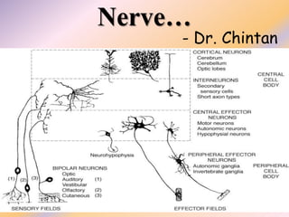
3. NERVE CELLS
- Nerve cell has five to seven processes called
dendrites that extend outward from the cell body
- Particularly in the cerebral and cerebellar cortex,
the dendrites have small knobby projections
called dendritic spines.
- A typical neuron also has a long fibrous axon that
originates from a somewhat thickened area of the
cell body, the axon hillock.
4. NERVE CELLS
- The first portion of the axon is called the initial
segment.
- The axon divides into terminal branches, each ending
in a number of synaptic knobs.
- The knobs are also called terminal buttons or axon
telodendria.
- They contain granules or vesicles in which the synaptic
transmitters secreted by the nerves are stored
5.
6.
7.
8. NERVE CELLS
- The axons of many neurons are myelinated, ie,
they acquire a sheath of myelin, a protein-lipid
complex that is covered around the axon.
- Outside the CNS, the myelin is produced by
Schwann cells, glia-like cells found along the
axon.
- Myelin forms when a Schwann cell wraps its
membrane around an axon up to 100 times
9. NERVE CELLS
- The myelin sheath envelopes the axon except at its
ending and at the nodes of Ranvier, periodic 1-um
constrictions that are about 1 mm apart.
- Not all mammalian neurons are myelinated; some
are unmyelinated,
- ie, are simply surrounded by Schwann cells without
the wrapping of the Schwann cell membrane around
the axon that produces myelin
10. NERVE CELLS
- In the CNS of mammals, most neurons are myelinated,
but the cells that form the myelin are
oligodendrogliocytes rather than Schwann cells
- Unlike the Schwann cell, which forms the myelin
between two nodes of Ranvier on a single neuron,
oligodendrogliocytes send off multiple processes that
form myelin on many neighboring axons.
- In multiple sclerosis, a crippling autoimmune disease,
there is patchy destruction of myelin in the CNS. The
loss of myelin is associated with delayed or blocked
conduction in the demyelinated axons.
11.
12.
13.
14. Axoplasmic Transport
- Nerve cells are secretory cells, but they differ from
other secretory cells in that the secretory zone is
generally at the end of the axon, far removed from
the cell body.
- There are few ribosomes in axons and nerve
terminals, and all necessary proteins are synthesized
in the endoplasmic reticulum and Golgi apparatus of
the cell body
- and then transported along the axon to the synaptic
knobs by the process of axoplasmic flow.
15. Axoplasmic Transport
- Thus, the cell body maintains the functional and
anatomic integrity of the axon; if the axon is cut,
the part distal to the cut degenerates (Wallerian
degeneration).
- Fast transport occurs at about 400 mm/d, and
slow anterograde transport occurs at 0.5-10
mm/d.
- Retrograde transport in the opposite direction
also occurs at about 200 mm/d.
16. Axoplasmic Transport
- Synaptic vesicles recycle in the membrane,
but some used vesicles are carried back to
the cell body and deposited in lysosomes.
- Some of the material taken up at the ending
by endocytosis, including nerve growth
factor and various viruses, is also
transported back to the cell body.
17. EXCITATION & CONDUCTION
- Nerve cells have a low threshold for excitation. The
stimulus may be electrical, chemical, or mechanical.
- Two types of physicochemical disturbances are
produced:
- local, nonpropagated potentials called, depending on
their location, synaptic, generator, or electrotonic
potentials;
- and propagated disturbances, the action potentials (or
nerve impulses).
- They are due to changes in the conduction of ions across
the cell membrane that are produced by alterations in
ion channels.
18.
19. Resting Membrane Potential
- When two electrodes are connected through a
suitable amplifier to a CRO and placed on the surface
of a single axon, no potential difference is observed.
- However, if one electrode is inserted into the
interior of the cell, a constant potential difference is
observed, with the inside negative relative to the
outside of the cell at rest.
- This resting membrane potential is found in almost
all cells. In neurons, it is usually about -70 mV.
20. Action Potential
- The first manifestation of the approaching action
potential is a beginning depolarization of the
membrane.
- After an initial 15 mV of depolarization, the rate of
depolarization increases. The point at which this change
in rate occurs is called the firing level or sometimes the
threshold.
- Thereafter, the tracing on the oscilloscope rapidly
reaches and overshoots the isopotential (zero potential)
line to approximately +35 mV. It then reverses and falls
rapidly toward the resting level.
21. Action Potential
- When repolarization is about 70% completed, the
rate of repolarization decreases and the tracing
approaches the resting level more slowly.
- The sharp rise and rapid fall are the spike potential
of the axon, and the slower fall at the end of the
process is the after-depolarization.
- After reaching the previous resting level, the tracing
overshoots slightly in the hyperpolarizing direction
to form the small but prolonged after-
hyperpolarization.
22.
23. "All-or-None" Law
- it is possible to determine the minimal intensity of
stimulating current (threshold intensity) that, acting
for a given duration, will just produce an action
potential.
- The threshold intensity varies with the duration; with
weak stimuli it is long, and with strong stimuli it is
short.
- Slowly rising currents fail to fire the nerve because
the nerve adapts to the applied stimulus, a process
called accommodation.
24. "All-or-None" Law
- Once threshold intensity is reached, a full-fledged action
potential is produced.
- Further increases in the intensity of a stimulus produce no
increment or other change in the action potential as long as the
other experimental conditions remain constant.
- The action potential fails to occur if the stimulus is
subthreshold in magnitude, and it occurs with a constant
amplitude and form regardless of the strength of the stimulus if
the stimulus is at or above threshold intensity.
- The action potential is therefore "all or none" in character and
is said to obey the all-or-none law.
25.
26. Saltatory Conduction
- The nerve cell membrane is polarized at rest, with positive
charges lined up along the outside of the membrane and
negative charges along the inside.
- During the action potential, this polarity is abolished and
for a brief period is actually reversed
- Conduction in myelinated axons - myelin is an effective
insulator, and current flow through it is negligible.
- Instead, depolarization in myelinated axons jumps from
one node of Ranvier - 50 times faster than the fastest
unmyelinated fibers.
27.
28.
29.
30. Orthodromic & Antidromic
- An axon can conduct in either direction. When an action
potential is initiated in the middle of it, two impulses
traveling in opposite directions are set up by
electrotonic depolarization on either side
- In a living animal, impulses normally pass in one
direction only, ie, from synaptic junctions or receptors
along axons to their termination. Such conduction is
called orthodromic.
- Conduction in the opposite direction is called
antidromic. Since synapses, unlike axons, permit
conduction in one direction only, any antidromic
impulses that are set up fail to pass the first synapse they
encounter
31.
32. The Nernst Potential
- The diffusion potential level across a membrane
that exactly opposes the net diffusion of a
particular ion through the membrane is called the
Nernst potential for that ion
- Nernst equation
- EMF (millivolts) = ± 61 log Concentration inside
Concentration outside
34. IONIC BASIS
- The cell membranes of nerves, like those of other
cells, contain many different types of ion channels.
Some of these are voltage-gated and others are
ligand-gated.
- It is the behavior of these channels, and particularly
Na+ and K+ channels, that explains the electrical
events in nerves.
- Na+ is actively transported out of neurons and
other cells and K+ is actively transported into cells.
35. IONIC BASIS
- K+ permeability at rest is greater than Na+
permeability.
- Therefore, K+ channels maintain the resting
membrane potential.
- With currents, some of the voltage-activated Na+
channels become active,
- and when the firing level is reached, the voltage-
activated Na+ channels overwhelm the K+ and other
channels and a spike potential results.
38. Erlanger and Gasser
Fiber
Type
Function Fiber
Diameter
(μm)
Conduction
Velocity
(m/s)
A α Proprioception; somatic
motor
12-20 70-120
β Touch, pressure 5-12 30-70
γ Motor to muscle spindles 3-6 15-30
δ Pain, touch, temperature 2-5 12-30
B Preganglionic autonomic <3 3-15
C Dorsal root Pain, temperature, some
mechano-reception, reflex
responses
0.4-1.2 0.5-2
Sympathetic Postganglionic
sympathetics
0.3-1.3 0.7-2.3
39. Numerical classification
Number Origin Fiber Type
Ia Muscle spindle,
annulospinal ending.
A α
Ib Golgi tendon organ. A α
II Muscle spindle, flower-
spray ending; touch,
pressure.
A β
III Pain and cold
receptors; some touch
receptors.
A δ
IV Pain, temperature, and
other receptors.
Dorsal root C
41. NEUROGLIA
- In addition to neurons, the nervous system contains glial
cells (neuroglia).
- The Schwann cells that invest axons in peripheral nerves
are classified as glia.
- In the CNS, there are three main types of neuroglia.
- Microglia consists of scavenger cells that resemble tissue
macrophages. They probably come from the bone
marrow and enter the nervous system from the
circulating blood vessels.
- Oligodendrogliocytes are involved in myelin formation
42. NEUROGLIA
- Astrocytes, which are found throughout the brain, are
of two subtypes.
- Fibrous astrocytes, which contain many intermediate
filaments, are found primarily in white matter.
- Protoplasmic astrocytes are found in gray matter and
have granular cytoplasm.
- Both types send processes to blood vessels, where
they induce capillaries to form the tight junctions that
form the blood-brain barrier.
- They also send processes that envelope synapses and
the surface of nerve cells.
43.
44. Nerve Injury
- Nerve injury is injury to nervous tissue.
- In 1941, Seddon introduced a classification of
nerve injuries based on
- three main types of nerve fiber injury
- and
- whether there is continuity of the nerve.
45. Neuropraxia
- This is the least severe form of nerve injury, with complete
recovery.
- In this case, the axon remains intact, but there is myelin
damage causing an interruption in conduction of the
impulse down the nerve fiber.
- Most commonly, this involves compression of the nerve or
disruption to the blood supply (ischemia).
- There is a temporary loss of function which is reversible
within hours to months of the injury (the average is 6–9
weeks).
46. Axonotmesis
- This is a more severe nerve injury with disruption of the
neuronal axon, but with maintenance of the epineurium
- Mainly seen in crush injury, strectching
- If the force creating the nerve damage is removed in a
timely fashion, the axon may regenerate, leading to
recovery – weeks to years
- Axonotmesis involves loss of the relative continuity of
the axon and its covering of myelin, but preservation of
the connective tissue framework of the nerve (the
encapsulating tissue, the epineurium and perineurium,
are preserved).
47. Neurotmesis
- Neurotmesis is the most severe lesion with no potential
of full recovery - severe contusion, stretch, laceration, or
Local Anesthetic Toxicity.
- The axon and encapsulating connective tissue lose their
continuity. The last (extreme) degree of neurotmesis is
transection,
- but most neurotmetic injuries do not produce gross loss
of continuity of the nerve but rather internal disruption
of the architecture of the nerve sufficient to involve
perineurium and endoneurium as well as axons and their
covering.
48.
49.
50.
51. Regeneration
- The processes that occur in peripheral regeneration
can be divided into the following major events:
Wallerian degeneration, axon regeneration/growth,
and nerve reinnervation.
- The proximal stump refers to the end of the injured
neuron that is still attached to the neuron cell body; it
is the part that regenerates.
- The distal stump refers to the end of the injured
neuron that is still attached to the end of the axon; it is
the part that will degenerate
52. Wallerian degeneration
- Wallerian degeneration is a process that occurs before
nerve regeneration and can be described as a cleaning or
clearing process that essentially prepares the distal
stump for reinnervation.
- Schwann cells are glial cells in the peripheral nervous
system that support neurons by forming myelin that
encases nerves.
- During Wallerian degeneration Schwann cells and
macrophages interact to remove debris, specifically
myelin and the damaged axon, from the distal injury site.
53. Wallerian degeneration
- anterograde or orthograde degeneration
- It occurs in the axon stump distal to a site of injury and
usually begins within 24–36 hours of a lesion.
- After injury, the axonal skeleton disintegrates, and the
axonal membrane breaks apart. The axonal
degeneration is followed by degradation of the myelin
sheath and infiltration by macrophages.
- The macrophages, accompanied by Schwann cells, serve
to clear the debris from the degeneration
54. Wallerian degeneration
- The nerve fiber's neurolemma does not degenerate and
remains as a hollow tube.
- Within 4 days of the injury, the distal end of the portion
of the nerve fiber proximal to the lesion sends out
sprouts towards those tubes and these sprouts are
attracted by growth factors produced by Schwann cells in
the tubes.
- If a sprout reaches the tube, it grows into it and
advances about 1 mm per day, eventually reaching and
reinnervating the target tissue.
55. Wallerian degeneration
- If the sprouts cannot reach the tube, for instance
because the gap is too wide or scar tissue has formed,
surgery can help to guide the sprouts into the tubes.
- This regeneration is much slower in the spinal cord than
in PNS
- Axonal injuries initially lead to acute axonal
degeneration (AAD), which is rapid separation of the
proximal (the part nearer the cell body) and distal ends
within 30 minutes of injury.
- Degeneration follows with swelling of the axolemma,
56. Wallerian degeneration
- Granular degeneration of the axonal cytoskeleton
and inner organelles occurs after axolemma
degradation.
- Early changes include accumulation of
mitochondria in the paranodal regions at the site
of injury.
- Endoplasmic reticulum degrades and
mitochondria swell up and eventually degenerate
57. Wallerian degeneration
- Myelin clearance is the next step in Wallerian
degeneration following axonal degeneration.
- The cleaning up of myelin debris is different for PNS and
CNS.
- PNS is much faster and efficient at clearing myelin debris
in comparison to CNS, and Schwann cells are the primary
cause of this difference
- Schwann cells continue to clear up the myelin debris by
degrading their own myelin, phagocytose extracellular
myelin and attract macrophages to myelin debris for
further phagocytosis
58.
59.
60.
61. Proximal Degeneration
- Schwann cells proliferate and the remaining connective
tissue basement membrane forms endoneurial tubes.
- At the neuronal cell body, a process called chromatolysis
occurs in which the nucleus migrates to the periphery of
the cell body and the endoplasmic reticulum breaks up
and disperses.
- Nerve damage causes the metabolic function of the cell
to change from that of producing molecules for synaptic
transmission to that of producing molecules for growth
and repair.
- Chromatolysis is reversed when the cell is prepared for
axon regeneration.
62. Regeneration
- Regeneration is rapid in PNS, allowing for rates of up
to 1 millimeter/day of regrowth.
- Grafts may also be needed to allow for appropriate
reinnervation. It is supported by Schwann cells
through growth factors release.
- CNS regeneration is much slower, and is almost absent
- cause for this could be the delay in clearing up myelin
debris.
- Myelin debris, present in CNS or PNS, contains several
inhibitory factors - The prolonged presence of myelin
debris in CNS could delay the regeneration