This document discusses neurophysiology and summarizes key aspects of nerve cells and signal transmission. It describes the basic anatomy of neurons including the cell body, dendrites, axon, and synaptic terminals. It explains how myelin sheaths insulate neurons and how synapses facilitate chemical transmission between neurons. It also summarizes how nerve impulses are generated through changes in ion permeability and the roles of sodium-potassium pumps in restoring polarization.
Synapse – Greek word –synaptein. Syn –together; aptein –clasp.
Synapse – Clasping of hands (as in hand shaking between two friends).
Site of functional continuity (transneuronal junctional complex) between two neurons.
Why need of synapse?
These slides contain the basic information and principle of nervous transduction, It also includes the information about the type of the neurons, structure of the neuron, resting and active membrane potential, synapes and events occurring in it, and introduction to the neurotransmitters.
Nerve Impulse is defined as a wave of electrical chemical changes across the neuron that helps in the generation of the action potential in response to the stimulus. This transmission of a nerve impulse across the neuron membrane as a result of a change in membrane potential is known as Nerve impulse conduction.
Mechanism of Nerve Impulse Conduction
Nerve impulse conduction is a major process occurring in the body responsible for organized functions of the body. So, for conduction of nerve impulse there are two mechanisms:
Continuous conduction
Saltatory conduction
Nerve muscle physiology /certified fixed orthodontic courses by Indian dental...Indian dental academy
The Indian Dental Academy is the Leader in continuing dental education , training dentists in all aspects of dentistry and offering a wide range of dental certified courses in different formats.
Indian dental academy provides dental crown & Bridge,rotary endodontics,fixed orthodontics,
Dental implants courses.for details pls visit www.indiandentalacademy.com ,or call
0091-9248678078
Neuron communication belongs to subject ANIMAL PHYSIOLOGY in course of zoology.
nerve communication.
how neuron communicate?
RESTING MEMBRANE POTENTIAL
Measurement of Membrane Potential
Nervous system forms an interconnecting fibers of communication network.
In the ‘hard-wiring’ of the nerves, the signals travel in the form of a flow of electrical current called nerve impulses.
The stimulus-response reactions afford internal constancy in the face of environmental changes.
basic nervous system-CNS-PNS -cell bodie- axon-dendron-grye matter- white mat...shailesh sangle
The nervous system is a complex network of cells, tissues, and organs that coordinates and regulates the body's responses to internal and external stimuli. It is responsible for the control and coordination of all the body's functions, including movement, sensation, thought, and behavior.
The nervous system can be divided into two main parts: the central nervous system (CNS) and the peripheral nervous system (PNS). The CNS consists of the brain and spinal cord, while the PNS consists of all the nerves that extend from the CNS to the rest of the body.
The nervous system is made up of different types of cells, including neurons and glial cells. Neurons are specialized cells that transmit signals through the body in the form of electrical impulses. Glial cells, on the other hand, support and protect the neurons and help maintain the proper functioning of the nervous system.
The nervous system is responsible for many vital functions, including:
Sensory processing: The nervous system receives sensory information from the environment and the body's internal organs, and processes and interprets this information to generate appropriate responses.
Motor control: The nervous system controls the muscles and other organs of the body to produce movement and other responses.
Cognitive functions: The nervous system is responsible for the processes of learning, memory, language, and other complex mental activities.
Autonomic functions: The nervous system regulates the body's automatic functions, such as breathing, heart rate, digestion, and other bodily processes that are not under conscious control.
Overall, the nervous system is a complex and intricate system that plays a critical role in maintaining the body's homeostasis and overall well-being.
IF YOU ARE INTRESTED TO SHOP MAMA EARTH'S PRODUCTES ....
Mamaearth is an Indian personal care brand that offers natural, toxin-free, and sustainable products for all age groups. Their product range includes skincare, haircare, baby care, and men's grooming products. The brand is focused on promoting a healthy and sustainable lifestyle, and all their products are cruelty-free and environmentally friendly.
COPY THAT LINK AND PASTE ON SEARCH BAR OF GOOGLE
https://ekaro.in/enkr20230328s23089700
CHOOSE YOUR AND ORDER NOW.......
thank you....
They are produced when high-velocity electrons collide with the metal plates, thereby giving the energy as the X-Rays and themselves absorbed by the metal plate.
The X-Ray beam travels through the air and comes in contact with the body tissues, and produces an image on a metal film.
Soft tissue like organs and skin, cannot absorb the high-energy rays, and the beam passes through them.
Dense materials inside our bodies, like bones, absorb the radiation.he X-Rays properties are given below:
They have a shorter wavelength of the electromagnetic spectrum.
Requires high voltage to produce X-Rays.
They are used to capture the human skeleton defects.
They travel in a straight line and do not carry an electric charge with them.
They are capable of travelling in a vacuum.Medical science recognizes different types of X-Rays. A few important types of X-Rays are given in the points below.
Standard Computed Tomography
Kidney, Ureter, and Bladder X-ray
Teeth and bones X-rays
Chest X-rays
Lungs X-rays
Abdomen X-rays
The following power point presentation talks about neural control and coordination in humans. In this, we study about neurons, the conduction of nerve impulse, about Central Nervous System and also about sense organs
Knee anatomy and clinical tests 2024.pdfvimalpl1234
This includes all relevant anatomy and clinical tests compiled from standard textbooks, Campbell,netter etc..It is comprehensive and best suited for orthopaedicians and orthopaedic residents.
TEST BANK for Operations Management, 14th Edition by William J. Stevenson, Ve...kevinkariuki227
TEST BANK for Operations Management, 14th Edition by William J. Stevenson, Verified Chapters 1 - 19, Complete Newest Version.pdf
TEST BANK for Operations Management, 14th Edition by William J. Stevenson, Verified Chapters 1 - 19, Complete Newest Version.pdf
Recomendações da OMS sobre cuidados maternos e neonatais para uma experiência pós-natal positiva.
Em consonância com os ODS – Objetivos do Desenvolvimento Sustentável e a Estratégia Global para a Saúde das Mulheres, Crianças e Adolescentes, e aplicando uma abordagem baseada nos direitos humanos, os esforços de cuidados pós-natais devem expandir-se para além da cobertura e da simples sobrevivência, de modo a incluir cuidados de qualidade.
Estas diretrizes visam melhorar a qualidade dos cuidados pós-natais essenciais e de rotina prestados às mulheres e aos recém-nascidos, com o objetivo final de melhorar a saúde e o bem-estar materno e neonatal.
Uma “experiência pós-natal positiva” é um resultado importante para todas as mulheres que dão à luz e para os seus recém-nascidos, estabelecendo as bases para a melhoria da saúde e do bem-estar a curto e longo prazo. Uma experiência pós-natal positiva é definida como aquela em que as mulheres, pessoas que gestam, os recém-nascidos, os casais, os pais, os cuidadores e as famílias recebem informação consistente, garantia e apoio de profissionais de saúde motivados; e onde um sistema de saúde flexível e com recursos reconheça as necessidades das mulheres e dos bebês e respeite o seu contexto cultural.
Estas diretrizes consolidadas apresentam algumas recomendações novas e já bem fundamentadas sobre cuidados pós-natais de rotina para mulheres e neonatos que recebem cuidados no pós-parto em unidades de saúde ou na comunidade, independentemente dos recursos disponíveis.
É fornecido um conjunto abrangente de recomendações para cuidados durante o período puerperal, com ênfase nos cuidados essenciais que todas as mulheres e recém-nascidos devem receber, e com a devida atenção à qualidade dos cuidados; isto é, a entrega e a experiência do cuidado recebido. Estas diretrizes atualizam e ampliam as recomendações da OMS de 2014 sobre cuidados pós-natais da mãe e do recém-nascido e complementam as atuais diretrizes da OMS sobre a gestão de complicações pós-natais.
O estabelecimento da amamentação e o manejo das principais intercorrências é contemplada.
Recomendamos muito.
Vamos discutir essas recomendações no nosso curso de pós-graduação em Aleitamento no Instituto Ciclos.
Esta publicação só está disponível em inglês até o momento.
Prof. Marcus Renato de Carvalho
www.agostodourado.com
Ozempic: Preoperative Management of Patients on GLP-1 Receptor Agonists Saeid Safari
Preoperative Management of Patients on GLP-1 Receptor Agonists like Ozempic and Semiglutide
ASA GUIDELINE
NYSORA Guideline
2 Case Reports of Gastric Ultrasound
Acute scrotum is a general term referring to an emergency condition affecting the contents or the wall of the scrotum.
There are a number of conditions that present acutely, predominantly with pain and/or swelling
A careful and detailed history and examination, and in some cases, investigations allow differentiation between these diagnoses. A prompt diagnosis is essential as the patient may require urgent surgical intervention
Testicular torsion refers to twisting of the spermatic cord, causing ischaemia of the testicle.
Testicular torsion results from inadequate fixation of the testis to the tunica vaginalis producing ischemia from reduced arterial inflow and venous outflow obstruction.
The prevalence of testicular torsion in adult patients hospitalized with acute scrotal pain is approximately 25 to 50 percent
The prostate is an exocrine gland of the male mammalian reproductive system
It is a walnut-sized gland that forms part of the male reproductive system and is located in front of the rectum and just below the urinary bladder
Function is to store and secrete a clear, slightly alkaline fluid that constitutes 10-30% of the volume of the seminal fluid that along with the spermatozoa, constitutes semen
A healthy human prostate measures (4cm-vertical, by 3cm-horizontal, 2cm ant-post ).
It surrounds the urethra just below the urinary bladder. It has anterior, median, posterior and two lateral lobes
It’s work is regulated by androgens which are responsible for male sex characteristics
Generalised disease of the prostate due to hormonal derangement which leads to non malignant enlargement of the gland (increase in the number of epithelial cells and stromal tissue)to cause compression of the urethra leading to symptoms (LUTS
Title: Sense of Taste
Presenter: Dr. Faiza, Assistant Professor of Physiology
Qualifications:
MBBS (Best Graduate, AIMC Lahore)
FCPS Physiology
ICMT, CHPE, DHPE (STMU)
MPH (GC University, Faisalabad)
MBA (Virtual University of Pakistan)
Learning Objectives:
Describe the structure and function of taste buds.
Describe the relationship between the taste threshold and taste index of common substances.
Explain the chemical basis and signal transduction of taste perception for each type of primary taste sensation.
Recognize different abnormalities of taste perception and their causes.
Key Topics:
Significance of Taste Sensation:
Differentiation between pleasant and harmful food
Influence on behavior
Selection of food based on metabolic needs
Receptors of Taste:
Taste buds on the tongue
Influence of sense of smell, texture of food, and pain stimulation (e.g., by pepper)
Primary and Secondary Taste Sensations:
Primary taste sensations: Sweet, Sour, Salty, Bitter, Umami
Chemical basis and signal transduction mechanisms for each taste
Taste Threshold and Index:
Taste threshold values for Sweet (sucrose), Salty (NaCl), Sour (HCl), and Bitter (Quinine)
Taste index relationship: Inversely proportional to taste threshold
Taste Blindness:
Inability to taste certain substances, particularly thiourea compounds
Example: Phenylthiocarbamide
Structure and Function of Taste Buds:
Composition: Epithelial cells, Sustentacular/Supporting cells, Taste cells, Basal cells
Features: Taste pores, Taste hairs/microvilli, and Taste nerve fibers
Location of Taste Buds:
Found in papillae of the tongue (Fungiform, Circumvallate, Foliate)
Also present on the palate, tonsillar pillars, epiglottis, and proximal esophagus
Mechanism of Taste Stimulation:
Interaction of taste substances with receptors on microvilli
Signal transduction pathways for Umami, Sweet, Bitter, Sour, and Salty tastes
Taste Sensitivity and Adaptation:
Decrease in sensitivity with age
Rapid adaptation of taste sensation
Role of Saliva in Taste:
Dissolution of tastants to reach receptors
Washing away the stimulus
Taste Preferences and Aversions:
Mechanisms behind taste preference and aversion
Influence of receptors and neural pathways
Impact of Sensory Nerve Damage:
Degeneration of taste buds if the sensory nerve fiber is cut
Abnormalities of Taste Detection:
Conditions: Ageusia, Hypogeusia, Dysgeusia (parageusia)
Causes: Nerve damage, neurological disorders, infections, poor oral hygiene, adverse drug effects, deficiencies, aging, tobacco use, altered neurotransmitter levels
Neurotransmitters and Taste Threshold:
Effects of serotonin (5-HT) and norepinephrine (NE) on taste sensitivity
Supertasters:
25% of the population with heightened sensitivity to taste, especially bitterness
Increased number of fungiform papillae
These lecture slides, by Dr Sidra Arshad, offer a quick overview of physiological basis of a normal electrocardiogram.
Learning objectives:
1. Define an electrocardiogram (ECG) and electrocardiography
2. Describe how dipoles generated by the heart produce the waveforms of the ECG
3. Describe the components of a normal electrocardiogram of a typical bipolar leads (limb II)
4. Differentiate between intervals and segments
5. Enlist some common indications for obtaining an ECG
Study Resources:
1. Chapter 11, Guyton and Hall Textbook of Medical Physiology, 14th edition
2. Chapter 9, Human Physiology - From Cells to Systems, Lauralee Sherwood, 9th edition
3. Chapter 29, Ganong’s Review of Medical Physiology, 26th edition
4. Electrocardiogram, StatPearls - https://www.ncbi.nlm.nih.gov/books/NBK549803/
5. ECG in Medical Practice by ABM Abdullah, 4th edition
6. ECG Basics, http://www.nataliescasebook.com/tag/e-c-g-basics
NVBDCP.pptx Nation vector borne disease control programSapna Thakur
NVBDCP was launched in 2003-2004 . Vector-Borne Disease: Disease that results from an infection transmitted to humans and other animals by blood-feeding arthropods, such as mosquitoes, ticks, and fleas. Examples of vector-borne diseases include Dengue fever, West Nile Virus, Lyme disease, and malaria.
Report Back from SGO 2024: What’s the Latest in Cervical Cancer?bkling
Are you curious about what’s new in cervical cancer research or unsure what the findings mean? Join Dr. Emily Ko, a gynecologic oncologist at Penn Medicine, to learn about the latest updates from the Society of Gynecologic Oncology (SGO) 2024 Annual Meeting on Women’s Cancer. Dr. Ko will discuss what the research presented at the conference means for you and answer your questions about the new developments.
HOT NEW PRODUCT! BIG SALES FAST SHIPPING NOW FROM CHINA!! EU KU DB BK substit...GL Anaacs
Contact us if you are interested:
Email / Skype : kefaya1771@gmail.com
Threema: PXHY5PDH
New BATCH Ku !!! MUCH IN DEMAND FAST SALE EVERY BATCH HAPPY GOOD EFFECT BIG BATCH !
Contact me on Threema or skype to start big business!!
Hot-sale products:
NEW HOT EUTYLONE WHITE CRYSTAL!!
5cl-adba precursor (semi finished )
5cl-adba raw materials
ADBB precursor (semi finished )
ADBB raw materials
APVP powder
5fadb/4f-adb
Jwh018 / Jwh210
Eutylone crystal
Protonitazene (hydrochloride) CAS: 119276-01-6
Flubrotizolam CAS: 57801-95-3
Metonitazene CAS: 14680-51-4
Payment terms: Western Union,MoneyGram,Bitcoin or USDT.
Deliver Time: Usually 7-15days
Shipping method: FedEx, TNT, DHL,UPS etc.Our deliveries are 100% safe, fast, reliable and discreet.
Samples will be sent for your evaluation!If you are interested in, please contact me, let's talk details.
We specializes in exporting high quality Research chemical, medical intermediate, Pharmaceutical chemicals and so on. Products are exported to USA, Canada, France, Korea, Japan,Russia, Southeast Asia and other countries.
HOT NEW PRODUCT! BIG SALES FAST SHIPPING NOW FROM CHINA!! EU KU DB BK substit...
Neurophysiology1
1. Kisii University, School of Health Sciences, Department of
Nursing NEUROPHYSIOLOGY By Nyaboga E
Esther Nyaboga Page 1
NEUROPHYSIOLOGY
Deals with the function of the nervous system.
Nerve:
Bundle of conducting fibres enclosed in a sheath called epineurium.
Its function is to transmit impulses between any part of the body and a nerve centre.
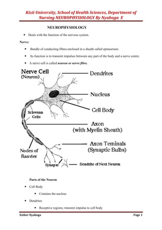
A nerve cell is called neuron or nerve fibre.
Parts of the Neuron
Cell Body
Contains the nucleus
Dendrites
Receptive regions; transmit impulse to cell body
2. Kisii University, School of Health Sciences, Department of
Nursing NEUROPHYSIOLOGY By Nyaboga E
Esther Nyaboga Page 2
Short, often highly branched
May be modified to form receptors
Axons
Transmit impulses away from cell body
Axon hillock; trigger zone
Where action potentials first develop
Presynaptic terminals (terminal boutons)
Contain neurotransmitter substance (NT)
Release of NT stimulates impulse in next neuron
Bundles of axons form nerves
In the peripheral nervous system, axons and dendrites are “wrapped” in specialized
cells called Schwann cells. During embryonic development, Schwann cells grow to
surround the neuron processes, enclosing them in several layers of Schwann cell
membrane.
These layers are the myelin sheath; myelin is a phospholipid that electrically
insulates neurons from one another.
The nuclei and cytoplasm of the Schwann cells are wrapped around the outside of the
myelin sheath and are called the neurolemma, which becomes very important if
nerves are damaged.
The Schwann cells are also believed to produce a chemical growth factor that
stimulates regeneration.
In the central nervous system, the myelin sheaths are formed by oligodendrocytes,
one of the neuroglia (glial cells), the specialized cells found only in the brain and
spinal cord.
Because no Schwann cells are present, however, there is no neurolemma, and
regeneration of neurons does not occur. This is why severing of the spinal cord, for
example, results in permanent loss of function.
SYNAPSES
The small gap or space between the axon of one neuron and the dendrites or cell body
of the next neuron is called the synapse.
3. Kisii University, School of Health Sciences, Department of
Nursing NEUROPHYSIOLOGY By Nyaboga E
Esther Nyaboga Page 3
Within the synaptic knob (terminal end) of the presynaptic axon is a chemical
neurotransmitter that is released into the synapse by the arrival of an electrical nerve
impulse. The neurotransmitter diffuses across the synapse, combines with specific
receptor sites on the cell membrane of the postsynaptic neuron, and there generates an
electrical impulse that is, in turn, carried by this neuron’s axon to the next synapse,
and so forth.
A chemical inactivator at the cell body or dendrite of the postsynaptic neuron
quickly inactivates the neurotransmitter. This prevents unwanted, continuous
impulses, unless a new impulse from the first neuron releases more neurotransmitter.
Many synapses are termed excitatory, because the neurotransmitter causes the
postsynaptic neuron to depolarize (become more negative outside as Na ions enter the
cell) and transmit an electrical impulse to another neuron, muscle cell, or gland. Some
synapses, however, are inhibitory, meaning that the neurotransmitter causes the
postsynaptic neuron to hyperpolarize (become even more positive outside as K ions
leave the cell or Cl ions enter the cell) and therefore not transmit an electrical
impulse.
Such inhibitory synapses are important, for example, for slowing the heart rate, and
for balancing the excitatory impulses transmitted to skeletal muscles. With respect to
the skeletal muscles, this inhibition prevents excessive contraction and is important
for coordination
Synaptic Functions of Neurons
As the impulses are transmitted from one neuron to the next:
Each impulse may be blocked in its transmission from one neuron to the next.
May be changed from one single impulse to repetitive impulses. Or
May be integrated with impulses from other neurons to cause highly intricate pattern
of impulses in successive neurons. All these functions can be classified as synaptic
functions of neurons.
Types of synapses:
1. Chemical synapse
Almost all synapses in the cns are chemical synapses. In these the first neuron
secretes at its nerve ending synapse a chemical substance called neurotransmitter
which acts on receptor proteins in the membrane of the next neuron to excite the
neuron, inhibit it or modify its sensitivity. Exa. of neurotransmitters are acetylcholine,
norepinephrine, epinephrine, histamine, serotonin etc.
2. Electrical synapse
4. Kisii University, School of Health Sciences, Department of
Nursing NEUROPHYSIOLOGY By Nyaboga E
Esther Nyaboga Page 4
Are characterized by direct open fluid channels that conduct electricity from one cell
to another. Most of these consist of small protein tubular structures called gap
junctions that allow free movement of ions from interior of one cell to interior of the
next.
‘one-way conduction’ at the chemical synapses
Chemical synapse transmit signals in one direction i.e. from one neuron that secretes
the transmitter substance called presynaptic neuron to the neuron on which transmitter
acts called postsynaptic neuron. This is the principle one-way conduction at chemical
synapses.
Electrical synapses often transmit signals in either direction.
The advantage of one-way conduction is that it allows signals to be directed towards
specific goals e.g. sensation, motor control, memory etc.
Physiologic Anatomy of the Synapse
Mitochondria at the prsynaptic neuron site
5. Kisii University, School of Health Sciences, Department of
Nursing NEUROPHYSIOLOGY By Nyaboga E
Esther Nyaboga Page 5
Presynaptic terminals/terminal knobs/synaptic knobs/boutons has two internal structures
important to the excitatory or inhibitory function of the synapse.
1. Transmitter vesicle: it contains the transmitter substance released to synaptic cleft
which excites or inhibits the postsynaptic neuron. It excites if the neuronal membrane
contains excitatory receptors & inhibits if the membrane contains inhibitory receptors.
2. Mitochondria: it provides ATP to supply energy for synthesis of new transmitter
substances.
Release and action of the neurotransmitter. How does it happen?
its membrane
into the cleft
in permeability characteristics of the postsynaptic neuronal membrane which leads to
excitation or inhibition of the postsynaptic neuron depending on neuronal receptor
characteristics.
Mechanism by which an action potential causes transmitter Release from the
presynaptic Terminal: Role of calcium ion.
large number of voltage-gated calcium channels
channels open and allow more calcium ions to flow into the terminal
into the synaptic cleft is directly related to the number of calcium ions that enter
The mechanism
It’s believed that when calcium ions enter the presynaptic terminal they bind with
special protein molecules on the inside surface of presynaptic membrane called
release sites. This binding causes release sites to open through the membrane
allowing a few transmitter vesicles to release their transmitter into a cleft after each
single action potential.
Action of the transmitter substance on the postsynaptic neuron: function of ‘receptor
proteins’
Postsynaptic membrane contain large number of receptor proteins. The molecules of these
receptors have two important components:
1. A binding component: protrudes outward from the membrane to synaptic cleft where it
binds the neurotransmitter coming from presynaptic terminal.
2. An ionophore component: passes all the way through the postsynaptic membrane to
the interior of the postsynaptic neuron
6. Kisii University, School of Health Sciences, Department of
Nursing NEUROPHYSIOLOGY By Nyaboga E
Esther Nyaboga Page 6
Types of ionophore :
1. An ion channel that allows passage of specified ions through the membrane
2. A secondary messanger activator that is not an ion channel, which is a molecule that
protrudes into the cell cytoplasm & activates one or more substances inside
postsynaptic neuron. These substance inturn serves as second messengers to increase
or decrease cellular function.
NERVE IMPULSE
The events of an electrical nerve impulse are the same as those of the electrical
impulse generated in muscle fibers. A neuron not carrying an impulse is in a state of
polarization, (resting state) with Na ions more abundant outside the cell, and K ions
and negative ions more abundant inside the cell. The neuron has a positive charge on
the outside of the cell membrane and a relative negative charge inside.
A stimulus (such as a neurotransmitter) makes the membrane very permeable to Na
ions, which rush into the cell. This brings about depolarization, a reversal of charges
on the membrane. The outside now has a negative charge, and the inside has a
positive charge.
As soon as depolarization takes place, the neuron membrane becomes very permeable
to K ions, which rush out of the cell. This restores the positive charge outside and the
negative charge inside, and is called repolarization.
(The term action potential refers to depolarization followed by repolarization.)
Then the sodium and potassium pumps return Na ions outside and K ions inside, and
the neuron is ready to respond to another stimulus and transmit another impulse.
Transmission of electrical impulses is very rapid. The presence of an insulating
myelin sheath increases the velocity of impulses since only the nodes of Ranvier
depolarize. This is called saltatory conduction.
At synapses, nerve impulse transmission changes from electrical to chemical and
depends on the release of neurotransmitters. Although diffusion across synapses is
slow, the synapses are so small that this does not significantly affect the velocity of
impulses.
Electrical Signals
Neurons produce electrical signals called action potentials ( = nerve impulse)
Nerve impulses transfer information from one part of body to another
e.g., receptor to CNS or CNS to effector
7. Kisii University, School of Health Sciences, Department of
Nursing NEUROPHYSIOLOGY By Nyaboga E
Esther Nyaboga Page 7
Electrical properties result from
ionic concentration differences across plasma membrane
permeability of membrane
Electrochemical Gradient of the Neuron Membrane
Electrical Gradient
Develops when there are more positive or negative charges (ions) on one side
of a membrane than on the other
Charges (ions) move toward the area of opposite charge
Positive toward negative and vice versa
Chemical Gradient
Develops when there are more ions of a substance in one area than in another
(e.g., more Na+
extracellularly than intracellularly)
Ions tend to move from an area of high concentration to an area of low
concentration; more to less (i.e., down their concentration gradient)
Electrochemical gradient
The sum of all electrical and chemical forces acting across the cell membrane
Resting Membrane Potential (RMP)
Nerve cell has an electrical potential, or voltage across its membrane of a –70 mV; (=
to 1/20th that of a flashlight battery (1.5 v)
The potential is generated by different concentrations of Na+
, K+
, Cl
, and protein
anions (A
)
But the ionic differences are the consequence of:
Differential permeability of the axon membrane to these ions
Operation of a membrane pump called the sodium-potassium pump
What Establishes the RMP?
Diffusion of Na+
and K+
down their concentration gradients
Na+
diffuses into the cell and K+
diffuses out of the cell
BUT, membrane is 75x’s more permeable to K+
than Na+
Thus, more K+
diffuses out than Na+
diffuses in
This increases the number of positive charges on the outside of the membrane
relative to the inside.
8. Kisii University, School of Health Sciences, Department of
Nursing NEUROPHYSIOLOGY By Nyaboga E
Esther Nyaboga Page 8
BUT, the Na+
-K+
pump carries 3 Na+
out for every 2 K+
in.
This is strange in that MORE K+
exited the cell than Na+
entered!
Pumping more + charges out than in also increases the number of + changes
on the outside of the membrane relative to the inside.
AND presence of anionic proteins (A-
) in the cytosol adds to the negativity of the
cytosolic side of the membrane
THEREFORE, the inside of the membrane is measured at a -70 mV (1 mv = one-
thousandth of a volt)
Changes in the Membrane Potential
Membrane potential is dynamic
Rises or falls in response to temporary changes in membrane permeability
Changes in membrane permeability result from the opening or closing of
membrane channels
Types of channels
Passive or leak channels - always open
Gated channels - open or close in response to specific stimuli;
Ligand-gated channels
Voltage-gated channels
Nongated (Leakage) channels
Many more of these for K+
and Cl-
than for Na+
.
So, at rest, more K+
and Cl-
are moving than Na+
.
How are they moving?
Protein repels Cl-
, so Cl-
moves out.
K+
are in higher concentration on inside than out, they diffuse out.
Always open and responsible for permeability when membrane is at rest.
Gated ion channels.
Gated ion channels open and close because of some sort of stimulus. When they
open, they change the permeability of the cell membrane.
Ligand-gated: open or close in response to ligand (a chemical) such as ACh
binding to receptor protein.
9. Kisii University, School of Health Sciences, Department of
Nursing NEUROPHYSIOLOGY By Nyaboga E
Esther Nyaboga Page 9
Acetylcholine (ACh) binds to acetylcholine receptor on a Na+
channel.
Channel opens, Na+
enters the cell.
Ligand-gated channels most abun- dant on dendrites and cell body;
areas where most synaptic commu-nication occurs
Voltage-gated:
open or close in response to small voltage changes across the cell membrane.
At rest, membrane is negative on the inside relative to the outside.
When cell is stimulated, that relative charge changes and voltage-gated ion channels
either open or close.
Most common voltage gated are Na+
, K+
, and Ca+2
Common on areas where action potentials develop
Axons of unipolar and multipolar neurons
Sarcolemma (including T-tubules) of skeletal muscle fibers and cardiac
muscle fibers
Local Potentials/Graded Potentials
Graded: of varying intensity; NOT all the same intensity
Changes in membrane potential that cannot spread far from site of stimulation
Can result in depolarization or hyperpolarization
Depolarization
Opening Na+
channels allows more + charges to enter thereby making interior
less negative (-70 mV -60mV); see next slide
RMP shifts toward O mV
Hyperpolarization
Opening of K+
channels allows more + charges to leave thereby making
interior more negative (-70 mV -80 mV)
RMP shifts away from O mV
Repolarization
Process of restoring membrane potential back to normal (RMP)
Degree of depolarization decreases with distance from stimulation site; called
decremental spread (see next slide)
10. Kisii University, School of Health Sciences, Department of
Nursing NEUROPHYSIOLOGY By Nyaboga E
Esther Nyaboga Page 10
Graded potentials occur on dendrites and cell bodies of neurons but also on gland
cells, sensory receptors, and muscle cell sarcolemma
Characteristics of local potentials:
1. A stimulus causes ion channels to open, resulting in increases permeability of the
membrane to Na+
, K+
or Cl-
.
2. Increased permeability of the membrane to Na+
results in depolarization. Increased
permeability to K+
or Cl-
results in hyperpolarization.
3. Local potentials are graded; that is, the size of the local potential is propotional to the
strength of the stimulus.
4. Local potentials are conducted in a decremental fashion, meaning that their magnitude
decreases as they spread over the plasma membrane. Local potentials cannot be
measures a few millimetres from the point of stimulation.
Action Potential: Resting State
Na+
and K+
channels are closed
Leakage accounts for small movements of Na+
and K+
Each Na+
channel has two voltage-regulated gates
Activation gates – closed in the resting state
Inactivation gates – open in the resting state
Action Potential: Depolarization Phase
11. Kisii University, School of Health Sciences, Department of
Nursing NEUROPHYSIOLOGY By Nyaboga E
Esther Nyaboga Page 11
Some stimulus opens Na+
gates and Na+
influx occurs
K+
gates are closed
Na+
influx causes a reversal of RMP
Interior of membrane now less negative (from -70 mV -55 mV)
Threshold – a critical level of depolarization (-55 to -50 mV)
At threshold, depolarization becomes self-generating
I.e., depolarization of one segment leads to depolarization in the next
If threshold is not reached, no action potential develops
Action Potential: Repolarization Phase
Sodium inactivation gates close
Membrane permeability to Na+
declines to resting levels
As sodium gates close, voltage-sensitive K+
gates open
K+
exits the cell and internal negativity of the resting neuron
is restored
Action Potential: Hyperpolarization
12. Kisii University, School of Health Sciences, Department of
Nursing NEUROPHYSIOLOGY By Nyaboga E
Esther Nyaboga Page 12
Potassium gates remain open, causing an excessive efflux of K+
This efflux causes hyperpolarization of the membrane (undershoot)
The neuron is insensitive to stimulus and depolarization during this time
Depolarization and Hyperpolarization
Phases of the Action Potential (figure below)
1 – RESTING STATE
RMP = -70 mV
2 – DEPOLARIZATION
Increased Na+
influx
Membrane Potential becomes less negative
If threshold is reached, depolarization continues
Peak reached at +30 mV
Total amplitude = 100 mV
3 – REPOLARIZATION
Decreased Na+
influx
Increased K+
efflux
Membrane Potential becomes more negative
4 – HYPERPOLARIZATION
13. Kisii University, School of Health Sciences, Department of
Nursing NEUROPHYSIOLOGY By Nyaboga E
Esther Nyaboga Page 13
Excess K+
efflux
Action Potential: Role of the Sodium-Potassium Pump
Repolarization
Restores the resting electrical conditions of the neuron
Does not restore the resting ionic conditions
Ionic redistribution back to resting conditions is restored by the sodium-potassium
pump
Speed of Impulse Conduction
Faster in myelinated than in non-myelinated
In myelinated axons, lipids act as insulation (the myelin sheath) forcing local currents
to jump from node to node
In myelinated neurons, speed is affected by:
Thickness of myelin sheath
Diameter of axons
14. Kisii University, School of Health Sciences, Department of
Nursing NEUROPHYSIOLOGY By Nyaboga E
Esther Nyaboga Page 14
Large-diameter conduct more rapidly than small-diameter. Large
diameter axons have greater surface area and more voltage-gated Na+
channels
Nerve Fiber Types
Type A: large-diameter (4-20 µm), heavily myelinated. Conduct at 15-120 m/s (= 300
mph).
Motor neurons supplying skeletal muscles and most sensory neurons carrying
info. about position, balance, delicate touch
Type B: medium-diameter (2-4 µm), lightly myelinated. Conduct at 3-15 m/s.
Sensory neurons carrying info. about temperature, pain, general touch,
pressure sensations
Type C: small-diameter (0.5-2 µm), unmyelinated. Conduct at 2 m/s or less.
Many sensory neurons and most ANS motor neurons to smooth muscle,
cardiac muscle, glands
Coding for Stimulus Intensity
All action potentials are alike (of the same amplitude) and are independent of stimulus
intensity.
The amplitude of the action potential is the same for a weak stimulus as it is
for a strong stimulus.
So how does one stimulus feel stronger than another?
Strong stimuli generate more action potentials than weaker stimuli.
More action potentials stimulate the release of more neurotransmitter from the
synaptic knob
The CNS determines stimulus intensity by the frequency of impulse transmission
15. Kisii University, School of Health Sciences, Department of
Nursing NEUROPHYSIOLOGY By Nyaboga E
Esther Nyaboga Page 15
Neuronal Pathways and Circuits
Organization of neurons in CNS varies in complexity
Convergent pathways: several neurons converge on a single postsynaptic
neuron. E.g., synthesis of data in brain.
Divergent pathways: the spread of information from one neuron to several
neurons. E.g., important information can be transmitted to many parts of the
brain.
Oscillating circuits: Arranged in circular fashion to allow action potentials to
cause a neuron in a farther along circuit to produce an action potential more
than once. Can be a single neuron or a group of neurons that are self
stimulating. Continue until neurons are fatigued or until inhibited by other
neurons. Respiration? Wake/sleep.
References
1. Arthur C Guyton and John E Hall (2015).textbook of Medical Physiology, 12th
edition, Elsevier Saunders.
2. Walter F Boron and Emile L Boulpaep (2015). Medical Physiology International
edition 2nd
edition, Saunders Elsevier.