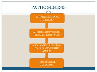
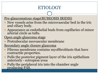
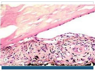
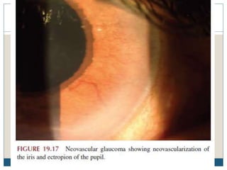
A R Y A . K . A
NEOVASCULAR GLAUCOMA
GLAUCOMA
Chronic, progressive optic neuropathy caused by a
group of ocular conditions, which lead to damage of
the optic nerve with loss of visual function.
The most common risk factor known is a raised
intraocular pressure.(N/l IOP:10-21mm Hg)
CLASSIFICATION
A. Congenital/developmental glaucomas
B. Primary adult glaucomas
a. POAG
b. PACG
C. Secondary glaucomas
a. Mechanism of rise in IOP
i. Secondary open angle glaucomas
ii. Secondary angle closure glaucoma
b. Causative primary disease
i. Lens induced
ii. Inflammatory
iii. Pigmentary
iv. Neovascular glaucoma
v. Traumatic glaucoma
NVG
Secondary Glaucoma due to fibrovascular membrane
on the surface of the iris and the angle of anterior
chamber.
Syn:Thrombotic glaucoma, hemorrhagic glaucoma,
rubeotic glaucoma.
Follows extensive retinal ischemia
PATHOGENESIS
NEOVASCULARISATION
ON IRIS AND IN THE
ANGLE
NEOVASCULAR
GLAUCOMA
ANGIOGENIC FACTORS
RELEASED & DIFFUSED
CHRONIC RETINAL
ISCHAEMIA
ETIOLOGY
Pre-glaucomatous stage(RUBEOSIS IRIDIS)
 New vessels arise from the microvascular bed in the iris
and ciliary body.
 Appearance as endothelial buds from capillaries of minor
arterial circle as tufts.
Open angle glaucoma stage
 Pretrabecular neovascular membrane
Secondary angle closure glaucoma
 Fibrous membrane contains myofibroblasts that have
contractile properties.
 Pulls the posterior pigment layer of the iris epithelium
anteriorly - ectropion uvea
 Pulls the peripheral iris into the chamber angle
producing PAS.
CAUSES
 Ischaemic Central retinal vein occlusion
 Proliferative Diabetic Retinopathy
 Sickle cell retinopathy
 Eales disease
 C/c intraocular inflammations
 Intraocular tumours
 Long standing RD
 CRAO
PRESENTATION
SYMPTOMS
 Sudden pain
 Headache
 Vomiting
 Redness
 Watering
 Defective vision
 Photophobia
SIGNS
 Ciliary injection
 Hazy cornea from epithelial
edema
 Deep anterior chamber
with moderate flare,
 Hyphema
 New vessels on the iris and
in the angle(Gonioscopy)
 Ectropion uveae
 Fixed dilated pupil
 Raised IOP
TREATMENT
 Panretinal photocoagulation
 Goniophotocoagulation
 Intra Vitreal Anti -VEGF
 Mydriatics
 Corticosteroids
 Filtering surgeries
 Cyclodestructive procedures
 Enucleation
 Glaucoma drainage device
CASE DISCUSSION
 69 years old female
 Presenting complaints:
 c/o DV of BE since long
 HOPI:
 Noticed DV(BE) since long.
 Diagnosed as CRVO(LE) and POAG(BE) 2 yrs back
 At present, increase in DV
 A/w severe headache
 h/o past illness
 T2 DM-10 yrs on OHA
 HTN- 20-25 yrs- on T.Amlo
 POAG(BE)- 2yrs
 Ischemic CRVO(LE)-2 yrs
 Family history: Nil significant
 Drug history
 1. T.Amlo 5mg OD
 2.T.Nebivolol 5mg 1-0-0
 Brimonidine e/d 1 BD(BE)
 Nepa e/d 1 BD
 T.Glimipride 2mg BD
 GENERAL EXAMINATION
 Conscious, oriented, Cooperative
 Poorly built and nourished
 No pallor, icterus, cyanosis, clubbing, LNE, pedal
edema
 PR: 66/’
 BP: 160/70 mm Hg
 CVS: S1 S2 heard, No murmurs
 Resp: NVBS B/L
 GIT: P/A-Soft, Nonender; No organomegaly
OCULAR EXAMINATION
RIGHT EYE LEFT EYE
Lid and adnexa N/l N/L
Conjunctiva N/l N/l
Cornea N/l size, shape, lustre
Corneal sensn-N/l
N/l size, shape, lustre
Corneal sensn-N/l
AC N/l depth and contents N/l depth and contents
Iris N/l colour and pattern NVI: 9-11 O’clock posn
Pupil N/l size and shape
DR-Brisk
CR- SLuggish
N/l size and shape
Gr. II RAPD
Lens IMSC IMSC
Vision-Vn 6/60 HM
Nv N24 N36
Head posture: N EOM-Full Orthophoric
 RIGHT EYE
 Media hazy
 Disc
 Defined
 CDR 0.7
 Nasalisation
 of vessels +
 Tesselated
fundus
 LEFT EYE
 Media hazy
 CDR 0.6
 Veins tortuous
and dilated
 Dot
hemorrhages in
macula
 Tesselated
fundus
NCT 16.3 37.6
Carotid doppler: diffuse wall thickening in all carotid
arteries
Imp: POAG(BE)
NVG(LE)
THANK YOU

Neovascular glaucoma

  • 1.
    A R YA . K . A NEOVASCULAR GLAUCOMA
  • 2.
    GLAUCOMA Chronic, progressive opticneuropathy caused by a group of ocular conditions, which lead to damage of the optic nerve with loss of visual function. The most common risk factor known is a raised intraocular pressure.(N/l IOP:10-21mm Hg)
  • 3.
    CLASSIFICATION A. Congenital/developmental glaucomas B.Primary adult glaucomas a. POAG b. PACG C. Secondary glaucomas a. Mechanism of rise in IOP i. Secondary open angle glaucomas ii. Secondary angle closure glaucoma b. Causative primary disease i. Lens induced ii. Inflammatory iii. Pigmentary iv. Neovascular glaucoma v. Traumatic glaucoma
  • 4.
    NVG Secondary Glaucoma dueto fibrovascular membrane on the surface of the iris and the angle of anterior chamber. Syn:Thrombotic glaucoma, hemorrhagic glaucoma, rubeotic glaucoma. Follows extensive retinal ischemia
  • 5.
    PATHOGENESIS NEOVASCULARISATION ON IRIS ANDIN THE ANGLE NEOVASCULAR GLAUCOMA ANGIOGENIC FACTORS RELEASED & DIFFUSED CHRONIC RETINAL ISCHAEMIA
  • 6.
    ETIOLOGY Pre-glaucomatous stage(RUBEOSIS IRIDIS) New vessels arise from the microvascular bed in the iris and ciliary body.  Appearance as endothelial buds from capillaries of minor arterial circle as tufts. Open angle glaucoma stage  Pretrabecular neovascular membrane Secondary angle closure glaucoma  Fibrous membrane contains myofibroblasts that have contractile properties.  Pulls the posterior pigment layer of the iris epithelium anteriorly - ectropion uvea  Pulls the peripheral iris into the chamber angle producing PAS.
  • 9.
    CAUSES  Ischaemic Centralretinal vein occlusion  Proliferative Diabetic Retinopathy  Sickle cell retinopathy  Eales disease  C/c intraocular inflammations  Intraocular tumours  Long standing RD  CRAO
  • 10.
    PRESENTATION SYMPTOMS  Sudden pain Headache  Vomiting  Redness  Watering  Defective vision  Photophobia SIGNS  Ciliary injection  Hazy cornea from epithelial edema  Deep anterior chamber with moderate flare,  Hyphema  New vessels on the iris and in the angle(Gonioscopy)  Ectropion uveae  Fixed dilated pupil  Raised IOP
  • 11.
    TREATMENT  Panretinal photocoagulation Goniophotocoagulation  Intra Vitreal Anti -VEGF  Mydriatics  Corticosteroids  Filtering surgeries  Cyclodestructive procedures  Enucleation  Glaucoma drainage device
  • 12.
    CASE DISCUSSION  69years old female  Presenting complaints:  c/o DV of BE since long  HOPI:  Noticed DV(BE) since long.  Diagnosed as CRVO(LE) and POAG(BE) 2 yrs back  At present, increase in DV  A/w severe headache
  • 13.
     h/o pastillness  T2 DM-10 yrs on OHA  HTN- 20-25 yrs- on T.Amlo  POAG(BE)- 2yrs  Ischemic CRVO(LE)-2 yrs  Family history: Nil significant  Drug history  1. T.Amlo 5mg OD  2.T.Nebivolol 5mg 1-0-0  Brimonidine e/d 1 BD(BE)  Nepa e/d 1 BD  T.Glimipride 2mg BD
  • 14.
     GENERAL EXAMINATION Conscious, oriented, Cooperative  Poorly built and nourished  No pallor, icterus, cyanosis, clubbing, LNE, pedal edema  PR: 66/’  BP: 160/70 mm Hg  CVS: S1 S2 heard, No murmurs  Resp: NVBS B/L  GIT: P/A-Soft, Nonender; No organomegaly
  • 15.
    OCULAR EXAMINATION RIGHT EYELEFT EYE Lid and adnexa N/l N/L Conjunctiva N/l N/l Cornea N/l size, shape, lustre Corneal sensn-N/l N/l size, shape, lustre Corneal sensn-N/l AC N/l depth and contents N/l depth and contents Iris N/l colour and pattern NVI: 9-11 O’clock posn Pupil N/l size and shape DR-Brisk CR- SLuggish N/l size and shape Gr. II RAPD Lens IMSC IMSC Vision-Vn 6/60 HM Nv N24 N36 Head posture: N EOM-Full Orthophoric
  • 16.
     RIGHT EYE Media hazy  Disc  Defined  CDR 0.7  Nasalisation  of vessels +  Tesselated fundus  LEFT EYE  Media hazy  CDR 0.6  Veins tortuous and dilated  Dot hemorrhages in macula  Tesselated fundus
  • 17.
    NCT 16.3 37.6 Carotiddoppler: diffuse wall thickening in all carotid arteries Imp: POAG(BE) NVG(LE)
  • 18.