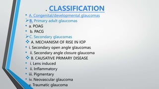
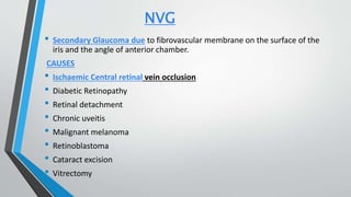
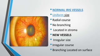
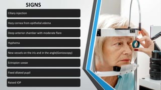
New Neovascular Glaucoma
Dr Muhammad Muneeb
Senior Registrar Ophthalmology
GLAUCOMA
•Chronic, progressive optic neuropathy caused by a group
of ocular conditions, which lead to damage of the optic
nerve with loss of visual function. The most common risk
factor known is a raised intraocular pressure.(N/l IOP:10-
21mm Hg)
. CLASSIFICATION
• A. Congenital/developmental glaucomas
B. Primary adult glaucomas
• a. POAG
• b. PACG
C. Secondary glaucomas
 A. MECHANISM OF RISE IN IOP
• i. Secondary open angle glaucomas
• ii. Secondary angle closure glaucoma
 B. CAUSATIVE PRIMARY DISEASE
• i. Lens induced
• ii. Inflammatory
• iii. Pigmentary
• iv. Neovascular glaucoma
• v. Traumatic glaucoma
NVG
• Secondary Glaucoma due to fibrovascular membrane on the surface of the
iris and the angle of anterior chamber.
CAUSES
• Ischaemic Central retinal vein occlusion
• Diabetic Retinopathy
• Retinal detachment
• Chronic uveitis
• Malignant melanoma
• Retinoblastoma
• Cataract excision
• Vitrectomy
PATHOGENESIS
CHRONIC RETINAL
ISCHAEMIA
ANGIOGENIC FACTORS
RELEASED & DIFFUSED
NEOVASCULARISATION ON
IRIS AND IN THE ANGLE
NEOVASCULAR
GLAUCOMA
ETIOLOGY
•PRE-GLAUCOMATOUS STAGE(RUBEOSIS IRIDIS)
•New vessels arise from the microvascular bed in the iris
and ciliary body.
•Appearance as endothelial buds from capillaries of minor
arterial circle as tufts.
•OPEN ANGLE GLAUCOMA STAGE
• Pretrabecular neovascular membrane
•SECONDARY ANGLE CLOSURE GLAUCOMA
•Fibrous membrane contains myofibroblasts that have
contractile properties.
•Pulls the posterior pigment layer of the iris epithelium
anteriorly - ectropion uvea
•Pulls the peripheral iris into the
chamber angle producing PAS
RUBEOSIS IRIDIS
•NORMAL IRIS VESSELS
•Uniform size
•Radial course
•No branching
• Located in stroma
•NEW VESSELS
• Irregular size
•Irregular course
•Branching Located on surface
PRESENTATION
SYMPTOMS
• Sudden pain
• Headache
• Vomiting
• Redness
• Watering
• Defective vision
• Photophobia SIGNS
SIGNS
Ciliary injection
Hazy cornea from epithelial edema
Deep anterior chamber with moderate flare
Hyphema
New vessels on the iris and in the angle(Gonioscopy)
Ectropion uveae
Fixed dilated pupil
Raised IOP
TREATMENT
• Panretinal photocoagulation
• Intra Vitreal Anti -VEGF
• Mydriatics
• Corticosteroids
• Filtering surgeries
• Cyclodestructive procedures
• Enucleation
PANRETINAL PHOTOCOAGULATION
• Performed by reducing stimulus for new vessel formation.
• in Diabetic retinopathy & Ischaemic CRVO.
• 200 -500 um spot size , 1500 -2000 burns
• Use of Argon laser
• To prevent the onset of NVG
INTRAVITREAL ANTI - VEGF
• . Bevacizumab (Avastin) at dose of 1.25 mg /0.05 ml .
• It inhibits the VEGF – receptor interaction and blocks
vascular permeability and angiogenesis.
FILTRATION SURGERIES
• Good visual potential, IOP not reduced by medical treatment and
if the neovascular membrane has regressed.
• To prevent pressure –induced injury to optic nerve and improve
vascular perfusion.
• To control pressures and preserve vision.
MEDICATIONS
•Mydriatics
•Corticosteroids
•Topical beta blocker
•Carbonic anhydrase inhibitors
CYCLODESTRUCTIVE
PROCEDURES
• In painful eyes ,poor visual
potential it is performed to destroy
secretory ciliary epithelium, reduces
aqueous secretion.
•Cyclocryotherapy is usually applied
at 60 degrees to 80 degrees using a
large-tip probe with its anterior
edge 2.5 mm posterior to the
limbus. Six to eight 60-second
freezes are placed over half of the
circumference of the ciliary body.
THANKYOU

Neovascular Glaucoma presentation made easy

  • 1.
    New Neovascular Glaucoma DrMuhammad Muneeb Senior Registrar Ophthalmology
  • 2.
    GLAUCOMA •Chronic, progressive opticneuropathy caused by a group of ocular conditions, which lead to damage of the optic nerve with loss of visual function. The most common risk factor known is a raised intraocular pressure.(N/l IOP:10- 21mm Hg)
  • 3.
    . CLASSIFICATION • A.Congenital/developmental glaucomas B. Primary adult glaucomas • a. POAG • b. PACG C. Secondary glaucomas  A. MECHANISM OF RISE IN IOP • i. Secondary open angle glaucomas • ii. Secondary angle closure glaucoma  B. CAUSATIVE PRIMARY DISEASE • i. Lens induced • ii. Inflammatory • iii. Pigmentary • iv. Neovascular glaucoma • v. Traumatic glaucoma
  • 6.
    NVG • Secondary Glaucomadue to fibrovascular membrane on the surface of the iris and the angle of anterior chamber. CAUSES • Ischaemic Central retinal vein occlusion • Diabetic Retinopathy • Retinal detachment • Chronic uveitis • Malignant melanoma • Retinoblastoma • Cataract excision • Vitrectomy
  • 7.
    PATHOGENESIS CHRONIC RETINAL ISCHAEMIA ANGIOGENIC FACTORS RELEASED& DIFFUSED NEOVASCULARISATION ON IRIS AND IN THE ANGLE NEOVASCULAR GLAUCOMA
  • 8.
    ETIOLOGY •PRE-GLAUCOMATOUS STAGE(RUBEOSIS IRIDIS) •Newvessels arise from the microvascular bed in the iris and ciliary body. •Appearance as endothelial buds from capillaries of minor arterial circle as tufts. •OPEN ANGLE GLAUCOMA STAGE • Pretrabecular neovascular membrane
  • 9.
    •SECONDARY ANGLE CLOSUREGLAUCOMA •Fibrous membrane contains myofibroblasts that have contractile properties. •Pulls the posterior pigment layer of the iris epithelium anteriorly - ectropion uvea •Pulls the peripheral iris into the chamber angle producing PAS
  • 10.
  • 11.
    •NORMAL IRIS VESSELS •Uniformsize •Radial course •No branching • Located in stroma •NEW VESSELS • Irregular size •Irregular course •Branching Located on surface
  • 12.
    PRESENTATION SYMPTOMS • Sudden pain •Headache • Vomiting • Redness • Watering • Defective vision • Photophobia SIGNS
  • 13.
    SIGNS Ciliary injection Hazy corneafrom epithelial edema Deep anterior chamber with moderate flare Hyphema New vessels on the iris and in the angle(Gonioscopy) Ectropion uveae Fixed dilated pupil Raised IOP
  • 14.
    TREATMENT • Panretinal photocoagulation •Intra Vitreal Anti -VEGF • Mydriatics • Corticosteroids • Filtering surgeries • Cyclodestructive procedures • Enucleation
  • 15.
    PANRETINAL PHOTOCOAGULATION • Performedby reducing stimulus for new vessel formation. • in Diabetic retinopathy & Ischaemic CRVO. • 200 -500 um spot size , 1500 -2000 burns • Use of Argon laser • To prevent the onset of NVG
  • 16.
    INTRAVITREAL ANTI -VEGF • . Bevacizumab (Avastin) at dose of 1.25 mg /0.05 ml . • It inhibits the VEGF – receptor interaction and blocks vascular permeability and angiogenesis.
  • 17.
    FILTRATION SURGERIES • Goodvisual potential, IOP not reduced by medical treatment and if the neovascular membrane has regressed. • To prevent pressure –induced injury to optic nerve and improve vascular perfusion. • To control pressures and preserve vision.
  • 18.
  • 19.
    CYCLODESTRUCTIVE PROCEDURES • In painfuleyes ,poor visual potential it is performed to destroy secretory ciliary epithelium, reduces aqueous secretion. •Cyclocryotherapy is usually applied at 60 degrees to 80 degrees using a large-tip probe with its anterior edge 2.5 mm posterior to the limbus. Six to eight 60-second freezes are placed over half of the circumference of the ciliary body.
  • 20.