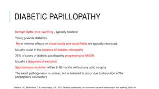
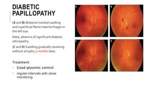
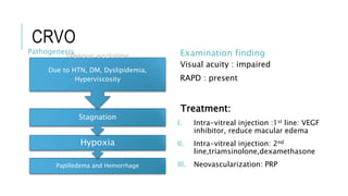
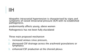
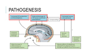
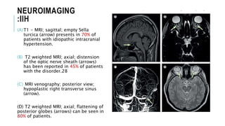
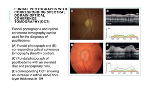
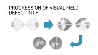
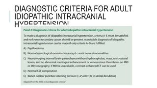
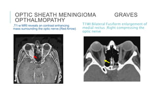
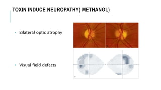
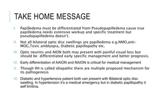
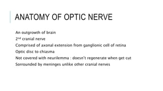
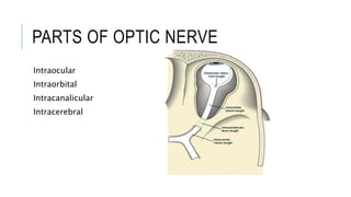
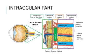
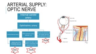
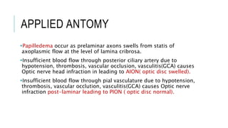
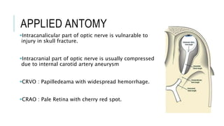
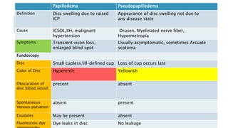
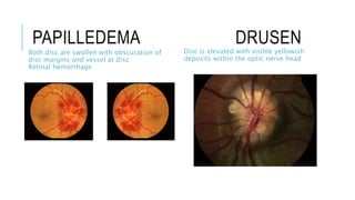
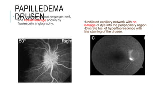
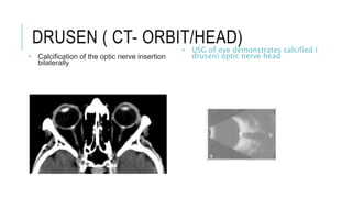
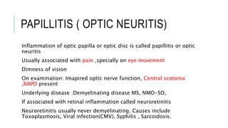
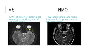
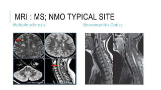
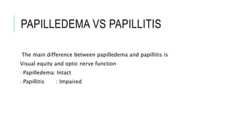
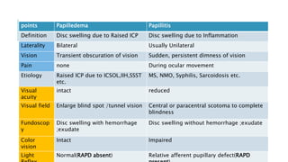
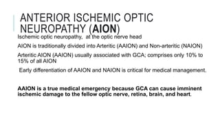
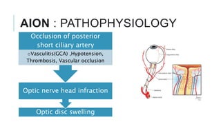
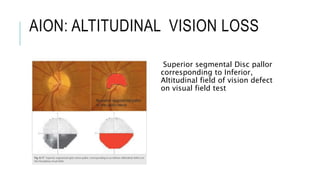
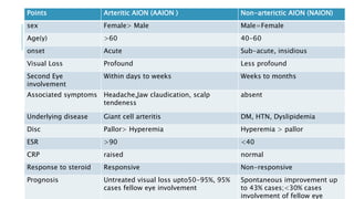
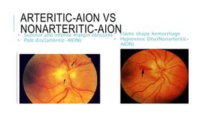
SlideShare a Scribd company logo
WELCOME
OPTIC DISC SWELLING
Dr. Tareq Esteak
Resident( Neurology)
NINS
OUTLINE
Anatomy of Optic Nerve
Blood supply of Optic Nerve
Applied anatomy
Pathogenesis
Differentials
ANATOMY OF OPTIC NERVE
An outgrowth of brain
2nd cranial nerve
Comprised of axonal extension from ganglionic cell of retina
Optic disc to chiasma
Not covered with neurilemma : doesn’t regenerate when get cut
Sorrounded by meninges unlike other cranial nerves
PARTS OF OPTIC NERVE
Intraocular
Intraorbital
Intracanalicular
Intracerebral
INTRAOCULAR PART
Lamina
Cribrosa
Meninges
Meninges
ARTERIAL SUPPLY:
OPTIC NERVE
Internal carotid
artery
Ophthalmic artery
Central Retinal Artery
anterior part of the
retrobulbar nerve via
centrifugal arterioles
Short posterior ciliary
artery(6-12)
Supply the Optic nerve
head in Centripetal and
segmental fashion
Arteries & arterioles of
pial sheeth derived
from ophthalmic artery
PION
CRA
O
AION
BLOOD SUPPLY OF OPTIC NERVE
VENOUS DRAINAGE
Optic nerve head : Central ratinal vein
Orbital part: Central retinal vein,Pial plexus
Intracranial part: Pial plexus<anterior cerebral vein
APPLIED ANTOMY
Papilledema occur as prelaminar axons swells from statis of
axoplasmic flow at the level of lamina cribrosa.
Insufficient blood flow through posterior ciliary artery due to
hypotension, thrombosis, vascular occlusion, vasculitis(GCA) causes
Optic nerve head infraction in leading to AION( optic disc swelled).
Insufficient blood flow through pial vasculature due to hypotension,
thrombosis, vascular occlution, vasculitis(GCA) causes Optic nerve
infraction post-laminar leading to PION ( optic disc normal).
APPLIED ANTOMY
Intracanalicular part of optic nerve is vulnarable to
injury in skull fracture.
Intracranial part of optic nerve is usually compressed
due to internal carotid artery aneurysm
CRVO : Papilledeama with widespread hemorrhage.
CRAO : Pale Retina with cherry red spot.
OPTIC DISC SWELLING
Optic disc swelling or disc edema may be defined as swelling of optic
nerve anterior to lamina cribrosa .
When found must be differentiated from Pseudopapilledema
Pseudopapilledema mostly congenital and physiological , doesn’t
need further workup
True Disc swelling is associated with numerous condition
DIFFERENTIALS OF OPTIC DISC
SWELLING
True disc swelling
Elevated ICP(Papilledema)
Optic Neuritits
Neuroretinitis
Anterior Ischemic Optic
Neuropathy(AION)
Diabetic papillopathy
Hypertensive retinopathy
Compressive neuropathy:
Meningioma ,Lymphangioma,
lymphoma
Pseudopapilledema
Drusen
Myelinated nerve fiber
Hypermetropia
Papilledema Pseudopapilledema
Definition Disc swelling due to raised
ICP
Appearance of disc swelling not due to
any disease state
Cause ICSOL,IIH, malignant
hypertension
Drusen, Myelinated nerve fiber,
Hypermetropia
Symptoms Transient vison loss,
enlarged blind spot
Usually asymptomatic, sometimes Arcuate
scotoma
Fundoscopy
Disc Small cupless/ill-defined cup Loss of cup occurs late
Color of Disc Hyperemic Yellowish
Obscuration of
disc blood vessel
present absent
Spontaneous
Venous pulsation
absent present
Exudates May be present absent
Fluorescein dye Dye leaks in disc No leakage
PAPILLEDEMA DRUSEN
Both disc are swollen with obscuration of
disc margins and vessel at disc
Retinal hemorrhage
Disc is elevated with visible yellowish
deposits within the optic nerve head
PAPILLEDEMA
DRUSEN
• Disc swelling, venous engorgement,
and vessel leakage shown by
fluorescein angiography, •Undilated capillary network with no
leakage of dye into the peripapillary region.
•Discrete foci of hyperfluorescence with
late staining of the drusen.
DRUSEN ( CT- ORBIT/HEAD)
• Calcification of the optic nerve insertion
bilaterally
• USG of eye demonstrates calcified (
drusen) optic nerve head
MYELINATED NERVE FIBER HYPERMETROPIA
• White, feathery ,Fanned out lesion
• Simulates disc edema
• Central retinal vessel crows in
small disc
• congested but no dialted vessels,
• Sponteneous venous pulsation
present
APPROACH
Optic disc selling
True Disc Swelling
Unilateral
1.Demyelinating : MS
2.Neuroretinitis: Viral,
Toxoplama, syphilis
3.Anterior Ischemic Optic
Neuropathy(AION)
4.Central Retinal Vein Occlusion
5.Compressive optic neuropathy
Bilateral
Papilledema
Malignant Hypertension
Diabetic papillpathy
Toxic Optic neuropathy
Demyelinating :NMO, Anti MOG
Hereditary optic neuropathy
Pseudopapilledema
PAPILLEDEMA
Once true disc swelling is confirmed , it should be determined
whether it is related to an optic neuropathy or raised ICP.
Papilledema is usually reserve for optic swelling secondary to raised
ICP
All other disc swelling is called optic disc swelling or disc edema.
Other of the major causes of disc edema other than papilledema is
Papillitis or optic neuritis,AION
PATHOGENEIS: PAPILLEDEMA
CAUSES OF PAPILLEDEMA
IIH( Idopathic Intracranial Hypertension)
Increased venous pressure : SSST ( superior Sagittal Sinus
Thrombosis)
ICSOL: Tumor ,Abscess, Large hematoma
Meningeal process: Infection, inflammation, neoplastic
Hydrocephalus
PAPILLEDEMA : GRADING ( THE FRISEN
SCALE)
NORMAL DISC : GRADE 0
GRADE 1
GRADE 2
GRADE 3
GRADE 4
GRADE 5
ANTERIOR OPTIC NEUROPATHIES
Etiology
Inflammatory .e.g.Optic
Neuritis(Papillitis)
Ischemic .e.g. AION
Compressive Optic neuropathy
Toxin induced.e.g. Methanol,
ethelene glycol
Heriditary.e.g. Leber’s optic
neuropathy
Infiltrative.e.g. sarcoisosis
Pathogenesis
Optic Disc Swelling
Inflammatory change
Local Optic nerve injury
e.g. Optic neuritis, Ischemia, Toxins
PAPILLITIS ( OPTIC NEURITIS)
Inflammation of optic papilla or optic disc is called papillitis or optic
neuritis
Usually associated with pain ,specially on eye movement
Dimness of vision
On examination: Imapired optic nerve function, Central scotoma
,RAPD present
Underlying disease :Demyelinating disease MS, NMO-SD,
If associated with retinal inflammation called neuroretinitis
Neuroretinitis usually never demyelinating. Causes include
Toxoplasmosis, Viral infection(CMV), Syphilis , Sarcoidosis.
MS NMO
T2WI- shows increased signal
intensity in small segmnt of
left optic nerve
T2WI-shows increased signal
intensity in large segment in
left optic nerve
MRI : MS; NMO TYPICAL SITE
Multiple sclerosis Neuromyelitis Optica
PAPILLEDEMA VS PAPILLITIS
The main difference between papilledema and papillitis is
Visual equity and optic nerve function
oPapilledema: Intact
oPapillitis : Impaired
points Papilledema Papillitis
Definition Disc swelling due to Raised ICP Disc swelling due to Inflammation
Laterality Bilateral Usually Unilateral
Vision Transient obscuration of vision Sudden, persistent dimness of vision
Pain none During ocular movement
Etiology Raised ICP due to ICSOL,IIH,SSST
etc.
MS, NMO, Syphilis, Sarcoidosis etc.
Visual
acuity
intact reduced
Visual field Enlarge blind spot /tunnel vision Central or paracentral scotoma to complete
blindness
Fundoscop
y
Disc swelling with hemorrhage
;exudate
Disc swelling without hemorrhage ;exudate
Color
vision
Intact Impaired
Light Normal(RAPD absent) Relative afferent pupillary defect(RAPD
ANTERIOR ISCHEMIC OPTIC
NEUROPATHY (AION)
Ischemic optic neuropathy, at the optic nerve head
AION is traditionally divided into Arteritic (AAION) and Non-arteritic (NAION)
Arteritic AION (AAION) usually associated with GCA; comprises only 10% to
15% of all AION
Early differentiation of AAION and NAION is critical for medical management.
AAION is a true medical emergency because GCA can cause imminent
ischemic damage to the fellow optic nerve, retina, brain, and heart.
AION : PATHOPHYSIOLOGY
Optic disc swelling
Optic nerve head infraction
Occlusion of posterior
short ciliary artery
oVasculitis(GCA) ,Hypotension,
Thrombosis, Vascular occlusion
AION: ALTITUDINAL VISION LOSS
Superior segmental Disc pallor
corresponding to Inferior,
Altitudinal field of vision defect
on visual field test
Points Arteritic AION (AAION ) Non-arterictic AION (NAION)
sex Female> Male Male=Female
Age(y) >60 40-60
onset Acute Sub-acute, insidious
Visual Loss Profound Less profound
Second Eye
involvement
Within days to weeks Weeks to months
Associated symptoms Headache,Jaw claudication, scalp
tendeness
absent
Underlying disease Giant cell arteritis DM, HTN, Dyslipidemia
Disc Pallor> Hyperemia Hyperemia > pallor
ESR >90 <40
CRP raised normal
Response to steroid Responsive Non-responsive
Prognosis Untreated visual loss upto50-95%, 95%
cases fellow eye involvement
Spontaneous improvement up
to 43% cases;<30% cases
involvement of fellow eye
ARTERITIC-AION VS
NONARTERITIC-AION
• Suoirior and inferior margin obscures
• Pale disc(arteritic-AION)
• Flame shape hemorrhage
• Hyperemic Disc(Nonarteritic-
AION)
TREATMENT APPROACH TO
AION(DUE TO GCA)
Koster, M.J., Warrington, K.J. and Kermani, T.A., 2016. Update on the epidemiology and treatment of giant cell
arteritis. Current Treatment Options in Rheumatology, 2(2), pp.138-152.
AION VS OPTIC NEURITIS
Why do we need to differentiate?
Similarities in presentation:
I. Unilateral dimness of vision
II. Painful optic neuropathy
III. Optic nerve dysfunction: field, acuity,color vision,RAPD,Optic disc
swelling
IV. May Respond to steroids
Points Optic Neuritis AION
Age of onset 20-30 >60
Pain 92% cases, exacerbate on eye
movement
Periorbital but no change with eye
movement
Associated Sensory disturbance, Limb
weakness,
Ataxia, Bowel bladder dysfunction,
Headache,Jaw claudication, scalp
tendeness
onset Insidious ,progresses of hours to
days
Sudden onset, often notice upon
awekenig
Disc swelling Present only one third of the
cases, in rest its retrobulbar
Present in most of the cases
Field defect Central scotoma Altitudinal
MRI Enhancement of optic nerve No enhancement of optic nerve
Recovery Within 2-4 weeks Over months
prognosis Good prognosis poor
LEBER’S HERIDITARY OPTIC NEUROPATHY(LHON)
• Mitocondrial disease
• Bilateral involvement of optic nerve
• May occur at any age
• Acute phase present with optic disc
swelling and hyperemia
• Ultimately patient develops optic
atrophy
• Associated with cardiac conduction
defects, Tremor, limb
weakness(LHON plus)
LEBER’S
HERIDITARY OPTIC
NEUROPATHY(LHON)
• Blilateral hyperemic disc
swelling
• Central scotomas;
Right>left
HYPERTENSIVE RETINOPATHY
excessive cerebral
blood flow
breakdown of the
blood–brain
barrier
secondary
vasogenic edema
Papilledema
Precapillaries in the
posterior fundus
get occluded
ischemic process
leading to
fibrinoid necrosis
cytotoxic edema
TREATMENT
It’s medical emergency
Must be managed promptly and
adequately to prevent further
target organ damage.
[AHA guideline,2017]
DIABETIC PAPILLOPATHY
Benign Optic disc swelling , typically bilateral
Young juvenile diabetics
No or minimal effects on visual acuity and visual fields are typically restricted.
Usually occur in the absence of diabetic retinopathy
36% of cases of diabetic papillopathy progressing to NAION.
Usually a diagnosis of exclution
Spontaneous resolution within 2-10 months without any optic atrophy
The exact pathogenesis is unclear, but is believed to occur due to disruption of the
peripapillary vasculature
Wallace, I.R., Mulholland, D.A. and Lindsay, J.R., 2012. Diabetic papillopathy: an uncommon cause of bilateral optic disc swelling. QJM: An
DIABETIC
PAPILLOPATHY
(A and B) Bilateral marked swelling
and superficial flame haemorrhages in
the left eye.
Note, absence of significant diabetic
retinopathy.
(C and D) Swelling gradually resolving
without atrophy 3 months later.
Treatment:
• Good glycemic control
• regular intervals with close
monitoring.
CRVO
Papilledema ;Flame shape
hemorrgahe in all qudrants
Macular hemorrhage
Setting sun Appearance
CRVO
Pathogenesis
Papilledema and Hemorrhage
Hypoxia
Stagnation
Venous occlution
Due to HTN, DM, Dyslipidemia,
Hyperviscosity
Examination finding
Visual acuity : impaired
RAPD : present
Treatment:
I. Intra-vitreal injection :1st line: VEGF
inhibitor, reduce macular edema
II. Intra-vitreal injection: 2nd
line,triamsinolone,dexamethasone
III. Neovascularization: PRP
IIH
Idiopathic intracranial hypertension is characterised by signs and
symptoms of raised intracranial pressure (ICP) with no established
pathogenesis.
predominantly affects young, obese women
Pathogenesis has not been fully elucidated
Three main proposed mechanism
1. increased venous sinus pressure,
2. decreased CSF drainage across the arachnoid granulations or
lymphatics
3. enhanced CSF production at the choroid plexus
PATHOGENESIS
NEUROIMAGING
:IIH
(A)T1 - MRI; sagittal; empty Sella
turcica (arrow) presents in 70% of
patients with idiopathic intracranial
hypertension.
(B) T2 weighted MRI; axial; distension
of the optic nerve sheath (arrows)
has been reported in 45% of patients
with the disorder.28
(C) MRI venography; posterior view;
hypoplastic right transverse sinus
(arrow).
(D) T2 weighted MRI; axial; flattening of
posterior globes (arrows) can be seen in
80% of patients.
FUNDAL PHOTOGRAPHS WITH
CORRESPONDING SPECTRAL
DOMAIN OPTICAL
COHERENCE
TOMOGRAPHY(OCT)
Fundal photographs and optical
coherence tomography can be
used for the diagnosis of
papilledema.
(A) Fundal photograph and (B)
corresponding optical coherence
tomography (healthy control).
(C) Fundal photograph of
papilloedema with an elevated
disc and peripapillary halo,
(D) corresponding OCT showing
an increase in retinal nerve fibre
layer thickness in IIH
PROGRESSION OF VISUAL FIELD
DEFECT IN IIH
DIAGNOSTIC CRITERIA FOR ADULT
IDIOPATHIC INTRACRANIAL
HYPERTENSION
CEREBRAL VENOUS SINUS
THROMBOSIS
CSF drains into Intracranial venous sinuses
CSF passively resorbrd into arachanoid granulations ,mostly withi
supiriror sagittal sinus
Incase of venous thrombosis CSF resorption decreased andvthe CSF
pressure increases
This raised ICP may ultimately give rise to papilledema
NCCT -head : Cord sign
Contrast CT –head : Empty
Delta sign
Figure : Sagittal sinus thrombosis.
(A) MR venogram showing absent venous
flow signal in the middle third of the
superior sagittal sinus (white arrows)
(B) Axial gadolinium-enhanced T1-weighted
MR scan showing an irregular filling defect of
the superior sagittal sinus (white arrow)
(C) Sagittal gadolinium-enhanced T1-
weighted MR scan also showing a focal filling
defect indicating thrombus (white arrow)
(D) Axial susceptibility-weighted imaging
showing low signal indicating blood products
(thrombus) in the superior sagittal sinus
(white arrow)
TREATMENT
Should be started as soon as the diagnosis of CVT is clearly confirmed
- Rapid anticoagulant therapy
- Treatment of underlying cause (eg, dehydration, sepsis, stopping
any prothrombotic medications)
- Control of seizures
- Management of intracranial hypertension if required.
COMPRESIVE OPTIC NEUROPATHY
• Lesion may compress or infiltrate the intraorbital, intracranial or prechiasmal optic nerve
• Large and intraorbital lesions often produce optic disc swelling
Presentation
o Progressive unilateral optic neuropathy
o Usually no pain with eye movement
o Headache occurs if raised ICP or involvement of Trigeminal nerve
o Proptosis is common in orbital lesion
o Cranial nerve palsys occur if lesion extending to orbital apex, superior orbital fissure or
cavernous sinus.
o Early disc change: Swelling ; Late change : Optic Atrophy
Compressive
Optic
neuropathy
Neoplastic
Intraorbital
Tumor(hemangioma,lympha
ngioma,Mets)
Optic nerve sheeth
meningioma
Chraniopharyngioma
Non-neoplastic
•Grave’s Ophthalmopathy
•Tolosa-Hunt syndrome
•Ophthalmic artery anyurism
OPTIC SHEATH MENINGIOMA GRAVES
OPTHALMOPATHY
,T1-w MRI reveals an contrast enhancing
mass surrounding the optic nerve.(Red Arrow)
T1WI:Bilateral Fusiform enlargement of
medial rectus ;Right compressing the
optic nerve
TOXIN INDUCED NEUROPATHY/ TOXIC
AMBLYOPIA
• Patient presents with sudden vison loss
• Abrupt onset central/cecocentral scotoma
• Associated nausea, vomiting, abdominal pain
• Metabolic acidosis is one of the hallmark
• Initially optic disc is swollen but eventually
patient develops optic atrophy
• Treatment:
I. Lavage
II. IV NaHCO3
III. Antidote: Ethanol, fomepizole
IV. Others: Folinic acid
V. Hemodialysis
III
IV
TOXIN INDUCE NEUROPATHY( METHANOL)
• Bilateral optic atrophy
• Visual field defects
TAKE HOME MESSAGE
I. Papilledema must be differentiated from Pseudopapilledema cause true
papilledema needs extensive workup and specific treatment but
pseudopapilledema doesn’t.
II. Not all bilateral optic disc swellings are papilledema e.g,NMO,anti-
MOG,Toxic amblyopia, Diabetic papillopathy etc.
III. Optic neuritis and AION both may present with painful visual loss but
should be differentiated early specific management and better prognosis.
IV. Early differentiation of AAION and NAION is critical for medical management
V. Though IIH is called idiopathic there are multiple proposed mechanism for
its pathogenesis
VI. Diabetic and hypertensive patient both can present with Bilateral optic disc
swelling. In hypertension it’s a medical emergency but in diabetic papillopathy it
self limiting.
THANK YOU ……..

More Related Content

What's hot

Anterior segment OCT & UBM
Anterior segment OCT & UBMAnterior segment OCT & UBM
Anterior segment OCT & UBM
Dinesh Madduri
 
Macular hole
Macular holeMacular hole
Macular hole
Laxmi Eye Institute
 
Pigmentary glaucoma - Dr Shylesh B Dabke
Pigmentary glaucoma - Dr Shylesh B DabkePigmentary glaucoma - Dr Shylesh B Dabke
Pigmentary glaucoma - Dr Shylesh B Dabke
Shylesh Dabke
 
Optic neuritis
Optic neuritisOptic neuritis
Optic neuritis
erameshita
 
Normal tension glaucoma
Normal tension glaucomaNormal tension glaucoma
Normal tension glaucoma
Laxmi Eye Institute
 
Target IOP
Target IOPTarget IOP
Target IOP
Dr Samarth Mishra
 
Clinical evaluation of optic disc changes
Clinical evaluation of optic disc changesClinical evaluation of optic disc changes
Clinical evaluation of optic disc changes
Dr Saurabh Kushwaha
 
Posterior capsular opacification
Posterior capsular opacificationPosterior capsular opacification
Posterior capsular opacification
Ashish Chaudhari
 
Branch retinal vein occlusion (BRVO)
Branch retinal vein occlusion (BRVO)Branch retinal vein occlusion (BRVO)
Branch retinal vein occlusion (BRVO)
NIKHIL GOTMARE
 
Retinal Vasculitis
Retinal VasculitisRetinal Vasculitis
Retinal Vasculitis
Sahil Thakur
 
Normal Tension Glaucoma
Normal Tension GlaucomaNormal Tension Glaucoma
Normal Tension Glaucoma
eknath87
 
Diagnosis of pre perimetric glaucoma
Diagnosis of pre perimetric glaucomaDiagnosis of pre perimetric glaucoma
Diagnosis of pre perimetric glaucoma
Sadhwini Harish
 
Optic neuritis
Optic neuritisOptic neuritis
Optic neuritis
Laya Pillai
 
Uveitic Glaucoma
Uveitic Glaucoma Uveitic Glaucoma
Uveitic Glaucoma
Visionary Ophthamology
 
Fundus examination
Fundus examinationFundus examination
Fundus examination
Samuel Ponraj
 
Fundus examination
Fundus   examinationFundus   examination
Fundus examination
DR.SOUMITRA DAS
 
Papilledema - Dr Shylesh Dabke
Papilledema - Dr Shylesh DabkePapilledema - Dr Shylesh Dabke
Papilledema - Dr Shylesh Dabke
Shylesh Dabke
 
Visual Field in Glaucoma
Visual Field in GlaucomaVisual Field in Glaucoma
Visual Field in Glaucoma
docsarsi
 
Visual Field Examination
Visual Field ExaminationVisual Field Examination
Visual Field Examination
Paavan Kalra
 
Cmv retinitis
Cmv retinitisCmv retinitis
Cmv retinitis
Dinesh Madduri
 

What's hot (20)

Anterior segment OCT & UBM
Anterior segment OCT & UBMAnterior segment OCT & UBM
Anterior segment OCT & UBM
 
Macular hole
Macular holeMacular hole
Macular hole
 
Pigmentary glaucoma - Dr Shylesh B Dabke
Pigmentary glaucoma - Dr Shylesh B DabkePigmentary glaucoma - Dr Shylesh B Dabke
Pigmentary glaucoma - Dr Shylesh B Dabke
 
Optic neuritis
Optic neuritisOptic neuritis
Optic neuritis
 
Normal tension glaucoma
Normal tension glaucomaNormal tension glaucoma
Normal tension glaucoma
 
Target IOP
Target IOPTarget IOP
Target IOP
 
Clinical evaluation of optic disc changes
Clinical evaluation of optic disc changesClinical evaluation of optic disc changes
Clinical evaluation of optic disc changes
 
Posterior capsular opacification
Posterior capsular opacificationPosterior capsular opacification
Posterior capsular opacification
 
Branch retinal vein occlusion (BRVO)
Branch retinal vein occlusion (BRVO)Branch retinal vein occlusion (BRVO)
Branch retinal vein occlusion (BRVO)
 
Retinal Vasculitis
Retinal VasculitisRetinal Vasculitis
Retinal Vasculitis
 
Normal Tension Glaucoma
Normal Tension GlaucomaNormal Tension Glaucoma
Normal Tension Glaucoma
 
Diagnosis of pre perimetric glaucoma
Diagnosis of pre perimetric glaucomaDiagnosis of pre perimetric glaucoma
Diagnosis of pre perimetric glaucoma
 
Optic neuritis
Optic neuritisOptic neuritis
Optic neuritis
 
Uveitic Glaucoma
Uveitic Glaucoma Uveitic Glaucoma
Uveitic Glaucoma
 
Fundus examination
Fundus examinationFundus examination
Fundus examination
 
Fundus examination
Fundus   examinationFundus   examination
Fundus examination
 
Papilledema - Dr Shylesh Dabke
Papilledema - Dr Shylesh DabkePapilledema - Dr Shylesh Dabke
Papilledema - Dr Shylesh Dabke
 
Visual Field in Glaucoma
Visual Field in GlaucomaVisual Field in Glaucoma
Visual Field in Glaucoma
 
Visual Field Examination
Visual Field ExaminationVisual Field Examination
Visual Field Examination
 
Cmv retinitis
Cmv retinitisCmv retinitis
Cmv retinitis
 

Similar to Optic Disc Swelling

optic nerve head swelling
optic nerve head swellingoptic nerve head swelling
optic nerve head swelling
Jehad Hassan
 
Papilledema
PapilledemaPapilledema
Papilledema
PRAKRITIYAGNAM
 
Ischemic optic neuropathy
Ischemic optic neuropathyIschemic optic neuropathy
Ischemic optic neuropathy
16divya
 
Optic disc swelling
Optic disc swellingOptic disc swelling
Optic disc swelling
Abdul Munim Khan Suri
 
Neuro ophthalmology RCSI
Neuro ophthalmology RCSINeuro ophthalmology RCSI
Neuro ophthalmology RCSI
OphthalmicDocs Chiong
 
Papilloedema and Optic Atrophy.ppt
Papilloedema and Optic Atrophy.pptPapilloedema and Optic Atrophy.ppt
Papilloedema and Optic Atrophy.ppt
AshrafIslam16
 
Ophthalmology 5th year, 7th lecture (Dr. Bakhtyar)
Ophthalmology 5th year, 7th lecture (Dr. Bakhtyar)Ophthalmology 5th year, 7th lecture (Dr. Bakhtyar)
Ophthalmology 5th year, 7th lecture (Dr. Bakhtyar)
College of Medicine, Sulaymaniyah
 
Optic Nerve
Optic NerveOptic Nerve
Optic Nerve
Aden University
 
Optic atrophy
Optic atrophyOptic atrophy
Optic atrophy
ChrisPius
 
Ischemic of optic neuropathy, Optic Neuropathy (Ischemic), Eye Stroke
Ischemic of optic neuropathy, Optic Neuropathy (Ischemic), Eye Stroke  Ischemic of optic neuropathy, Optic Neuropathy (Ischemic), Eye Stroke
Ischemic of optic neuropathy, Optic Neuropathy (Ischemic), Eye Stroke
Mahavir Mohire
 
Papilledema - Optic Nerve Head Swelling
Papilledema - Optic Nerve Head Swelling Papilledema - Optic Nerve Head Swelling
Papilledema - Optic Nerve Head Swelling
Shreeji Shrestha
 
approaches to pale disc
approaches to pale discapproaches to pale disc
approaches to pale disc
Prajakta Matey
 
Papilloedema.pptx
Papilloedema.pptxPapilloedema.pptx
Papilloedema.pptx
dratulkranand
 
Approach to Pain ophthalmoplegia.
Approach to Pain ophthalmoplegia.Approach to Pain ophthalmoplegia.
Approach to Pain ophthalmoplegia.
tintus123
 
Painful Ophthalmoplegia
Painful Ophthalmoplegia   Painful Ophthalmoplegia
Painful Ophthalmoplegia
Junaid Naina
 
Optic Nerve and pupil tutorial
Optic Nerve and pupil tutorialOptic Nerve and pupil tutorial
Optic Nerve and pupil tutorial
Ahmed Osama Hashem
 
Ischemic Optic Neuropathy 1.pptx
Ischemic Optic Neuropathy 1.pptxIschemic Optic Neuropathy 1.pptx
Ischemic Optic Neuropathy 1.pptx
rameshbhandari32
 
Neuro-opthalmology
Neuro-opthalmologyNeuro-opthalmology
Neuro-opthalmology
Sameen Jawed
 
Neuro ophthalmologic causes of headache
Neuro ophthalmologic causes of headacheNeuro ophthalmologic causes of headache
Neuro ophthalmologic causes of headache
neurophq8
 
Optic nerve Clinical significance
Optic nerve Clinical significance Optic nerve Clinical significance
Optic nerve Clinical significance
KafrELShiekh University
 

Similar to Optic Disc Swelling (20)

optic nerve head swelling
optic nerve head swellingoptic nerve head swelling
optic nerve head swelling
 
Papilledema
PapilledemaPapilledema
Papilledema
 
Ischemic optic neuropathy
Ischemic optic neuropathyIschemic optic neuropathy
Ischemic optic neuropathy
 
Optic disc swelling
Optic disc swellingOptic disc swelling
Optic disc swelling
 
Neuro ophthalmology RCSI
Neuro ophthalmology RCSINeuro ophthalmology RCSI
Neuro ophthalmology RCSI
 
Papilloedema and Optic Atrophy.ppt
Papilloedema and Optic Atrophy.pptPapilloedema and Optic Atrophy.ppt
Papilloedema and Optic Atrophy.ppt
 
Ophthalmology 5th year, 7th lecture (Dr. Bakhtyar)
Ophthalmology 5th year, 7th lecture (Dr. Bakhtyar)Ophthalmology 5th year, 7th lecture (Dr. Bakhtyar)
Ophthalmology 5th year, 7th lecture (Dr. Bakhtyar)
 
Optic Nerve
Optic NerveOptic Nerve
Optic Nerve
 
Optic atrophy
Optic atrophyOptic atrophy
Optic atrophy
 
Ischemic of optic neuropathy, Optic Neuropathy (Ischemic), Eye Stroke
Ischemic of optic neuropathy, Optic Neuropathy (Ischemic), Eye Stroke  Ischemic of optic neuropathy, Optic Neuropathy (Ischemic), Eye Stroke
Ischemic of optic neuropathy, Optic Neuropathy (Ischemic), Eye Stroke
 
Papilledema - Optic Nerve Head Swelling
Papilledema - Optic Nerve Head Swelling Papilledema - Optic Nerve Head Swelling
Papilledema - Optic Nerve Head Swelling
 
approaches to pale disc
approaches to pale discapproaches to pale disc
approaches to pale disc
 
Papilloedema.pptx
Papilloedema.pptxPapilloedema.pptx
Papilloedema.pptx
 
Approach to Pain ophthalmoplegia.
Approach to Pain ophthalmoplegia.Approach to Pain ophthalmoplegia.
Approach to Pain ophthalmoplegia.
 
Painful Ophthalmoplegia
Painful Ophthalmoplegia   Painful Ophthalmoplegia
Painful Ophthalmoplegia
 
Optic Nerve and pupil tutorial
Optic Nerve and pupil tutorialOptic Nerve and pupil tutorial
Optic Nerve and pupil tutorial
 
Ischemic Optic Neuropathy 1.pptx
Ischemic Optic Neuropathy 1.pptxIschemic Optic Neuropathy 1.pptx
Ischemic Optic Neuropathy 1.pptx
 
Neuro-opthalmology
Neuro-opthalmologyNeuro-opthalmology
Neuro-opthalmology
 
Neuro ophthalmologic causes of headache
Neuro ophthalmologic causes of headacheNeuro ophthalmologic causes of headache
Neuro ophthalmologic causes of headache
 
Optic nerve Clinical significance
Optic nerve Clinical significance Optic nerve Clinical significance
Optic nerve Clinical significance
 

More from Tareq Esteak

Clinical presentation on multiple sclerosis
Clinical presentation on multiple sclerosisClinical presentation on multiple sclerosis
Clinical presentation on multiple sclerosis
Tareq Esteak
 
Case presentation on Cavernous sinus thrombosis
Case presentation on Cavernous sinus thrombosisCase presentation on Cavernous sinus thrombosis
Case presentation on Cavernous sinus thrombosis
Tareq Esteak
 
Neurosyphilis
NeurosyphilisNeurosyphilis
Neurosyphilis
Tareq Esteak
 
Cerebellum
CerebellumCerebellum
Cerebellum
Tareq Esteak
 
Neuromyelitis optica
Neuromyelitis opticaNeuromyelitis optica
Neuromyelitis optica
Tareq Esteak
 
Multiple Sclerosis
Multiple SclerosisMultiple Sclerosis
Multiple Sclerosis
Tareq Esteak
 

More from Tareq Esteak (6)

Clinical presentation on multiple sclerosis
Clinical presentation on multiple sclerosisClinical presentation on multiple sclerosis
Clinical presentation on multiple sclerosis
 
Case presentation on Cavernous sinus thrombosis
Case presentation on Cavernous sinus thrombosisCase presentation on Cavernous sinus thrombosis
Case presentation on Cavernous sinus thrombosis
 
Neurosyphilis
NeurosyphilisNeurosyphilis
Neurosyphilis
 
Cerebellum
CerebellumCerebellum
Cerebellum
 
Neuromyelitis optica
Neuromyelitis opticaNeuromyelitis optica
Neuromyelitis optica
 
Multiple Sclerosis
Multiple SclerosisMultiple Sclerosis
Multiple Sclerosis
 

Recently uploaded

Hemodialysis: Chapter 4, Dialysate Circuit - Dr.Gawad
Hemodialysis: Chapter 4, Dialysate Circuit - Dr.GawadHemodialysis: Chapter 4, Dialysate Circuit - Dr.Gawad
Hemodialysis: Chapter 4, Dialysate Circuit - Dr.Gawad
NephroTube - Dr.Gawad
 
Top 10 Best Ayurvedic Kidney Stone Syrups in India
Top 10 Best Ayurvedic Kidney Stone Syrups in IndiaTop 10 Best Ayurvedic Kidney Stone Syrups in India
Top 10 Best Ayurvedic Kidney Stone Syrups in India
Swastik Ayurveda
 
Identification and nursing management of congenital malformations .pptx
Identification and nursing management of congenital malformations .pptxIdentification and nursing management of congenital malformations .pptx
Identification and nursing management of congenital malformations .pptx
MGM SCHOOL/COLLEGE OF NURSING
 
Local Advanced Lung Cancer: Artificial Intelligence, Synergetics, Complex Sys...
Local Advanced Lung Cancer: Artificial Intelligence, Synergetics, Complex Sys...Local Advanced Lung Cancer: Artificial Intelligence, Synergetics, Complex Sys...
Local Advanced Lung Cancer: Artificial Intelligence, Synergetics, Complex Sys...
Oleg Kshivets
 
Aortic Association CBL Pilot April 19 – 20 Bern
Aortic Association CBL Pilot April 19 – 20 BernAortic Association CBL Pilot April 19 – 20 Bern
Aortic Association CBL Pilot April 19 – 20 Bern
suvadeepdas911
 
8 Surprising Reasons To Meditate 40 Minutes A Day That Can Change Your Life.pptx
8 Surprising Reasons To Meditate 40 Minutes A Day That Can Change Your Life.pptx8 Surprising Reasons To Meditate 40 Minutes A Day That Can Change Your Life.pptx
8 Surprising Reasons To Meditate 40 Minutes A Day That Can Change Your Life.pptx
Holistified Wellness
 
Top-Vitamin-Supplement-Brands-in-India List
Top-Vitamin-Supplement-Brands-in-India ListTop-Vitamin-Supplement-Brands-in-India List
Top-Vitamin-Supplement-Brands-in-India List
SwisschemDerma
 
Role of Mukta Pishti in the Management of Hyperthyroidism
Role of Mukta Pishti in the Management of HyperthyroidismRole of Mukta Pishti in the Management of Hyperthyroidism
Role of Mukta Pishti in the Management of Hyperthyroidism
Dr. Jyothirmai Paindla
 
Histololgy of Female Reproductive System.pptx
Histololgy of Female Reproductive System.pptxHistololgy of Female Reproductive System.pptx
Histololgy of Female Reproductive System.pptx
AyeshaZaid1
 
Part II - Body Grief: Losing parts of ourselves and our identity before, duri...
Part II - Body Grief: Losing parts of ourselves and our identity before, duri...Part II - Body Grief: Losing parts of ourselves and our identity before, duri...
Part II - Body Grief: Losing parts of ourselves and our identity before, duri...
bkling
 
Adhd Medication Shortage Uk - trinexpharmacy.com
Adhd Medication Shortage Uk - trinexpharmacy.comAdhd Medication Shortage Uk - trinexpharmacy.com
Adhd Medication Shortage Uk - trinexpharmacy.com
reignlana06
 
TEST BANK For Community Health Nursing A Canadian Perspective, 5th Edition by...
TEST BANK For Community Health Nursing A Canadian Perspective, 5th Edition by...TEST BANK For Community Health Nursing A Canadian Perspective, 5th Edition by...
TEST BANK For Community Health Nursing A Canadian Perspective, 5th Edition by...
Donc Test
 
Chapter 11 Nutrition and Chronic Diseases.pptx
Chapter 11 Nutrition and Chronic Diseases.pptxChapter 11 Nutrition and Chronic Diseases.pptx
Chapter 11 Nutrition and Chronic Diseases.pptx
Earlene McNair
 
Journal Article Review on Rasamanikya
Journal Article Review on RasamanikyaJournal Article Review on Rasamanikya
Journal Article Review on Rasamanikya
Dr. Jyothirmai Paindla
 
#cALL# #gIRLS# In Dehradun ꧁❤8107221448❤꧂#cALL# #gIRLS# Service In Dehradun W...
#cALL# #gIRLS# In Dehradun ꧁❤8107221448❤꧂#cALL# #gIRLS# Service In Dehradun W...#cALL# #gIRLS# In Dehradun ꧁❤8107221448❤꧂#cALL# #gIRLS# Service In Dehradun W...
#cALL# #gIRLS# In Dehradun ꧁❤8107221448❤꧂#cALL# #gIRLS# Service In Dehradun W...
chandankumarsmartiso
 
Light House Retreats: Plant Medicine Retreat Europe
Light House Retreats: Plant Medicine Retreat EuropeLight House Retreats: Plant Medicine Retreat Europe
Light House Retreats: Plant Medicine Retreat Europe
Lighthouse Retreat
 
The Best Ayurvedic Antacid Tablets in India
The Best Ayurvedic Antacid Tablets in IndiaThe Best Ayurvedic Antacid Tablets in India
The Best Ayurvedic Antacid Tablets in India
Swastik Ayurveda
 
Integrating Ayurveda into Parkinson’s Management: A Holistic Approach
Integrating Ayurveda into Parkinson’s Management: A Holistic ApproachIntegrating Ayurveda into Parkinson’s Management: A Holistic Approach
Integrating Ayurveda into Parkinson’s Management: A Holistic Approach
Ayurveda ForAll
 
CHEMOTHERAPY_RDP_CHAPTER 3_ANTIFUNGAL AGENT.pdf
CHEMOTHERAPY_RDP_CHAPTER 3_ANTIFUNGAL AGENT.pdfCHEMOTHERAPY_RDP_CHAPTER 3_ANTIFUNGAL AGENT.pdf
CHEMOTHERAPY_RDP_CHAPTER 3_ANTIFUNGAL AGENT.pdf
rishi2789
 
Muscles of Mastication by Dr. Rabia Inam Gandapore.pptx
Muscles of Mastication by Dr. Rabia Inam Gandapore.pptxMuscles of Mastication by Dr. Rabia Inam Gandapore.pptx
Muscles of Mastication by Dr. Rabia Inam Gandapore.pptx
Dr. Rabia Inam Gandapore
 

Recently uploaded (20)

Hemodialysis: Chapter 4, Dialysate Circuit - Dr.Gawad
Hemodialysis: Chapter 4, Dialysate Circuit - Dr.GawadHemodialysis: Chapter 4, Dialysate Circuit - Dr.Gawad
Hemodialysis: Chapter 4, Dialysate Circuit - Dr.Gawad
 
Top 10 Best Ayurvedic Kidney Stone Syrups in India
Top 10 Best Ayurvedic Kidney Stone Syrups in IndiaTop 10 Best Ayurvedic Kidney Stone Syrups in India
Top 10 Best Ayurvedic Kidney Stone Syrups in India
 
Identification and nursing management of congenital malformations .pptx
Identification and nursing management of congenital malformations .pptxIdentification and nursing management of congenital malformations .pptx
Identification and nursing management of congenital malformations .pptx
 
Local Advanced Lung Cancer: Artificial Intelligence, Synergetics, Complex Sys...
Local Advanced Lung Cancer: Artificial Intelligence, Synergetics, Complex Sys...Local Advanced Lung Cancer: Artificial Intelligence, Synergetics, Complex Sys...
Local Advanced Lung Cancer: Artificial Intelligence, Synergetics, Complex Sys...
 
Aortic Association CBL Pilot April 19 – 20 Bern
Aortic Association CBL Pilot April 19 – 20 BernAortic Association CBL Pilot April 19 – 20 Bern
Aortic Association CBL Pilot April 19 – 20 Bern
 
8 Surprising Reasons To Meditate 40 Minutes A Day That Can Change Your Life.pptx
8 Surprising Reasons To Meditate 40 Minutes A Day That Can Change Your Life.pptx8 Surprising Reasons To Meditate 40 Minutes A Day That Can Change Your Life.pptx
8 Surprising Reasons To Meditate 40 Minutes A Day That Can Change Your Life.pptx
 
Top-Vitamin-Supplement-Brands-in-India List
Top-Vitamin-Supplement-Brands-in-India ListTop-Vitamin-Supplement-Brands-in-India List
Top-Vitamin-Supplement-Brands-in-India List
 
Role of Mukta Pishti in the Management of Hyperthyroidism
Role of Mukta Pishti in the Management of HyperthyroidismRole of Mukta Pishti in the Management of Hyperthyroidism
Role of Mukta Pishti in the Management of Hyperthyroidism
 
Histololgy of Female Reproductive System.pptx
Histololgy of Female Reproductive System.pptxHistololgy of Female Reproductive System.pptx
Histololgy of Female Reproductive System.pptx
 
Part II - Body Grief: Losing parts of ourselves and our identity before, duri...
Part II - Body Grief: Losing parts of ourselves and our identity before, duri...Part II - Body Grief: Losing parts of ourselves and our identity before, duri...
Part II - Body Grief: Losing parts of ourselves and our identity before, duri...
 
Adhd Medication Shortage Uk - trinexpharmacy.com
Adhd Medication Shortage Uk - trinexpharmacy.comAdhd Medication Shortage Uk - trinexpharmacy.com
Adhd Medication Shortage Uk - trinexpharmacy.com
 
TEST BANK For Community Health Nursing A Canadian Perspective, 5th Edition by...
TEST BANK For Community Health Nursing A Canadian Perspective, 5th Edition by...TEST BANK For Community Health Nursing A Canadian Perspective, 5th Edition by...
TEST BANK For Community Health Nursing A Canadian Perspective, 5th Edition by...
 
Chapter 11 Nutrition and Chronic Diseases.pptx
Chapter 11 Nutrition and Chronic Diseases.pptxChapter 11 Nutrition and Chronic Diseases.pptx
Chapter 11 Nutrition and Chronic Diseases.pptx
 
Journal Article Review on Rasamanikya
Journal Article Review on RasamanikyaJournal Article Review on Rasamanikya
Journal Article Review on Rasamanikya
 
#cALL# #gIRLS# In Dehradun ꧁❤8107221448❤꧂#cALL# #gIRLS# Service In Dehradun W...
#cALL# #gIRLS# In Dehradun ꧁❤8107221448❤꧂#cALL# #gIRLS# Service In Dehradun W...#cALL# #gIRLS# In Dehradun ꧁❤8107221448❤꧂#cALL# #gIRLS# Service In Dehradun W...
#cALL# #gIRLS# In Dehradun ꧁❤8107221448❤꧂#cALL# #gIRLS# Service In Dehradun W...
 
Light House Retreats: Plant Medicine Retreat Europe
Light House Retreats: Plant Medicine Retreat EuropeLight House Retreats: Plant Medicine Retreat Europe
Light House Retreats: Plant Medicine Retreat Europe
 
The Best Ayurvedic Antacid Tablets in India
The Best Ayurvedic Antacid Tablets in IndiaThe Best Ayurvedic Antacid Tablets in India
The Best Ayurvedic Antacid Tablets in India
 
Integrating Ayurveda into Parkinson’s Management: A Holistic Approach
Integrating Ayurveda into Parkinson’s Management: A Holistic ApproachIntegrating Ayurveda into Parkinson’s Management: A Holistic Approach
Integrating Ayurveda into Parkinson’s Management: A Holistic Approach
 
CHEMOTHERAPY_RDP_CHAPTER 3_ANTIFUNGAL AGENT.pdf
CHEMOTHERAPY_RDP_CHAPTER 3_ANTIFUNGAL AGENT.pdfCHEMOTHERAPY_RDP_CHAPTER 3_ANTIFUNGAL AGENT.pdf
CHEMOTHERAPY_RDP_CHAPTER 3_ANTIFUNGAL AGENT.pdf
 
Muscles of Mastication by Dr. Rabia Inam Gandapore.pptx
Muscles of Mastication by Dr. Rabia Inam Gandapore.pptxMuscles of Mastication by Dr. Rabia Inam Gandapore.pptx
Muscles of Mastication by Dr. Rabia Inam Gandapore.pptx
 

Optic Disc Swelling

  • 2. OPTIC DISC SWELLING Dr. Tareq Esteak Resident( Neurology) NINS
  • 3. OUTLINE Anatomy of Optic Nerve Blood supply of Optic Nerve Applied anatomy Pathogenesis Differentials
  • 4. ANATOMY OF OPTIC NERVE An outgrowth of brain 2nd cranial nerve Comprised of axonal extension from ganglionic cell of retina Optic disc to chiasma Not covered with neurilemma : doesn’t regenerate when get cut Sorrounded by meninges unlike other cranial nerves
  • 5. PARTS OF OPTIC NERVE Intraocular Intraorbital Intracanalicular Intracerebral
  • 7. ARTERIAL SUPPLY: OPTIC NERVE Internal carotid artery Ophthalmic artery Central Retinal Artery anterior part of the retrobulbar nerve via centrifugal arterioles Short posterior ciliary artery(6-12) Supply the Optic nerve head in Centripetal and segmental fashion Arteries & arterioles of pial sheeth derived from ophthalmic artery PION CRA O AION
  • 8. BLOOD SUPPLY OF OPTIC NERVE
  • 9. VENOUS DRAINAGE Optic nerve head : Central ratinal vein Orbital part: Central retinal vein,Pial plexus Intracranial part: Pial plexus<anterior cerebral vein
  • 10. APPLIED ANTOMY Papilledema occur as prelaminar axons swells from statis of axoplasmic flow at the level of lamina cribrosa. Insufficient blood flow through posterior ciliary artery due to hypotension, thrombosis, vascular occlusion, vasculitis(GCA) causes Optic nerve head infraction in leading to AION( optic disc swelled). Insufficient blood flow through pial vasculature due to hypotension, thrombosis, vascular occlution, vasculitis(GCA) causes Optic nerve infraction post-laminar leading to PION ( optic disc normal).
  • 11. APPLIED ANTOMY Intracanalicular part of optic nerve is vulnarable to injury in skull fracture. Intracranial part of optic nerve is usually compressed due to internal carotid artery aneurysm CRVO : Papilledeama with widespread hemorrhage. CRAO : Pale Retina with cherry red spot.
  • 12. OPTIC DISC SWELLING Optic disc swelling or disc edema may be defined as swelling of optic nerve anterior to lamina cribrosa . When found must be differentiated from Pseudopapilledema Pseudopapilledema mostly congenital and physiological , doesn’t need further workup True Disc swelling is associated with numerous condition
  • 13. DIFFERENTIALS OF OPTIC DISC SWELLING True disc swelling Elevated ICP(Papilledema) Optic Neuritits Neuroretinitis Anterior Ischemic Optic Neuropathy(AION) Diabetic papillopathy Hypertensive retinopathy Compressive neuropathy: Meningioma ,Lymphangioma, lymphoma Pseudopapilledema Drusen Myelinated nerve fiber Hypermetropia
  • 14. Papilledema Pseudopapilledema Definition Disc swelling due to raised ICP Appearance of disc swelling not due to any disease state Cause ICSOL,IIH, malignant hypertension Drusen, Myelinated nerve fiber, Hypermetropia Symptoms Transient vison loss, enlarged blind spot Usually asymptomatic, sometimes Arcuate scotoma Fundoscopy Disc Small cupless/ill-defined cup Loss of cup occurs late Color of Disc Hyperemic Yellowish Obscuration of disc blood vessel present absent Spontaneous Venous pulsation absent present Exudates May be present absent Fluorescein dye Dye leaks in disc No leakage
  • 15. PAPILLEDEMA DRUSEN Both disc are swollen with obscuration of disc margins and vessel at disc Retinal hemorrhage Disc is elevated with visible yellowish deposits within the optic nerve head
  • 16. PAPILLEDEMA DRUSEN • Disc swelling, venous engorgement, and vessel leakage shown by fluorescein angiography, •Undilated capillary network with no leakage of dye into the peripapillary region. •Discrete foci of hyperfluorescence with late staining of the drusen.
  • 17. DRUSEN ( CT- ORBIT/HEAD) • Calcification of the optic nerve insertion bilaterally • USG of eye demonstrates calcified ( drusen) optic nerve head
  • 18. MYELINATED NERVE FIBER HYPERMETROPIA • White, feathery ,Fanned out lesion • Simulates disc edema • Central retinal vessel crows in small disc • congested but no dialted vessels, • Sponteneous venous pulsation present
  • 19. APPROACH Optic disc selling True Disc Swelling Unilateral 1.Demyelinating : MS 2.Neuroretinitis: Viral, Toxoplama, syphilis 3.Anterior Ischemic Optic Neuropathy(AION) 4.Central Retinal Vein Occlusion 5.Compressive optic neuropathy Bilateral Papilledema Malignant Hypertension Diabetic papillpathy Toxic Optic neuropathy Demyelinating :NMO, Anti MOG Hereditary optic neuropathy Pseudopapilledema
  • 20. PAPILLEDEMA Once true disc swelling is confirmed , it should be determined whether it is related to an optic neuropathy or raised ICP. Papilledema is usually reserve for optic swelling secondary to raised ICP All other disc swelling is called optic disc swelling or disc edema. Other of the major causes of disc edema other than papilledema is Papillitis or optic neuritis,AION
  • 22. CAUSES OF PAPILLEDEMA IIH( Idopathic Intracranial Hypertension) Increased venous pressure : SSST ( superior Sagittal Sinus Thrombosis) ICSOL: Tumor ,Abscess, Large hematoma Meningeal process: Infection, inflammation, neoplastic Hydrocephalus
  • 23. PAPILLEDEMA : GRADING ( THE FRISEN SCALE)
  • 24. NORMAL DISC : GRADE 0
  • 30. ANTERIOR OPTIC NEUROPATHIES Etiology Inflammatory .e.g.Optic Neuritis(Papillitis) Ischemic .e.g. AION Compressive Optic neuropathy Toxin induced.e.g. Methanol, ethelene glycol Heriditary.e.g. Leber’s optic neuropathy Infiltrative.e.g. sarcoisosis Pathogenesis Optic Disc Swelling Inflammatory change Local Optic nerve injury e.g. Optic neuritis, Ischemia, Toxins
  • 31. PAPILLITIS ( OPTIC NEURITIS) Inflammation of optic papilla or optic disc is called papillitis or optic neuritis Usually associated with pain ,specially on eye movement Dimness of vision On examination: Imapired optic nerve function, Central scotoma ,RAPD present Underlying disease :Demyelinating disease MS, NMO-SD, If associated with retinal inflammation called neuroretinitis Neuroretinitis usually never demyelinating. Causes include Toxoplasmosis, Viral infection(CMV), Syphilis , Sarcoidosis.
  • 32. MS NMO T2WI- shows increased signal intensity in small segmnt of left optic nerve T2WI-shows increased signal intensity in large segment in left optic nerve
  • 33. MRI : MS; NMO TYPICAL SITE Multiple sclerosis Neuromyelitis Optica
  • 34. PAPILLEDEMA VS PAPILLITIS The main difference between papilledema and papillitis is Visual equity and optic nerve function oPapilledema: Intact oPapillitis : Impaired
  • 35. points Papilledema Papillitis Definition Disc swelling due to Raised ICP Disc swelling due to Inflammation Laterality Bilateral Usually Unilateral Vision Transient obscuration of vision Sudden, persistent dimness of vision Pain none During ocular movement Etiology Raised ICP due to ICSOL,IIH,SSST etc. MS, NMO, Syphilis, Sarcoidosis etc. Visual acuity intact reduced Visual field Enlarge blind spot /tunnel vision Central or paracentral scotoma to complete blindness Fundoscop y Disc swelling with hemorrhage ;exudate Disc swelling without hemorrhage ;exudate Color vision Intact Impaired Light Normal(RAPD absent) Relative afferent pupillary defect(RAPD
  • 36. ANTERIOR ISCHEMIC OPTIC NEUROPATHY (AION) Ischemic optic neuropathy, at the optic nerve head AION is traditionally divided into Arteritic (AAION) and Non-arteritic (NAION) Arteritic AION (AAION) usually associated with GCA; comprises only 10% to 15% of all AION Early differentiation of AAION and NAION is critical for medical management. AAION is a true medical emergency because GCA can cause imminent ischemic damage to the fellow optic nerve, retina, brain, and heart.
  • 37. AION : PATHOPHYSIOLOGY Optic disc swelling Optic nerve head infraction Occlusion of posterior short ciliary artery oVasculitis(GCA) ,Hypotension, Thrombosis, Vascular occlusion
  • 38. AION: ALTITUDINAL VISION LOSS Superior segmental Disc pallor corresponding to Inferior, Altitudinal field of vision defect on visual field test
  • 39. Points Arteritic AION (AAION ) Non-arterictic AION (NAION) sex Female> Male Male=Female Age(y) >60 40-60 onset Acute Sub-acute, insidious Visual Loss Profound Less profound Second Eye involvement Within days to weeks Weeks to months Associated symptoms Headache,Jaw claudication, scalp tendeness absent Underlying disease Giant cell arteritis DM, HTN, Dyslipidemia Disc Pallor> Hyperemia Hyperemia > pallor ESR >90 <40 CRP raised normal Response to steroid Responsive Non-responsive Prognosis Untreated visual loss upto50-95%, 95% cases fellow eye involvement Spontaneous improvement up to 43% cases;<30% cases involvement of fellow eye
  • 40. ARTERITIC-AION VS NONARTERITIC-AION • Suoirior and inferior margin obscures • Pale disc(arteritic-AION) • Flame shape hemorrhage • Hyperemic Disc(Nonarteritic- AION)
  • 41. TREATMENT APPROACH TO AION(DUE TO GCA) Koster, M.J., Warrington, K.J. and Kermani, T.A., 2016. Update on the epidemiology and treatment of giant cell arteritis. Current Treatment Options in Rheumatology, 2(2), pp.138-152.
  • 42. AION VS OPTIC NEURITIS Why do we need to differentiate? Similarities in presentation: I. Unilateral dimness of vision II. Painful optic neuropathy III. Optic nerve dysfunction: field, acuity,color vision,RAPD,Optic disc swelling IV. May Respond to steroids
  • 43. Points Optic Neuritis AION Age of onset 20-30 >60 Pain 92% cases, exacerbate on eye movement Periorbital but no change with eye movement Associated Sensory disturbance, Limb weakness, Ataxia, Bowel bladder dysfunction, Headache,Jaw claudication, scalp tendeness onset Insidious ,progresses of hours to days Sudden onset, often notice upon awekenig Disc swelling Present only one third of the cases, in rest its retrobulbar Present in most of the cases Field defect Central scotoma Altitudinal MRI Enhancement of optic nerve No enhancement of optic nerve Recovery Within 2-4 weeks Over months prognosis Good prognosis poor
  • 44. LEBER’S HERIDITARY OPTIC NEUROPATHY(LHON) • Mitocondrial disease • Bilateral involvement of optic nerve • May occur at any age • Acute phase present with optic disc swelling and hyperemia • Ultimately patient develops optic atrophy • Associated with cardiac conduction defects, Tremor, limb weakness(LHON plus)
  • 45. LEBER’S HERIDITARY OPTIC NEUROPATHY(LHON) • Blilateral hyperemic disc swelling • Central scotomas; Right>left
  • 46. HYPERTENSIVE RETINOPATHY excessive cerebral blood flow breakdown of the blood–brain barrier secondary vasogenic edema Papilledema Precapillaries in the posterior fundus get occluded ischemic process leading to fibrinoid necrosis cytotoxic edema
  • 47. TREATMENT It’s medical emergency Must be managed promptly and adequately to prevent further target organ damage. [AHA guideline,2017]
  • 48. DIABETIC PAPILLOPATHY Benign Optic disc swelling , typically bilateral Young juvenile diabetics No or minimal effects on visual acuity and visual fields are typically restricted. Usually occur in the absence of diabetic retinopathy 36% of cases of diabetic papillopathy progressing to NAION. Usually a diagnosis of exclution Spontaneous resolution within 2-10 months without any optic atrophy The exact pathogenesis is unclear, but is believed to occur due to disruption of the peripapillary vasculature Wallace, I.R., Mulholland, D.A. and Lindsay, J.R., 2012. Diabetic papillopathy: an uncommon cause of bilateral optic disc swelling. QJM: An
  • 49. DIABETIC PAPILLOPATHY (A and B) Bilateral marked swelling and superficial flame haemorrhages in the left eye. Note, absence of significant diabetic retinopathy. (C and D) Swelling gradually resolving without atrophy 3 months later. Treatment: • Good glycemic control • regular intervals with close monitoring.
  • 50. CRVO Papilledema ;Flame shape hemorrgahe in all qudrants Macular hemorrhage Setting sun Appearance
  • 51. CRVO Pathogenesis Papilledema and Hemorrhage Hypoxia Stagnation Venous occlution Due to HTN, DM, Dyslipidemia, Hyperviscosity Examination finding Visual acuity : impaired RAPD : present Treatment: I. Intra-vitreal injection :1st line: VEGF inhibitor, reduce macular edema II. Intra-vitreal injection: 2nd line,triamsinolone,dexamethasone III. Neovascularization: PRP
  • 52. IIH Idiopathic intracranial hypertension is characterised by signs and symptoms of raised intracranial pressure (ICP) with no established pathogenesis. predominantly affects young, obese women Pathogenesis has not been fully elucidated Three main proposed mechanism 1. increased venous sinus pressure, 2. decreased CSF drainage across the arachnoid granulations or lymphatics 3. enhanced CSF production at the choroid plexus
  • 54. NEUROIMAGING :IIH (A)T1 - MRI; sagittal; empty Sella turcica (arrow) presents in 70% of patients with idiopathic intracranial hypertension. (B) T2 weighted MRI; axial; distension of the optic nerve sheath (arrows) has been reported in 45% of patients with the disorder.28 (C) MRI venography; posterior view; hypoplastic right transverse sinus (arrow). (D) T2 weighted MRI; axial; flattening of posterior globes (arrows) can be seen in 80% of patients.
  • 55. FUNDAL PHOTOGRAPHS WITH CORRESPONDING SPECTRAL DOMAIN OPTICAL COHERENCE TOMOGRAPHY(OCT) Fundal photographs and optical coherence tomography can be used for the diagnosis of papilledema. (A) Fundal photograph and (B) corresponding optical coherence tomography (healthy control). (C) Fundal photograph of papilloedema with an elevated disc and peripapillary halo, (D) corresponding OCT showing an increase in retinal nerve fibre layer thickness in IIH
  • 56. PROGRESSION OF VISUAL FIELD DEFECT IN IIH
  • 57. DIAGNOSTIC CRITERIA FOR ADULT IDIOPATHIC INTRACRANIAL HYPERTENSION
  • 58.
  • 59. CEREBRAL VENOUS SINUS THROMBOSIS CSF drains into Intracranial venous sinuses CSF passively resorbrd into arachanoid granulations ,mostly withi supiriror sagittal sinus Incase of venous thrombosis CSF resorption decreased andvthe CSF pressure increases This raised ICP may ultimately give rise to papilledema
  • 60.
  • 61. NCCT -head : Cord sign Contrast CT –head : Empty Delta sign
  • 62. Figure : Sagittal sinus thrombosis. (A) MR venogram showing absent venous flow signal in the middle third of the superior sagittal sinus (white arrows) (B) Axial gadolinium-enhanced T1-weighted MR scan showing an irregular filling defect of the superior sagittal sinus (white arrow) (C) Sagittal gadolinium-enhanced T1- weighted MR scan also showing a focal filling defect indicating thrombus (white arrow) (D) Axial susceptibility-weighted imaging showing low signal indicating blood products (thrombus) in the superior sagittal sinus (white arrow)
  • 63. TREATMENT Should be started as soon as the diagnosis of CVT is clearly confirmed - Rapid anticoagulant therapy - Treatment of underlying cause (eg, dehydration, sepsis, stopping any prothrombotic medications) - Control of seizures - Management of intracranial hypertension if required.
  • 64. COMPRESIVE OPTIC NEUROPATHY • Lesion may compress or infiltrate the intraorbital, intracranial or prechiasmal optic nerve • Large and intraorbital lesions often produce optic disc swelling Presentation o Progressive unilateral optic neuropathy o Usually no pain with eye movement o Headache occurs if raised ICP or involvement of Trigeminal nerve o Proptosis is common in orbital lesion o Cranial nerve palsys occur if lesion extending to orbital apex, superior orbital fissure or cavernous sinus. o Early disc change: Swelling ; Late change : Optic Atrophy
  • 66. OPTIC SHEATH MENINGIOMA GRAVES OPTHALMOPATHY ,T1-w MRI reveals an contrast enhancing mass surrounding the optic nerve.(Red Arrow) T1WI:Bilateral Fusiform enlargement of medial rectus ;Right compressing the optic nerve
  • 67. TOXIN INDUCED NEUROPATHY/ TOXIC AMBLYOPIA • Patient presents with sudden vison loss • Abrupt onset central/cecocentral scotoma • Associated nausea, vomiting, abdominal pain • Metabolic acidosis is one of the hallmark • Initially optic disc is swollen but eventually patient develops optic atrophy • Treatment: I. Lavage II. IV NaHCO3 III. Antidote: Ethanol, fomepizole IV. Others: Folinic acid V. Hemodialysis III IV
  • 68. TOXIN INDUCE NEUROPATHY( METHANOL) • Bilateral optic atrophy • Visual field defects
  • 69. TAKE HOME MESSAGE I. Papilledema must be differentiated from Pseudopapilledema cause true papilledema needs extensive workup and specific treatment but pseudopapilledema doesn’t. II. Not all bilateral optic disc swellings are papilledema e.g,NMO,anti- MOG,Toxic amblyopia, Diabetic papillopathy etc. III. Optic neuritis and AION both may present with painful visual loss but should be differentiated early specific management and better prognosis. IV. Early differentiation of AAION and NAION is critical for medical management V. Though IIH is called idiopathic there are multiple proposed mechanism for its pathogenesis VI. Diabetic and hypertensive patient both can present with Bilateral optic disc swelling. In hypertension it’s a medical emergency but in diabetic papillopathy it self limiting.