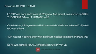
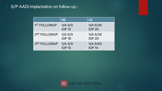
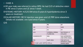
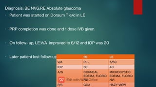
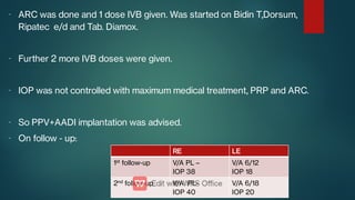
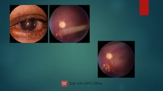
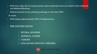
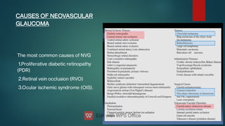
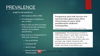
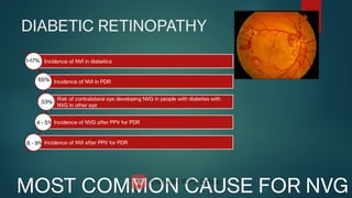
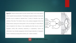
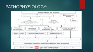
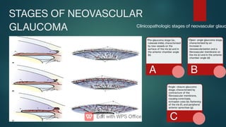
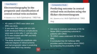
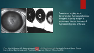
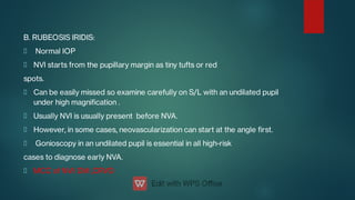
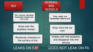
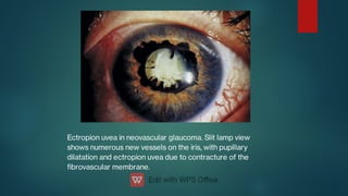
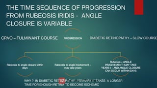
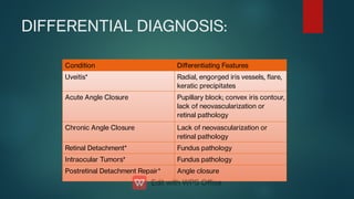
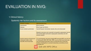
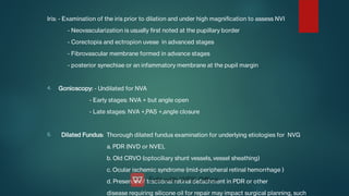
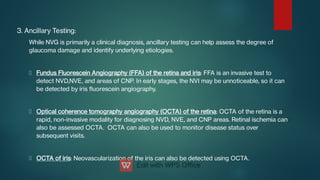
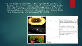
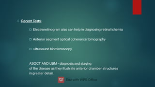
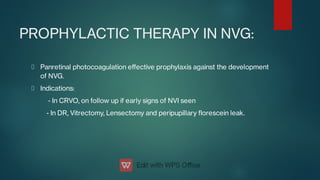
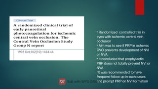
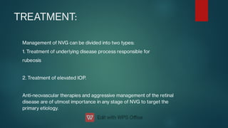
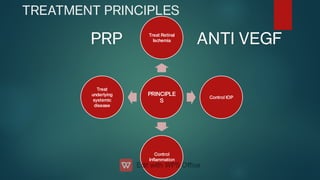
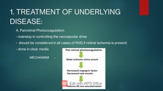
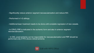
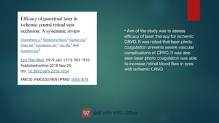
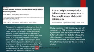
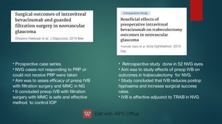
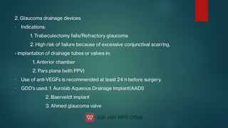
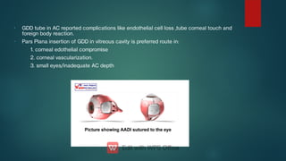
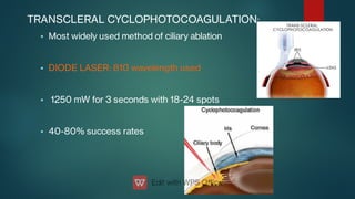
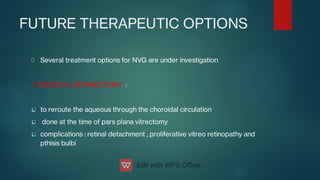
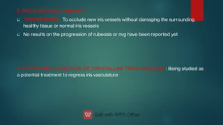
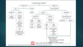
NEOVASCULAR
GLAUCOMA
PRESENTER: DR.HARSHA BHAMIDIPATI
MODERATORS: DR.DEEPAK ANTHWAL
DR. VENUGOPAL REDDY
CASE 1:
A 67 years female was referred from glaucoma department.
C/O Defective vision in LE since 1 week.
SYSTEMIC HISTORY: K/C/O DM since 4 years on treatment.
OCULAR HSTORY:BE K/C/O POAG on medication since 2 years but stopped
medication 6 months back , RE operated for TRAB.
O/E
RE LE
V/A 6/12 PL +
IOP 15 49
A/S LOW BLEB, PI
AT 12 O’CLOCK
360 DEGREE
NVI
GOINIOSCOPY OPEN NVA +,
OCCLUDABLE
P/S CDR 0.8, IR
LOSS
CRVO, GOA
-
-
-
Diagnosis: LE NVG post CRVO.
LE IVB 1 dose was given and PRP was done
Patient was started on Dorsum e/d, Bidin T e/d and Cap. IOPAR-SR in LE
On next follow ups:
RE LE
1ST
FOLLOWUP V/A 6/6P
IOP 17
V/A PL+,
REGRESSING
NVI
IOP 38
2ND
FOLLOWUP V/A 6/6P
IOP 15
V/A PL+,
REGRESSING
NVI
IOP 39
CASE 2
A 66 years male was referred to retina. Patient was diagnosed as
RE ITBRVO 1.5 months elsewhere.
SYSTEMIC HISTORY : NIL
OCULAR HISTORY: NIL
O/E:
RE LE
V/A HM 6/18
IOP 49 18
A/S CONGESTION,
CORNEAL
EDEMA ,FLORID
NVI
IMC
GONIOSCOPY NVA, OPEN OPEN
P/S NO VIEW D/T
CORNEAL
EDEMA
CDR 0.5
-
-
-
-
Diagnosis: RE NVG post ITBRVO
LE ARC was done and 1 dose IVB given
Patient was started on BIDIN T,DORSUM e/d and IOPAR-SR in RE
On next follow up:
On this visit accessible PRP was done.
RE LE
V/A HM 6/12
IOP 28 19
A/S CONGESTION,
CORNEAL
EDEMA
REDUCING,
REGRESSING
NVI
IMC
P/S CDR 0.9,CRVO CDR 0.5
On further follow up
RE LE
V/A HM+
IOP 24
Regressing NVI at 10 &
6 o clock
V/A 6/12
IOP 20
CASE 3:
A 47 years male patient was referred from glaucoma department with C/O
defective vision BE and for evaluation of BE PDR with RE VH.
SYSTEMIC HISTORY: K/C/O DM since 11 years on treatment, HTN since 2
years on treatment.
OCULAR HISTORY: NIL
O/E:
RE LE
1. V/A 5/60 5/60
2. IOP 14 mmHG 44 mmHG
3. ANTERIOR
SEGMENT
No NVI NVI +,Ectropion
uvea+
4.
GONIOSCOPY
OPEN ANGLE NVA + with PAS
5. POSTERIOR
SEGMENT
VH, no view PDR
-
-
-
-
Diagnosis: BE PDR, LE NVG
LE PRP was done and 1 dose of IVB given. And patient was started on BIDIN
T , DORSUM E/D and T. DIAMOX in LE
On follow-up, LE regression of NVI was seen but IOP was 48mmHG. Ripatec
E/D was added.
IOP was not in control even with maximum medical treatment, PRP and IVB.
So he was advised for AADI implantation with PPV in LE
- S/P AADI implantation on follow-up :
RE LE
1ST
FOLLOWUP V/A 6/9
IOP 12
V/A 6/36
IOP 20
2ND
FOLLOWUP V/A 6/9
IOP 18
V/A 6/36
IOP 20
3RD
FOLLOWUP V/A 6/9
IOP 15
V/A 6/60
IOP 14
CASE 4:
A 60 year male was referred to retina OPD. He had C/O of defective vision
in RE since 3 years and LE since 1 week.
SYSTEMIC HISTORY: K/C/O DM since 8 years & hyperlipidemia since 3
years on treatment
OCULAR HISTORY: RE IV injection was given and LE PRP done elsewhere.
Details not available. one eyed since 4 years.
O/E
RE LE
V/A PL - 6/60
IOP 54 24
A/S CORNEAL EDEMA,
FLORID NVI,
NVI FROM 9 -11
O’CLOCK
GONIOSCOPY NVA NVA SUPERIORLY
AND OPEN ANGLE.
P/S HAZY, GOA PDR WITH VH.
INFERIOR LASER
MARKS SEEN
-
-
-
-
Diagnosis: BE NVG,RE Absolute glaucoma
Patient was started on Dorsum T e/d in LE
PRP completion was done and 1 dose IVB given.
On follow- up, LE:V/A improved to 6/12 and IOP was 20
Later patient lost follow-up and came after 3 months, on this visit:
RE LE
V/A PL - 5/60
IOP 50 40
A/S CORNEAL
EDEMA, FLORID
NVI
MICROCYSTIC
EDEMA, FLORID
NVI
P/S GOA HAZY VIEW
-
-
-
-
-
ARC was done and 1 dose IVB given. Was started on Bidin T,Dorsum,
Ripatec e/d and Tab. Diamox.
Further 2 more IVB doses were given.
IOP was not controlled with maximum medical treatment, PRP and ARC.
So PPV+AADI implantation was advised.
On follow - up:
RE LE
1st
follow-up V/A PL –
IOP 38
V/A 6/12
IOP 18
2nd
follow-up V/A PL-
IOP 40
V/A 6/18
IOP 20
INTRODUCTION
Neovascular glaucoma (NVG) is severe form of secondary glaucoma
characterized by the proliferation of brovascular tissue in the anterior
chamber, which subsequently causes elevation in intraocular pressure
(IOP).
In 1906, G.Coats rst demonstrated new blood vessels growth on iris
surface in prior CRVO cases
Also known as:
1.Hemorrhagic glaucoma
2.Congestive glaucoma
3.Thrombotic glaucoma
This is a case report in 43 year old female with Carotid Cavernous Fistula.
It is noted that IOP elevation was related to new vessels and associated connective
tissue growth and proposed the term NEOVASACULAR GLAUCOMA which was early
termed as hemorrhagic glaucoma.
1.
2.
3.
4.
NVG has a high risk of causing severe vision impairment and can result in both unilateral
and bilateral blindness.
Retinal ischemia is the underlying etiology in more than 95%
of cases.
NVG makes approximately 3.9% of all glaucoma's.
RISK FACTORS FOR NVI:
RETINAL ISCHEMIA
SURGICAL CAUSES
TUMORS
OCULAR INFLAMATORY DISEASES
The most common causes of NVG
1.Proliferative diabetic retinopathy
(PDR)
2.Retinal vein occlusion (RVO)
3.Ocular ischemic syndrome (OIS).
CAUSES OF NEOVASCULAR
GLAUCOMA
PREVALENCE
A.








DIABETIC RETINOPATHY:
NVI incidence 65% in PDR.
5 % blindness in diabetics is
due to NVG.
Risk for NVI and NVG increases
in DR patients after vitrectomy,
cataract and posterior
capsulotomy.
Risk factors for development of
NVG post vitrectomy:
Pre op NVI/NVA
increased IOP
HbA1C levels
Retinal tamponade used in
retinal surgery.
DIABETIC RETINOPATHY
MOST COMMON CAUSE FOR NVG
Incidence of NVI in diabetics
Incidence of NVI in PDR
Risk of contralateral eye developing NVG in people with diabetes with
NVG in other eye
Incidence of NVG after PPV for PDR
Incidence of NVI after PPV for PDR
1-17%
65%
33%
4 - 5%
8 - 9%



B. CENTRAL RETINAL VIEN OCCLUSION:
90 DAY GLAUCOMA (develops in 3 months of onset)
incidence of NVG following CRVO - 16%
45% ischemic CRVO develop NVG due to retinal capillary non perfusion





Study done in 2019
Retrospective study done in 646 CRVO
patients
To determine risk factors for NVG
development
13% developed NVG
Risk factors for NVG : RAPD,Poor V/A and
H/O HTN
Rong AJ, Swaminathan SS, Vanner EA, Parrish RK 2nd. Predictors of Neovascular Glaucoma in Central Retinal Vein
Occlusion. Am J Ophthalmol. 2019 Aug;204:62-69. doi: 10.1016/j.ajo.2019.02.038. Epub 2019 Mar 9. PMID: 30862502;
PMCID: PMC6642681.
•
•
Data was analyzed on 1108
patients
The study showed probability
of conversion of nonischemic
to ischemic CRVO at 6 months
and 18 months was 13.2% and
18.6 5 respectively
•
•
Study was done in 93 eyes with PDR
treated by argon laser
This study showed PRP was more
effective in controlling NVI in diabetics
than in CRVO

C. Ocular Ischemic Syndrome:
Develop NVG in 50% cases



Retrospective study in 25 OIS
patients
To determine risk factors for NVG
Risk factors: severity of carotid
artery stenosis and duration of
time from symptoms to diagnosis
CAROTID OCCLUSIVE DISEASE
A stenosis of
90% or more
of the common
or internal
carotid arteries
Reduces the perfusion
pressure within the
central retinal artery by
approximately 50%
Poor collateral
circulation between the
internal and external
carotid arteries or
between the two
internal carotid arteries
Well-
developed
collateral
circulation
OIS
OIS
May not
develop OIS
In most cases , well developed
collateral circulation is present –
hence NVG is less common
D. Chronic RD
•
•
•
•
Schulz reported RD as one of the common cause of NVI (23%)
Persistent RD in post-vitrectomy case often causes NVI.
RD due to Coats disease, x-linked Retinoschisis & Marfan’s syndrome are reported with
presenting feature of NVI.
Old settled RD must be considered when no obvious cause of NVI is found. These eyes
present with low IOP.
E. Ocular inamatory disease:
•
•
•
•
Low grade uveitis like Fuchs heterochromic iridocyclitis – frequently associated with NVA
and rarely NVI
NVG is hence not reported with this condition.
Gartner and Henkind mentioned endophthalmitis and sympathetic ophthalmitis as causes
of rubeosis
But no studies provide enough details regarding the pathogenesis of rubeosis iridis in
inammatory conditions
F. Tumors and Radiation:
A)


B)



RETINOBLASTOMA:
Howard & Ellsworth (1965) noted association of RB & NVI.
Signicant correlation was found between extent of tumor involvement & occurrence of NVI.
MALIGNANT MELANOMA:
0.5%-15%
NVG may occur due to RD, retinal hypoxia or tumor necrosis.
Irradiation doesn't seem to decrease the incidence of NVG in these cases.
THEORIES OF
NEOVASCULOGENESIS:
A. RETINAL HYPOXIA:
Diminished perfusion of retina
↓
Capillary non perfusion areas causing retinal ischemia
↓
New vessels formation on retina, optic disc, iris and anterior
chamber angle




B. Angiogenesis Factor:
Presence of angiogenic factors:- regulation of normal development of retinal blood
vessels
VEGF - Potent angiogenic stimulator, major role in normal and pathologic
angiogenesis. Major source: MULLER CELLS
FOUR isoforms: VEGF 121, VEGF 165, VEGF 189, VEGF 206
VEGF has ability to induce vascular permeability, also known as: Vascular Permeability
factor



Aim was to evaluate VEGF family in
aqeous humor of patients with NVG
Comparitive study between 22 NVG
cases and 20 contols
it concluded that high concentrations of
VEGF family except for VEGF B were
found in aqeous humor of patients with
NVG



Aim was to assess effectiveness
of intra ocular anti VEGF
medications alone or with other
types of conventional therapies
it concluded that Anti VEGF as an
adjunct to conventional treatment
could help reduce IOP in NVG in
short term
No evidence seen on its effect for
long term.



Aim: To determine timing of neovascular regression
after intra vitreal Avastin given as initial therapy for
eyes with high risk PDR with or without CSME
Done in 24 PDR eyes
It concluded that majority of new vessels
completely regressed within 48 hours after IVB.
The effect occured within 48 hours and was
mtained for atleast 2 weeks.
THIS STUDY SUPPORTED
ANGIOGENIC FACTOR THEORY
OF NEOVASCULARIZATION
THIS STUDY SUPPORTED
ANGIOGENIC FACTOR THEORY
OF NEOVASCULARIZATION



C. Vasoinhibitory Factors:
Ocular tissues produce substances inhibiting neovascularisation
Sources of vasoinhibitory substances: LENS and VITREOUS
RPE cells also release vaso inhibitory substances.
• Study showed lens may play an active
role in prevention of iris
neovascularisation.
PATHOPHYSIOLOGY:
STAGES OF NEOVASCULAR
GLAUCOMA
•Pre-glaucoma stage (i.e.,
rubeosis iridis), characterized
by new vessels on the
surface of the iris (a) and in
the anterior chamber angle
(b).
A
•Open- angle glaucoma stage,
characterized by an
increase in
neovascularization and a
brovascular membrane on
the iris (c) and in the anterior
chamber angle (d).
B
•Angle- closure glaucoma
stage, characterized by
contracture of the
brovascular membrane,
causing corectopia,
ectropion uvea (e), attening
of the iris (f), and peripheral
anterior synechiae (g).
C
Clinicopathologic stages of neovascular glauc



A. PRE RUBEOSIS:
Predisposing factor, such as diabetic retinopathy or CRVO have risk of
developing rubeosis iridis and chance for progression to neovascular
glaucoma.
In PDR,rubeosis iridis is reported to occur in approximately one- half of the
cases.
Risk for rubeosis iridis and NVG in patients
with diabetic retinopathy is greatly increased when arteriolar or
capillary nonperfusion is present or after vitrectomy or lensectomy,




On iris FFA - peripupillary leakage correlates with the presence of abnormal iris
vessels.
Iris FFA can give idea of risk for rubeosis iridis after vitrectomy in DR
On S/L -pupillary margin of iris: First site for neovascularisation earliest
biomicroscopic evidence of anterior segment rubeosis.
Gonioscopy is also important - check for NVA



Incidence of NVI and NVG in eyes with CRVO is correlated with the extent
of retinal capillary nonperfusion.
FFA is the most direct method of evaluating CNP areas
RAPD also indicates an increased risk for NVI after CRVO, and infrared
pupillometry is an objective method of documenting this nding
- Prospective study in 20 CRVO cases and 20
controls
- It was noted that marked reduction in venous
velocity was seen in CRV in affected eyes
- Suggesting that colour doppler ow imaging
has role on CRVO diagnosing and also help in
differentiation of ischemic and non ischemic.
- Non invasive, easy to perform and well tolerated
FUNCTIONAL
TESTS
MORPHOLOGICA
L TESTS
FFA IN ISCHEMIC CRVO
FFA IN NON ISCHEMIC CRVO
•
•
•
45 cases of CRVO were taken and
ERG was done.
It was seen that cases with b/a ratio
0.84 developed NVG,no complications
were seen in b/a ratio >1 and b/a ratio
0.73 were treated with PRP and NVG
developed in none.
The study showed b/a amplitude ratio
reected degree of retinal ischemia
and had prognostic value in predicting
which cases NVG may develop
•
•
•
Aim was to evaluate usefulness of 30 Hz
icker ERG in predicting outcome in
patients with CRVO
76 CRVO cases underwent 30 Hz icker
ERG.
It concluded that 30 Hzicker ERG
provides useful information about CRVO
by computing the probability of NVI
Fluorescein angiographic
demonstrates uorescein leakage
along the pupillary margin. In
subsequent frames, the area of
uorescein leakage enlarges.
(From Reiss GR,Sipperley JO. Glaucoma associated with retinal disorders and retinal surgery. In:Tasman W, Jaeger EA, eds.
Duane’s Clinical Ophthalmology. Vol 3. Philadelphia,PA: Lippincott Williams & Wilkins:chap 54E.)
B. RUBEOSIS IRIDIS:
Normal IOP
NVI starts from the pupillary margin as tiny tufts or red
spots.
Can be easily missed so examine carefully on S/L with an undilated pupil
under high magnication .
Usually NVI is usually present before NVA.
However, in some cases, neovascularization can start at the angle rst.
Gonioscopy in an undilated pupil is essential in all high-risk
cases to diagnose early NVA.
MCC of NVI: DM ,CRVO
NVI
Thin , tortuous , arborising,
fenestrated, irregular size
and course
Randomly oriented on
the surface of iris
Arise near the
pupillary margin
NORMAL
IRIS
VESSELS
Thick, radial , non
fenestrated, Uniform size
Arise from the iris
root
Visible until mid periphery
and disappear into the
stroma
LEAKS ON FA
LEAKS ON FA DOES NOT LEAK ON FA
DOES NOT LEAK ON FA
Slitlamp view of the iris
showing
rubeosis with tortuous
vessels on the surface of the
iris.
The rubeosis iridis begins intrastromally and then develops on
the
surface of the iris
C. OPEN ANGLE GLAUCOMA:
New vessels from the pupillary margin grow radially on the surface of the iris.
These vessels grow toward the angle, cross the ciliary body band and scleral spur.
A brovascular membrane, invisible on gonioscopy, accompanies the new vessels,
blocks the trabecular meshwork, and raises the IOP.
Incidence of NVG in patients with diabetes and rubeosis iridis ranges from 13% to
41%.
However, incidence of NVI in CRVO is probably signicantly higher.
NVI is more orid in this stage.
S/L often reveals an inammatory reaction and sometimes a hyphema
On Gonioscopy: AC angle-open, NVA ++.
The IOP is elevated and may rise suddenly, causing the patient to present with acute-
onset glaucoma.
Hallmark- brovascular membrane that covers the anterior chamber angle
Chronic inammatory changes are also typically seen on histologic examination.
brovascular
membrane,
inammation
and
hemorrhage
neovascular
tissue found
in the
trabecular
spaces
obstruction
of the
trabecular
meshwork
IOP elevation
in eyes with
neovascular
glaucoma.
Results in
glaucoma in
this stage
This stage is often responsive to medical IOP-lowering therapy
since the trabecular meshwork may still be partially functional
Hyphema in neovascular glaucoma from CRVO.
A: hyphema is present,with rubeosis. B: Fundus
in the same eye shows diffuse intraretinal
hemorrhages along the vascular arcades and
macula.
From Scruggs BA, Quist TS, Syed NA, Alward WLM. Neovascular
Glaucoma. EyeRounds.org. Posted May 22, 2018.
NEOVASCULARIZATION OF
ANGLE
D. CLOSED ANGLE GLAUCOMA
Myobroblasts in the brovascular membrane proliferate and contract to form PAS
Contracture of these brovascular membrane causes alterations of iris and anterior
chamber angle causing angle closure.
This leads to angle-closure glaucoma.
Glaucoma in this stage is typically severe and usually requires surgical
intervention.
The clinical features of advanced stage of NVG are as
follows:
• Severe decrease in visual acuity
• Eye pain
• Conjunctival and episcleral congestion
• Corneal edema
• Aqueous are
• Very high IOP
• Florid NVI and angle
• Ectropion Uveae
• Synechial angle closure on gonioscopy
Ectropion uvea in neovascular glaucoma. Slit lamp view
shows numerous new vessels on the iris, with pupillary
dilatation and ectropion uvea due to contracture of the
brovascular membrane.
THE TIME SEQUENCE OF PROGRESSION
FROM RUBEOSIS IRIDIS - ANGLE
CLOSURE IS VARIABLE
PROGRESSION
Rubeosis to angle closure within
days
Rubeosis to angle involvement –
may take years
Rubeosis – ANGLE
INVOLVEMENT (MAY TAKE
YEARS ) – AND ANGLE CLOSURE
CAN OCCUR WITHIN DAYS
CRVO – FULMINANT COURSE
CRVO – FULMINANT COURSE DIABETIC RETINOPATHY – SLOW COURSE
DIABETIC RETINOPATHY – SLOW COURSE
WHY ? IN DIABETIC RETINOPATHY , PERHAPS IT TAKES A LONGER
TIME FOR ENOUGH RETINA TO BECOME ISCHEMIC
DIFFERENTIAL DIAGNOSIS:
Condition Differentiating Features
Uveitis* Radial, engorged iris vessels, are,
keratic precipitates
Acute Angle Closure Pupillary block; convex iris contour,
lack of neovascularization or
retinal pathology
Chronic Angle Closure Lack of neovascularization or
retinal pathology
Retinal Detachment* Fundus pathology
Intraocular Tumors* Fundus pathology
Postretinal Detachment Repair* Angle closure
EVALUATION IN NVG:
1. Clinical history:
Systemic risk factors and its assessment.
Systemic condition Recommended tests
Hypertension Blood pressure
Diabetes Blood sugar and HbA1c
Ocular ischemic syndromere Carotid Doppler (retrobulbar vessels, intra, and extracranial)
Magnetic resonance and computed tomographic angiography Carotid
intra-arterial digital subtraction angiography (selectively and with
extreme caution)
Carotid cavernous stula/tumor
metastasis Uveitis, retinal
vasculitis, blood dyscrasis
CT scan/magnetic resonance imaging/MR venogram/PET scan HLA B
27 Hemogram+ESR, CRP, ANA, VDRL, serum protein electrophoresis,
and immunoelectrophoresis (rules out hyperviscosity syndromes
Ancillary investigations to exclude tubeculosis, sarcoidosis, blood
dyscrasias, etc.
Past ocular history for possible conditions that can lead to the development of NVG,
including PDR or CRVO.
Assess what previous treatments the patient has had for underlying retinal ischemic
disorders (PRP or anti VEGF)
Whether the patient has ever been diagnosed with high IOP or glaucoma in the past.
Assess the onset and duration of disease.
Based on symptoms: Early-asymptomatic, late stages- pain, decreased vision,
headache.
1.
1.
2.
3.
2. Examination:
VISUAL ACUITY: most cases poor D/T corneal edema or underlying retinal disease
PUPIL EXAMINATION: for RAPD, studies showed CRVO cases with RAPD are at more risk of
NVI formation
IOP: initially normal, as angle starts to close, IOP rises.
On Slit Lamp Biomicroscopy:
Conjunctiva and Cornea: - Conjunctival hyperamia
- Corneal edema: in acute IOP rise
- Cornea can be clear in cases with longstanding high IOP
Anterior Chamber: - hyphema due to NVI,
- mild AC reaction
4.
5.
Iris: - Examination of the iris prior to dilation and under high magnication to assess NVI
- Neovascularization is usually rst noted at the pupillary border
- Corectopia and ectropion uveae in advanced stages
- Fibrovascular membrane formed in advance stages
- posterior synechiae or an infammatory membrane at the pupil margin
Gonioscopy: - Undilated for NVA
- Early stages: NVA + but angle open
- Late stages: NVA +,PAS +,angle closure
Dilated Fundus: Thorough dilated fundus examination for underlying etiologies for NVG
a. PDR (NVD or NVE),
b. Old CRVO (optociliary shunt vessels, vessel sheathing)
c. Ocular ischemic syndrome (mid-peripheral retinal hemorrhage )
d. Presence of tractional retinal detachment in PDR or other
disease requiring silicone oil for repair may impact surgical planning, such
3. Ancillary Testing:
While NVG is primarily a clinical diagnosis, ancillary testing can help assess the degree of
glaucoma damage and identify underlying etiologies.
Fundus Fluorescein Angiography (FFA) of the retina and iris: FFA is an invasive test to
detect NVD,NVE, and areas of CNP. In early stages, the NVI may be unnoticeable, so it can
be detected by iris uorescein angiography.
Optical coherence tomography angiography (OCTA) of the retina: OCTA of the retina is a
rapid, non-invasive modality for diagnosing NVD, NVE, and CNP areas. Retinal ischemia can
also be assessed OCTA. OCTA can also be used to monitor disease status over
subsequent visits.
OCTA of iris: Neovascularization of the iris can also be detected using OCTA.
B-scan: Detection of lesions of the posterior segment of the eyes. B scan can
detect the presence of vitreous hemorrhage and additional brous proliferation,
tractional or rhegmatogenous retinal detachment. Mass lesions like choroidal
melanoma, ciliary body melanoma, and retinoblastoma can be detected. Detect
retinal detachment in the advanced stages of ROP with NVG



Recent Tests:
Electroretinogram also can help in diagnosing retinal ichemia
Anterior segment optical coherence tomography
ultrasound biomicroscopy.
ASOCT AND UBM - diagnosis and staging
of the disease as they illustrate anterior chamber structures
in greater detail.
PROPHYLACTIC THERAPY IN NVG:
Panretinal photocoagulation effective prophylaxis against the development
of NVG.
Indications:
- In CRVO, on follow up if early signs of NVI seen
- In DR, Vitrectomy, Lensectomy and peripupillary orescein leak.
•
•
•
•
Randomized controlled trial in
eyes with ischemic central vein
occlusion
Aim was to see if PRP in ischemic
CVO prevents development of NVI
or NVA.
It concluded that prophylactic
PRP does not totally prevent NVI or
NVA
It was recommended to have
frequent follow up in such cases
and prompt PRP on NVI formation
TREATMENT:
Management of NVG can be divided into two types:
1. Treatment of underlying disease process responsible for
rubeosis
2. Treatment of elevated IOP.
Anti-neovascular therapies and aggressive management of the retinal
disease are of utmost importance in any stage of NVG to target the
primary etiology.
TREATMENT PRINCIPLES
PRINCIPLE
S
Treat Retinal
Ischemia
Control IOP
Control
Inammation
Treat
underlying
systemic
disease
PRP
PRP ANTI VEGF
ANTI VEGF
1. TREATMENT OF UNDERLYING
DISEASE:
A. Panretinal Photocoagulation:
- mainstay in controlling the neovascular drive
- should be considered in all cases of NVG if retinal ischemia is present.
- done in clear media
MECHANISM →
- Signicantly reduce anterior segment neovascularization and reduce NVI.
- Performed in 1–3 sittings.
- Additional laser treatment needs to be done until complete regression of new vessels.
- In CRVO, PRP is indicated in the ischemic form and also in anterior segment
neovascularization.
- In OIS, uveal ischemia can be responsible for neovascularization and PRP should be
performed only if FFA shows retinal ischemia
• Aim of the study was to assess
efficacy of laser therapy for ischemic
CRVO. It was noted that laser photo
coagulation prevents severe vascular
complications of CRVO. It was also
seen laser photo coagulation was able
to increase retinal blood ow in eyes
with ischemic CRVO.
Hamanaka T, Omata T, Akabane N, Yajima T, Ishida N(2012) Retinal
Photocoagulation Density in the Treatment of Neovascular Glaucoma due to
Diabetic Retinopathy. J Clinic Experiment Ophthalmol S4:007. doi:10.4172/2155-
9570.S4-007
Study was done in 80 vitrectomy eyes with
previtrectomy PRP and compared with 402
eyes without PRP. Study showed that PRP
before vitrectomy for diabetic retinopathy
did not prevent postoperative rubeosis
iridis.In these cases, intraocular PRP at the
time of vitrectomy may be the procedure of
choice.
Retrospective cross sectional study was done in
11 eyes with new on-set neovascular glaucoma
cases without PRP and with UWFA completed.
Aim was to study patterns of non perfusion in
neovascular glaucoma cases. Ischemic index was
found to be 91% in far periphery 77% in mid
periphery and 42% at the posterior pole. There
was a statistically signicant difference between
the ischemic index in far periphery, posterior Pole
and mid periphery. The study concluded that this
knowledge can be used for further guide
treatment and understand risk of NVG.
B. Panretinal Cryotherapy
- Done in cloudy media where PRP not possible.
- In these cases transscleral panretinal cryotherapy can be combined with cyclocryotherapy.
- It controls the IOP and reduce or abolish the neovascularization.
Anterior Retinal Cryotherapy
8 cryo application in each quadrant – anterior to the equator in two rows
Each application for 8 seconds
•
•
•
•
•
•
Prospective study, the effect of anterior
retinal cryoablation (ARC) in the management
of neovascular glaucoma (NVG)
74 eyes were studied.
Following ARC, pain relief with regression of
anterior chamber inammatory reaction was
observed in 95% of the patients.
Regression of neovascularization of the iris
was documented in 93.5%.
Intraocular pressure control was achieved in
82.3%
ARC is strongly recommended in NVG,
especially in eyes with media opacities and as
a preliminary procedure for ltering surgery
or drainage implant surgery.
C. Anti VEGF Agents
- Anti-VEGF injection leads to regression of anterior and posterior segment
neovascularization and reduction of IOP.
- Anti-VEGF agents used: bevacizumab (Avastin), ranibizumab, and aibercept
- IV Bevacizumab causes regression of NVI and NVA within 24–48 h compared to PRP,
which takes 2–3 weeks.
- Avastin effective in reducing intraocular inammation and pain, and IOP lowering has
been noted in the open-angle stage.
- anti-VEGFs are used only as an adjunct to PRP, medical therapy, and surgery
- The most frequent recommendation is the combination of IV anti VEGF and PRP.
- PRP with adjunctive use of intravitreal anti-VEGF agent along with antiglaucoma
medications may be sufficient to control IOP in the open-angle stage of NVG.
- Surgical intervention for IOP lowering is often required in advanced stage with
synechial angle closure.
- repeated intravitreal anti-VEGF injections may cause both transient as well as
sustained elevation of IOP
•
•
•
•
Retrospective study done in 34
NVG eyes.
Aim was to assess long term
effects of IVB in NVG
Study concluded IVB has
favorable effect in controlling IOP
and pain in as it decreases
angiogenesis.
It can be used as an adjunctive
treatment
•
•
•
Prospective study done in newly
diagnosed NVG.
Aim was to investigate initial results
regarding treatment of NVG with IV
aibercept.
Study concluded that IV aibercept
may be an effective treatment for
stage 1 and 2 NVG. Results in rapid
regression of NVI and NVA as well as
IOP control.
•
•
•
Purpose was to summarize reports of both
transient and sustained IOP elevation due to
anti VEGF injections
Repeated IV anti‑VEGF injections may
cause both transient as well as sustained
elevation of IOP.
Regular monitoring needed
1.
2.
3.
D. Gene Therapy : Future Therapeutic strategies
Various pharmacological agents that modulate the angiogenesis cascade now
offer great potential as therapeutic agents for NVG.
Pigment epithelium derived factor (PEDF) overexpression inhibits retinal
inammation and neovascularization in mouse models.
VEGF trap (EYLEA, Bayer), a modied soluble VEGF receptor analog binds VEGF
more tightly than all other anti-VEGF therapies.
Small interfering RNA (siRNA) targeting VEGF expression has been
downregulated in vitro and in vivo, leading to .inhibition of retinal
neovascularization.
2. CONTROL OF IOP
A.
-
-
Medical management
- Sufficient to control the pressure in the OAG stage if treatment is
initiated early
AGM of rst choice: Beta blockers, topical carbonic anhydrase inhibitors,
alpha‑2 adrenergic agonists.
PG analouges are not of much use in closed angles and have risk to
increase inammation.
-
-
-
-
Pilocar is C/I as they may increases inammation and worsen synechial angle
closure due to miosis .
Atropine: cycloplegia.
Topical corticosteroids: reduce inammation and pain
Hyperosmotic agents and oral carbonic anhydrase inhibitors: for temporary
control high IOP and help clear corneal edema
MEDICAL THERAPY
ANTIGLAUCOMA MEDICATIONS -
B
BLOCKERS
ALPHA 2
AGONISTS
PILOCARPINE
PROSTAGLANDI
N ANALOGUES
CARBONIC
ANHYDRASE
INHIBITORS
X
•
•
TOPICAL
STEROIDS – TO
REDUCE
INFLAMMATION
ATROPINE 1 % -
TO REDUCE
OCULAR
CONGESTION
B. Surgical management
Glaucoma surgery indications:
1. Open‑angle stage of NVG to control IOP until PRP takes effect
2. Synechial angle‑closure stage for long‑term IOP control.
Surgical interventions for NVG:
1. trabeculectomy with antimetabolites
2. Glaucoma Drainage Devices
3. Cyclodestructive procedures.
1.
-
-
-
TRABECULECTOMY
- In NVG success of trabeculectomy alone is limited due inammation, high risk for
intraoperative bleeding and postoperative progression of the brovascular
membrane.
PRP with anti- VEGF, reduces NVI and inammation, increasing success rate of
trabeculectomy with 5-FU/MMC.
Prognostic factors for surgical failure: young age and vitrectomized eyes.
Surgical intervention should be planned within a week of injection of anti‑VEGF.
•
•
•
•
Prospective case series.
NVG cases not responding to PRP or
could not receive PRP were taken
Aim was to asses efficacy of preop IVB
with ltration surgery and MMC in NG
It concluded preop IVB with ltration
surgery with MMC is safe and effective
method to control IOP
•
•
•
•
Retrospective study done in 52 NVG eyes.
Aim was to study effects of preop IVB on
outcomes in trabeculectomy for NVG.
Study concluded that IVB reduces postop
hyphaema and increase surgical success
rates.
IVB is effective adjuunct to TRAB in NVG
-
-
-
2. Glaucoma drainage devices
Indications:
1. Trabeculectomy fails/Refractory glaucoma
2. High risk of failure because of excessive conjunctival scarring.
- implantation of drainage tubes or valves in:
1. Anterior chamber
2. Pars plana (with PPV)
Use of anti‑VEGFs is recommended at least 24 h before surgery.
GDD’s used: 1. Aurolab Aqueous Drainage Implant(AADI)
2. Baerveldt implant
3. Ahmed glaucoma valve
-
-
GDD tube in AC reported complications like endothelial cell loss ,tube corneal touch and
foreign body reaction.
Pars Plana insertion of GDD in vitreous cavity is preferred route in:
1. corneal edothelial compromise
2. corneal vascularization.
3. small eyes/inadequate AC depth
Kader MA, Dabke SB, Shukla AG, Reddy V, Abdul Khadar SM, Maheshwari D, Ramakrishnan R. Pars plana Aurolab
aqueous drainage implantation for refractory glaucoma: Outcome of a new modied technique. Indian J Ophthalmol. 2022
Mar;70(3):839-845. doi: 10.4103/ijo.IJO_1791_21. PMID: 35225526; PMCID: PMC9114538.
•
•
•
•
This study was done in 18
eyes with pp AGV and 18 eyes
with ac AGV
purpose of study was to
compare changes in corneal
endothelium cells after pars
plana AGV and anterior
chamber AGV
It concluded that endothelial
cell damage in ppAGV was less
than acAGV.
ppAGV can be preferred as it
has less endothelial cellloss
-
-
-
-
-
3. Cyclodestructive procedures:
Effective option in eyes with refractory NVG having poor visual prognosis.
Procedure destroys the ciliary body to reduce aqueous humor production.
Effective option to control IOP in poor visual prognosis
Transscleral cyclophotocoagulation or endocyclophotocoagulation (Limbal
or Pars Plana approach) may have a lower complication rate than
cyclocryotherapy.
painful blind eyes: retrobulbar alcohol injection




TRANSCLERAL CYCLOPHOTOCOAGULATION:
Most widely used method of ciliary ablation
DIODE LASER: 810 wavelength used
1250 mW for 3 seconds with 18-24 spots
40-80% success rates
FUTURE THERAPEUTIC OPTIONS



Several treatment options for NVG are under investigation
1. SURGICAL RETINECTOMY :
to reroute the aqueous through the choroidal circulation
done at the time of pars plana vitrectomy
complications : retinal detachment , proliferative vitreo retinopathy and
pthisis bulbi


2. PHOTODYNAMIC THERAPY :
VERTEPORFIN - To occlude new iris vessels without damaging the surrounding
healthy tissue or normal iris vessels
No results on the progression of rubeosis or nvg have been reported yet
3. INTRAVITREAL INJECTION OF CRYSTALLINE TRIAMCINOLONE : Being studied as
a potential treatment to regress iris vasculature
THANK YOU
THANK YOU

Neovascular glaucoma retinal aspects....

  • 1.
  • 2.
    CASE 1: A 67years female was referred from glaucoma department. C/O Defective vision in LE since 1 week. SYSTEMIC HISTORY: K/C/O DM since 4 years on treatment. OCULAR HSTORY:BE K/C/O POAG on medication since 2 years but stopped medication 6 months back , RE operated for TRAB. O/E RE LE V/A 6/12 PL + IOP 15 49 A/S LOW BLEB, PI AT 12 O’CLOCK 360 DEGREE NVI GOINIOSCOPY OPEN NVA +, OCCLUDABLE P/S CDR 0.8, IR LOSS CRVO, GOA
  • 3.
    - - - Diagnosis: LE NVGpost CRVO. LE IVB 1 dose was given and PRP was done Patient was started on Dorsum e/d, Bidin T e/d and Cap. IOPAR-SR in LE On next follow ups: RE LE 1ST FOLLOWUP V/A 6/6P IOP 17 V/A PL+, REGRESSING NVI IOP 38 2ND FOLLOWUP V/A 6/6P IOP 15 V/A PL+, REGRESSING NVI IOP 39
  • 5.
    CASE 2 A 66years male was referred to retina. Patient was diagnosed as RE ITBRVO 1.5 months elsewhere. SYSTEMIC HISTORY : NIL OCULAR HISTORY: NIL O/E: RE LE V/A HM 6/18 IOP 49 18 A/S CONGESTION, CORNEAL EDEMA ,FLORID NVI IMC GONIOSCOPY NVA, OPEN OPEN P/S NO VIEW D/T CORNEAL EDEMA CDR 0.5
  • 6.
    - - - - Diagnosis: RE NVGpost ITBRVO LE ARC was done and 1 dose IVB given Patient was started on BIDIN T,DORSUM e/d and IOPAR-SR in RE On next follow up: On this visit accessible PRP was done. RE LE V/A HM 6/12 IOP 28 19 A/S CONGESTION, CORNEAL EDEMA REDUCING, REGRESSING NVI IMC P/S CDR 0.9,CRVO CDR 0.5
  • 7.
    On further followup RE LE V/A HM+ IOP 24 Regressing NVI at 10 & 6 o clock V/A 6/12 IOP 20
  • 8.
    CASE 3: A 47years male patient was referred from glaucoma department with C/O defective vision BE and for evaluation of BE PDR with RE VH. SYSTEMIC HISTORY: K/C/O DM since 11 years on treatment, HTN since 2 years on treatment. OCULAR HISTORY: NIL O/E: RE LE 1. V/A 5/60 5/60 2. IOP 14 mmHG 44 mmHG 3. ANTERIOR SEGMENT No NVI NVI +,Ectropion uvea+ 4. GONIOSCOPY OPEN ANGLE NVA + with PAS 5. POSTERIOR SEGMENT VH, no view PDR
  • 9.
    - - - - Diagnosis: BE PDR,LE NVG LE PRP was done and 1 dose of IVB given. And patient was started on BIDIN T , DORSUM E/D and T. DIAMOX in LE On follow-up, LE regression of NVI was seen but IOP was 48mmHG. Ripatec E/D was added. IOP was not in control even with maximum medical treatment, PRP and IVB. So he was advised for AADI implantation with PPV in LE
  • 10.
    - S/P AADIimplantation on follow-up : RE LE 1ST FOLLOWUP V/A 6/9 IOP 12 V/A 6/36 IOP 20 2ND FOLLOWUP V/A 6/9 IOP 18 V/A 6/36 IOP 20 3RD FOLLOWUP V/A 6/9 IOP 15 V/A 6/60 IOP 14
  • 11.
    CASE 4: A 60year male was referred to retina OPD. He had C/O of defective vision in RE since 3 years and LE since 1 week. SYSTEMIC HISTORY: K/C/O DM since 8 years & hyperlipidemia since 3 years on treatment OCULAR HISTORY: RE IV injection was given and LE PRP done elsewhere. Details not available. one eyed since 4 years. O/E RE LE V/A PL - 6/60 IOP 54 24 A/S CORNEAL EDEMA, FLORID NVI, NVI FROM 9 -11 O’CLOCK GONIOSCOPY NVA NVA SUPERIORLY AND OPEN ANGLE. P/S HAZY, GOA PDR WITH VH. INFERIOR LASER MARKS SEEN
  • 12.
    - - - - Diagnosis: BE NVG,REAbsolute glaucoma Patient was started on Dorsum T e/d in LE PRP completion was done and 1 dose IVB given. On follow- up, LE:V/A improved to 6/12 and IOP was 20 Later patient lost follow-up and came after 3 months, on this visit: RE LE V/A PL - 5/60 IOP 50 40 A/S CORNEAL EDEMA, FLORID NVI MICROCYSTIC EDEMA, FLORID NVI P/S GOA HAZY VIEW
  • 13.
    - - - - - ARC was doneand 1 dose IVB given. Was started on Bidin T,Dorsum, Ripatec e/d and Tab. Diamox. Further 2 more IVB doses were given. IOP was not controlled with maximum medical treatment, PRP and ARC. So PPV+AADI implantation was advised. On follow - up: RE LE 1st follow-up V/A PL – IOP 38 V/A 6/12 IOP 18 2nd follow-up V/A PL- IOP 40 V/A 6/18 IOP 20
  • 15.
    INTRODUCTION Neovascular glaucoma (NVG)is severe form of secondary glaucoma characterized by the proliferation of brovascular tissue in the anterior chamber, which subsequently causes elevation in intraocular pressure (IOP). In 1906, G.Coats rst demonstrated new blood vessels growth on iris surface in prior CRVO cases Also known as: 1.Hemorrhagic glaucoma 2.Congestive glaucoma 3.Thrombotic glaucoma
  • 16.
    This is acase report in 43 year old female with Carotid Cavernous Fistula. It is noted that IOP elevation was related to new vessels and associated connective tissue growth and proposed the term NEOVASACULAR GLAUCOMA which was early termed as hemorrhagic glaucoma.
  • 17.
    1. 2. 3. 4. NVG has ahigh risk of causing severe vision impairment and can result in both unilateral and bilateral blindness. Retinal ischemia is the underlying etiology in more than 95% of cases. NVG makes approximately 3.9% of all glaucoma's. RISK FACTORS FOR NVI: RETINAL ISCHEMIA SURGICAL CAUSES TUMORS OCULAR INFLAMATORY DISEASES
  • 18.
    The most commoncauses of NVG 1.Proliferative diabetic retinopathy (PDR) 2.Retinal vein occlusion (RVO) 3.Ocular ischemic syndrome (OIS). CAUSES OF NEOVASCULAR GLAUCOMA
  • 19.
    PREVALENCE A.         DIABETIC RETINOPATHY: NVI incidence65% in PDR. 5 % blindness in diabetics is due to NVG. Risk for NVI and NVG increases in DR patients after vitrectomy, cataract and posterior capsulotomy. Risk factors for development of NVG post vitrectomy: Pre op NVI/NVA increased IOP HbA1C levels Retinal tamponade used in retinal surgery.
  • 20.
    DIABETIC RETINOPATHY MOST COMMONCAUSE FOR NVG MOST COMMON CAUSE FOR NVG Incidence of NVI in diabetics Incidence of NVI in PDR Risk of contralateral eye developing NVG in people with diabetes with NVG in other eye Incidence of NVG after PPV for PDR Incidence of NVI after PPV for PDR 1-17% 65% 33% 4 - 5% 8 - 9%
  • 21.
       B. CENTRAL RETINALVIEN OCCLUSION: 90 DAY GLAUCOMA (develops in 3 months of onset) incidence of NVG following CRVO - 16% 45% ischemic CRVO develop NVG due to retinal capillary non perfusion      Study done in 2019 Retrospective study done in 646 CRVO patients To determine risk factors for NVG development 13% developed NVG Risk factors for NVG : RAPD,Poor V/A and H/O HTN Rong AJ, Swaminathan SS, Vanner EA, Parrish RK 2nd. Predictors of Neovascular Glaucoma in Central Retinal Vein Occlusion. Am J Ophthalmol. 2019 Aug;204:62-69. doi: 10.1016/j.ajo.2019.02.038. Epub 2019 Mar 9. PMID: 30862502; PMCID: PMC6642681.
  • 22.
    • • Data was analyzedon 1108 patients The study showed probability of conversion of nonischemic to ischemic CRVO at 6 months and 18 months was 13.2% and 18.6 5 respectively • • Study was done in 93 eyes with PDR treated by argon laser This study showed PRP was more effective in controlling NVI in diabetics than in CRVO
  • 23.
     C. Ocular IschemicSyndrome: Develop NVG in 50% cases    Retrospective study in 25 OIS patients To determine risk factors for NVG Risk factors: severity of carotid artery stenosis and duration of time from symptoms to diagnosis
  • 24.
  • 25.
    A stenosis of 90%or more of the common or internal carotid arteries Reduces the perfusion pressure within the central retinal artery by approximately 50% Poor collateral circulation between the internal and external carotid arteries or between the two internal carotid arteries Well- developed collateral circulation OIS OIS May not develop OIS In most cases , well developed collateral circulation is present – hence NVG is less common
  • 26.
    D. Chronic RD • • • • Schulzreported RD as one of the common cause of NVI (23%) Persistent RD in post-vitrectomy case often causes NVI. RD due to Coats disease, x-linked Retinoschisis & Marfan’s syndrome are reported with presenting feature of NVI. Old settled RD must be considered when no obvious cause of NVI is found. These eyes present with low IOP.
  • 27.
    E. Ocular inamatorydisease: • • • • Low grade uveitis like Fuchs heterochromic iridocyclitis – frequently associated with NVA and rarely NVI NVG is hence not reported with this condition. Gartner and Henkind mentioned endophthalmitis and sympathetic ophthalmitis as causes of rubeosis But no studies provide enough details regarding the pathogenesis of rubeosis iridis in inammatory conditions
  • 28.
    F. Tumors andRadiation: A)   B)    RETINOBLASTOMA: Howard & Ellsworth (1965) noted association of RB & NVI. Signicant correlation was found between extent of tumor involvement & occurrence of NVI. MALIGNANT MELANOMA: 0.5%-15% NVG may occur due to RD, retinal hypoxia or tumor necrosis. Irradiation doesn't seem to decrease the incidence of NVG in these cases.
  • 29.
    THEORIES OF NEOVASCULOGENESIS: A. RETINALHYPOXIA: Diminished perfusion of retina ↓ Capillary non perfusion areas causing retinal ischemia ↓ New vessels formation on retina, optic disc, iris and anterior chamber angle
  • 30.
        B. Angiogenesis Factor: Presenceof angiogenic factors:- regulation of normal development of retinal blood vessels VEGF - Potent angiogenic stimulator, major role in normal and pathologic angiogenesis. Major source: MULLER CELLS FOUR isoforms: VEGF 121, VEGF 165, VEGF 189, VEGF 206 VEGF has ability to induce vascular permeability, also known as: Vascular Permeability factor
  • 31.
       Aim was toevaluate VEGF family in aqeous humor of patients with NVG Comparitive study between 22 NVG cases and 20 contols it concluded that high concentrations of VEGF family except for VEGF B were found in aqeous humor of patients with NVG    Aim was to assess effectiveness of intra ocular anti VEGF medications alone or with other types of conventional therapies it concluded that Anti VEGF as an adjunct to conventional treatment could help reduce IOP in NVG in short term No evidence seen on its effect for long term.
  • 32.
       Aim: To determinetiming of neovascular regression after intra vitreal Avastin given as initial therapy for eyes with high risk PDR with or without CSME Done in 24 PDR eyes It concluded that majority of new vessels completely regressed within 48 hours after IVB. The effect occured within 48 hours and was mtained for atleast 2 weeks. THIS STUDY SUPPORTED ANGIOGENIC FACTOR THEORY OF NEOVASCULARIZATION THIS STUDY SUPPORTED ANGIOGENIC FACTOR THEORY OF NEOVASCULARIZATION
  • 33.
       C. Vasoinhibitory Factors: Oculartissues produce substances inhibiting neovascularisation Sources of vasoinhibitory substances: LENS and VITREOUS RPE cells also release vaso inhibitory substances. • Study showed lens may play an active role in prevention of iris neovascularisation.
  • 35.
  • 36.
    STAGES OF NEOVASCULAR GLAUCOMA •Pre-glaucomastage (i.e., rubeosis iridis), characterized by new vessels on the surface of the iris (a) and in the anterior chamber angle (b). A •Open- angle glaucoma stage, characterized by an increase in neovascularization and a brovascular membrane on the iris (c) and in the anterior chamber angle (d). B •Angle- closure glaucoma stage, characterized by contracture of the brovascular membrane, causing corectopia, ectropion uvea (e), attening of the iris (f), and peripheral anterior synechiae (g). C Clinicopathologic stages of neovascular glauc
  • 37.
       A. PRE RUBEOSIS: Predisposingfactor, such as diabetic retinopathy or CRVO have risk of developing rubeosis iridis and chance for progression to neovascular glaucoma. In PDR,rubeosis iridis is reported to occur in approximately one- half of the cases. Risk for rubeosis iridis and NVG in patients with diabetic retinopathy is greatly increased when arteriolar or capillary nonperfusion is present or after vitrectomy or lensectomy,
  • 38.
        On iris FFA- peripupillary leakage correlates with the presence of abnormal iris vessels. Iris FFA can give idea of risk for rubeosis iridis after vitrectomy in DR On S/L -pupillary margin of iris: First site for neovascularisation earliest biomicroscopic evidence of anterior segment rubeosis. Gonioscopy is also important - check for NVA
  • 39.
       Incidence of NVIand NVG in eyes with CRVO is correlated with the extent of retinal capillary nonperfusion. FFA is the most direct method of evaluating CNP areas RAPD also indicates an increased risk for NVI after CRVO, and infrared pupillometry is an objective method of documenting this nding - Prospective study in 20 CRVO cases and 20 controls - It was noted that marked reduction in venous velocity was seen in CRV in affected eyes - Suggesting that colour doppler ow imaging has role on CRVO diagnosing and also help in differentiation of ischemic and non ischemic. - Non invasive, easy to perform and well tolerated
  • 40.
  • 41.
    FFA IN ISCHEMICCRVO FFA IN ISCHEMIC CRVO FFA IN NON ISCHEMIC CRVO FFA IN NON ISCHEMIC CRVO
  • 42.
    • • • 45 cases ofCRVO were taken and ERG was done. It was seen that cases with b/a ratio 0.84 developed NVG,no complications were seen in b/a ratio >1 and b/a ratio 0.73 were treated with PRP and NVG developed in none. The study showed b/a amplitude ratio reected degree of retinal ischemia and had prognostic value in predicting which cases NVG may develop • • • Aim was to evaluate usefulness of 30 Hz icker ERG in predicting outcome in patients with CRVO 76 CRVO cases underwent 30 Hz icker ERG. It concluded that 30 Hzicker ERG provides useful information about CRVO by computing the probability of NVI
  • 43.
    Fluorescein angiographic demonstrates uoresceinleakage along the pupillary margin. In subsequent frames, the area of uorescein leakage enlarges. (From Reiss GR,Sipperley JO. Glaucoma associated with retinal disorders and retinal surgery. In:Tasman W, Jaeger EA, eds. Duane’s Clinical Ophthalmology. Vol 3. Philadelphia,PA: Lippincott Williams & Wilkins:chap 54E.)
  • 44.
    B. RUBEOSIS IRIDIS: NormalIOP NVI starts from the pupillary margin as tiny tufts or red spots. Can be easily missed so examine carefully on S/L with an undilated pupil under high magnication . Usually NVI is usually present before NVA. However, in some cases, neovascularization can start at the angle rst. Gonioscopy in an undilated pupil is essential in all high-risk cases to diagnose early NVA. MCC of NVI: DM ,CRVO
  • 45.
    NVI Thin , tortuous, arborising, fenestrated, irregular size and course Randomly oriented on the surface of iris Arise near the pupillary margin NORMAL IRIS VESSELS Thick, radial , non fenestrated, Uniform size Arise from the iris root Visible until mid periphery and disappear into the stroma LEAKS ON FA LEAKS ON FA DOES NOT LEAK ON FA DOES NOT LEAK ON FA
  • 46.
    Slitlamp view ofthe iris showing rubeosis with tortuous vessels on the surface of the iris. The rubeosis iridis begins intrastromally and then develops on the surface of the iris
  • 47.
    C. OPEN ANGLEGLAUCOMA: New vessels from the pupillary margin grow radially on the surface of the iris. These vessels grow toward the angle, cross the ciliary body band and scleral spur. A brovascular membrane, invisible on gonioscopy, accompanies the new vessels, blocks the trabecular meshwork, and raises the IOP. Incidence of NVG in patients with diabetes and rubeosis iridis ranges from 13% to 41%. However, incidence of NVI in CRVO is probably signicantly higher.
  • 48.
    NVI is moreorid in this stage. S/L often reveals an inammatory reaction and sometimes a hyphema On Gonioscopy: AC angle-open, NVA ++. The IOP is elevated and may rise suddenly, causing the patient to present with acute- onset glaucoma. Hallmark- brovascular membrane that covers the anterior chamber angle Chronic inammatory changes are also typically seen on histologic examination.
  • 49.
    brovascular membrane, inammation and hemorrhage neovascular tissue found in the trabecular spaces obstruction ofthe trabecular meshwork IOP elevation in eyes with neovascular glaucoma. Results in glaucoma in this stage This stage is often responsive to medical IOP-lowering therapy since the trabecular meshwork may still be partially functional
  • 50.
    Hyphema in neovascularglaucoma from CRVO. A: hyphema is present,with rubeosis. B: Fundus in the same eye shows diffuse intraretinal hemorrhages along the vascular arcades and macula. From Scruggs BA, Quist TS, Syed NA, Alward WLM. Neovascular Glaucoma. EyeRounds.org. Posted May 22, 2018. NEOVASCULARIZATION OF ANGLE
  • 51.
    D. CLOSED ANGLEGLAUCOMA Myobroblasts in the brovascular membrane proliferate and contract to form PAS Contracture of these brovascular membrane causes alterations of iris and anterior chamber angle causing angle closure. This leads to angle-closure glaucoma. Glaucoma in this stage is typically severe and usually requires surgical intervention.
  • 52.
    The clinical featuresof advanced stage of NVG are as follows: • Severe decrease in visual acuity • Eye pain • Conjunctival and episcleral congestion • Corneal edema • Aqueous are • Very high IOP • Florid NVI and angle • Ectropion Uveae • Synechial angle closure on gonioscopy
  • 53.
    Ectropion uvea inneovascular glaucoma. Slit lamp view shows numerous new vessels on the iris, with pupillary dilatation and ectropion uvea due to contracture of the brovascular membrane.
  • 54.
    THE TIME SEQUENCEOF PROGRESSION FROM RUBEOSIS IRIDIS - ANGLE CLOSURE IS VARIABLE PROGRESSION Rubeosis to angle closure within days Rubeosis to angle involvement – may take years Rubeosis – ANGLE INVOLVEMENT (MAY TAKE YEARS ) – AND ANGLE CLOSURE CAN OCCUR WITHIN DAYS CRVO – FULMINANT COURSE CRVO – FULMINANT COURSE DIABETIC RETINOPATHY – SLOW COURSE DIABETIC RETINOPATHY – SLOW COURSE WHY ? IN DIABETIC RETINOPATHY , PERHAPS IT TAKES A LONGER TIME FOR ENOUGH RETINA TO BECOME ISCHEMIC
  • 55.
    DIFFERENTIAL DIAGNOSIS: Condition DifferentiatingFeatures Uveitis* Radial, engorged iris vessels, are, keratic precipitates Acute Angle Closure Pupillary block; convex iris contour, lack of neovascularization or retinal pathology Chronic Angle Closure Lack of neovascularization or retinal pathology Retinal Detachment* Fundus pathology Intraocular Tumors* Fundus pathology Postretinal Detachment Repair* Angle closure
  • 56.
    EVALUATION IN NVG: 1.Clinical history: Systemic risk factors and its assessment. Systemic condition Recommended tests Hypertension Blood pressure Diabetes Blood sugar and HbA1c Ocular ischemic syndromere Carotid Doppler (retrobulbar vessels, intra, and extracranial) Magnetic resonance and computed tomographic angiography Carotid intra-arterial digital subtraction angiography (selectively and with extreme caution) Carotid cavernous stula/tumor metastasis Uveitis, retinal vasculitis, blood dyscrasis CT scan/magnetic resonance imaging/MR venogram/PET scan HLA B 27 Hemogram+ESR, CRP, ANA, VDRL, serum protein electrophoresis, and immunoelectrophoresis (rules out hyperviscosity syndromes Ancillary investigations to exclude tubeculosis, sarcoidosis, blood dyscrasias, etc.
  • 57.
    Past ocular historyfor possible conditions that can lead to the development of NVG, including PDR or CRVO. Assess what previous treatments the patient has had for underlying retinal ischemic disorders (PRP or anti VEGF) Whether the patient has ever been diagnosed with high IOP or glaucoma in the past. Assess the onset and duration of disease. Based on symptoms: Early-asymptomatic, late stages- pain, decreased vision, headache.
  • 58.
    1. 1. 2. 3. 2. Examination: VISUAL ACUITY:most cases poor D/T corneal edema or underlying retinal disease PUPIL EXAMINATION: for RAPD, studies showed CRVO cases with RAPD are at more risk of NVI formation IOP: initially normal, as angle starts to close, IOP rises. On Slit Lamp Biomicroscopy: Conjunctiva and Cornea: - Conjunctival hyperamia - Corneal edema: in acute IOP rise - Cornea can be clear in cases with longstanding high IOP Anterior Chamber: - hyphema due to NVI, - mild AC reaction
  • 59.
    4. 5. Iris: - Examinationof the iris prior to dilation and under high magnication to assess NVI - Neovascularization is usually rst noted at the pupillary border - Corectopia and ectropion uveae in advanced stages - Fibrovascular membrane formed in advance stages - posterior synechiae or an infammatory membrane at the pupil margin Gonioscopy: - Undilated for NVA - Early stages: NVA + but angle open - Late stages: NVA +,PAS +,angle closure Dilated Fundus: Thorough dilated fundus examination for underlying etiologies for NVG a. PDR (NVD or NVE), b. Old CRVO (optociliary shunt vessels, vessel sheathing) c. Ocular ischemic syndrome (mid-peripheral retinal hemorrhage ) d. Presence of tractional retinal detachment in PDR or other disease requiring silicone oil for repair may impact surgical planning, such
  • 60.
    3. Ancillary Testing: WhileNVG is primarily a clinical diagnosis, ancillary testing can help assess the degree of glaucoma damage and identify underlying etiologies. Fundus Fluorescein Angiography (FFA) of the retina and iris: FFA is an invasive test to detect NVD,NVE, and areas of CNP. In early stages, the NVI may be unnoticeable, so it can be detected by iris uorescein angiography. Optical coherence tomography angiography (OCTA) of the retina: OCTA of the retina is a rapid, non-invasive modality for diagnosing NVD, NVE, and CNP areas. Retinal ischemia can also be assessed OCTA. OCTA can also be used to monitor disease status over subsequent visits. OCTA of iris: Neovascularization of the iris can also be detected using OCTA.
  • 61.
    B-scan: Detection oflesions of the posterior segment of the eyes. B scan can detect the presence of vitreous hemorrhage and additional brous proliferation, tractional or rhegmatogenous retinal detachment. Mass lesions like choroidal melanoma, ciliary body melanoma, and retinoblastoma can be detected. Detect retinal detachment in the advanced stages of ROP with NVG
  • 62.
       Recent Tests: Electroretinogram alsocan help in diagnosing retinal ichemia Anterior segment optical coherence tomography ultrasound biomicroscopy. ASOCT AND UBM - diagnosis and staging of the disease as they illustrate anterior chamber structures in greater detail.
  • 63.
    PROPHYLACTIC THERAPY INNVG: Panretinal photocoagulation effective prophylaxis against the development of NVG. Indications: - In CRVO, on follow up if early signs of NVI seen - In DR, Vitrectomy, Lensectomy and peripupillary orescein leak.
  • 64.
    • • • • Randomized controlled trialin eyes with ischemic central vein occlusion Aim was to see if PRP in ischemic CVO prevents development of NVI or NVA. It concluded that prophylactic PRP does not totally prevent NVI or NVA It was recommended to have frequent follow up in such cases and prompt PRP on NVI formation
  • 65.
    TREATMENT: Management of NVGcan be divided into two types: 1. Treatment of underlying disease process responsible for rubeosis 2. Treatment of elevated IOP. Anti-neovascular therapies and aggressive management of the retinal disease are of utmost importance in any stage of NVG to target the primary etiology.
  • 66.
    TREATMENT PRINCIPLES PRINCIPLE S Treat Retinal Ischemia ControlIOP Control Inammation Treat underlying systemic disease PRP PRP ANTI VEGF ANTI VEGF
  • 67.
    1. TREATMENT OFUNDERLYING DISEASE: A. Panretinal Photocoagulation: - mainstay in controlling the neovascular drive - should be considered in all cases of NVG if retinal ischemia is present. - done in clear media MECHANISM →
  • 68.
    - Signicantly reduceanterior segment neovascularization and reduce NVI. - Performed in 1–3 sittings. - Additional laser treatment needs to be done until complete regression of new vessels. - In CRVO, PRP is indicated in the ischemic form and also in anterior segment neovascularization. - In OIS, uveal ischemia can be responsible for neovascularization and PRP should be performed only if FFA shows retinal ischemia
  • 69.
    • Aim ofthe study was to assess efficacy of laser therapy for ischemic CRVO. It was noted that laser photo coagulation prevents severe vascular complications of CRVO. It was also seen laser photo coagulation was able to increase retinal blood ow in eyes with ischemic CRVO.
  • 70.
    Hamanaka T, OmataT, Akabane N, Yajima T, Ishida N(2012) Retinal Photocoagulation Density in the Treatment of Neovascular Glaucoma due to Diabetic Retinopathy. J Clinic Experiment Ophthalmol S4:007. doi:10.4172/2155- 9570.S4-007
  • 71.
    Study was donein 80 vitrectomy eyes with previtrectomy PRP and compared with 402 eyes without PRP. Study showed that PRP before vitrectomy for diabetic retinopathy did not prevent postoperative rubeosis iridis.In these cases, intraocular PRP at the time of vitrectomy may be the procedure of choice. Retrospective cross sectional study was done in 11 eyes with new on-set neovascular glaucoma cases without PRP and with UWFA completed. Aim was to study patterns of non perfusion in neovascular glaucoma cases. Ischemic index was found to be 91% in far periphery 77% in mid periphery and 42% at the posterior pole. There was a statistically signicant difference between the ischemic index in far periphery, posterior Pole and mid periphery. The study concluded that this knowledge can be used for further guide treatment and understand risk of NVG.
  • 72.
    B. Panretinal Cryotherapy -Done in cloudy media where PRP not possible. - In these cases transscleral panretinal cryotherapy can be combined with cyclocryotherapy. - It controls the IOP and reduce or abolish the neovascularization.
  • 74.
    Anterior Retinal Cryotherapy 8cryo application in each quadrant – anterior to the equator in two rows Each application for 8 seconds
  • 75.
    • • • • • • Prospective study, theeffect of anterior retinal cryoablation (ARC) in the management of neovascular glaucoma (NVG) 74 eyes were studied. Following ARC, pain relief with regression of anterior chamber inammatory reaction was observed in 95% of the patients. Regression of neovascularization of the iris was documented in 93.5%. Intraocular pressure control was achieved in 82.3% ARC is strongly recommended in NVG, especially in eyes with media opacities and as a preliminary procedure for ltering surgery or drainage implant surgery.
  • 77.
    C. Anti VEGFAgents - Anti-VEGF injection leads to regression of anterior and posterior segment neovascularization and reduction of IOP. - Anti-VEGF agents used: bevacizumab (Avastin), ranibizumab, and aibercept - IV Bevacizumab causes regression of NVI and NVA within 24–48 h compared to PRP, which takes 2–3 weeks. - Avastin effective in reducing intraocular inammation and pain, and IOP lowering has been noted in the open-angle stage. - anti-VEGFs are used only as an adjunct to PRP, medical therapy, and surgery
  • 78.
    - The mostfrequent recommendation is the combination of IV anti VEGF and PRP. - PRP with adjunctive use of intravitreal anti-VEGF agent along with antiglaucoma medications may be sufficient to control IOP in the open-angle stage of NVG. - Surgical intervention for IOP lowering is often required in advanced stage with synechial angle closure. - repeated intravitreal anti-VEGF injections may cause both transient as well as sustained elevation of IOP
  • 79.
    • • • • Retrospective study donein 34 NVG eyes. Aim was to assess long term effects of IVB in NVG Study concluded IVB has favorable effect in controlling IOP and pain in as it decreases angiogenesis. It can be used as an adjunctive treatment • • • Prospective study done in newly diagnosed NVG. Aim was to investigate initial results regarding treatment of NVG with IV aibercept. Study concluded that IV aibercept may be an effective treatment for stage 1 and 2 NVG. Results in rapid regression of NVI and NVA as well as IOP control.
  • 80.
    • • • Purpose was tosummarize reports of both transient and sustained IOP elevation due to anti VEGF injections Repeated IV anti‑VEGF injections may cause both transient as well as sustained elevation of IOP. Regular monitoring needed
  • 81.
    1. 2. 3. D. Gene Therapy: Future Therapeutic strategies Various pharmacological agents that modulate the angiogenesis cascade now offer great potential as therapeutic agents for NVG. Pigment epithelium derived factor (PEDF) overexpression inhibits retinal inammation and neovascularization in mouse models. VEGF trap (EYLEA, Bayer), a modied soluble VEGF receptor analog binds VEGF more tightly than all other anti-VEGF therapies. Small interfering RNA (siRNA) targeting VEGF expression has been downregulated in vitro and in vivo, leading to .inhibition of retinal neovascularization.
  • 82.
    2. CONTROL OFIOP A. - - Medical management - Sufficient to control the pressure in the OAG stage if treatment is initiated early AGM of rst choice: Beta blockers, topical carbonic anhydrase inhibitors, alpha‑2 adrenergic agonists. PG analouges are not of much use in closed angles and have risk to increase inammation.
  • 83.
    - - - - Pilocar is C/Ias they may increases inammation and worsen synechial angle closure due to miosis . Atropine: cycloplegia. Topical corticosteroids: reduce inammation and pain Hyperosmotic agents and oral carbonic anhydrase inhibitors: for temporary control high IOP and help clear corneal edema
  • 84.
    MEDICAL THERAPY ANTIGLAUCOMA MEDICATIONS- B BLOCKERS ALPHA 2 AGONISTS PILOCARPINE PROSTAGLANDI N ANALOGUES CARBONIC ANHYDRASE INHIBITORS X • • TOPICAL STEROIDS – TO REDUCE INFLAMMATION ATROPINE 1 % - TO REDUCE OCULAR CONGESTION
  • 85.
    B. Surgical management Glaucomasurgery indications: 1. Open‑angle stage of NVG to control IOP until PRP takes effect 2. Synechial angle‑closure stage for long‑term IOP control. Surgical interventions for NVG: 1. trabeculectomy with antimetabolites 2. Glaucoma Drainage Devices 3. Cyclodestructive procedures.
  • 86.
    1. - - - TRABECULECTOMY - In NVGsuccess of trabeculectomy alone is limited due inammation, high risk for intraoperative bleeding and postoperative progression of the brovascular membrane. PRP with anti- VEGF, reduces NVI and inammation, increasing success rate of trabeculectomy with 5-FU/MMC. Prognostic factors for surgical failure: young age and vitrectomized eyes. Surgical intervention should be planned within a week of injection of anti‑VEGF.
  • 87.
    • • • • Prospective case series. NVGcases not responding to PRP or could not receive PRP were taken Aim was to asses efficacy of preop IVB with ltration surgery and MMC in NG It concluded preop IVB with ltration surgery with MMC is safe and effective method to control IOP • • • • Retrospective study done in 52 NVG eyes. Aim was to study effects of preop IVB on outcomes in trabeculectomy for NVG. Study concluded that IVB reduces postop hyphaema and increase surgical success rates. IVB is effective adjuunct to TRAB in NVG
  • 88.
    - - - 2. Glaucoma drainagedevices Indications: 1. Trabeculectomy fails/Refractory glaucoma 2. High risk of failure because of excessive conjunctival scarring. - implantation of drainage tubes or valves in: 1. Anterior chamber 2. Pars plana (with PPV) Use of anti‑VEGFs is recommended at least 24 h before surgery. GDD’s used: 1. Aurolab Aqueous Drainage Implant(AADI) 2. Baerveldt implant 3. Ahmed glaucoma valve
  • 89.
    - - GDD tube inAC reported complications like endothelial cell loss ,tube corneal touch and foreign body reaction. Pars Plana insertion of GDD in vitreous cavity is preferred route in: 1. corneal edothelial compromise 2. corneal vascularization. 3. small eyes/inadequate AC depth
  • 90.
    Kader MA, DabkeSB, Shukla AG, Reddy V, Abdul Khadar SM, Maheshwari D, Ramakrishnan R. Pars plana Aurolab aqueous drainage implantation for refractory glaucoma: Outcome of a new modied technique. Indian J Ophthalmol. 2022 Mar;70(3):839-845. doi: 10.4103/ijo.IJO_1791_21. PMID: 35225526; PMCID: PMC9114538.
  • 93.
    • • • • This study wasdone in 18 eyes with pp AGV and 18 eyes with ac AGV purpose of study was to compare changes in corneal endothelium cells after pars plana AGV and anterior chamber AGV It concluded that endothelial cell damage in ppAGV was less than acAGV. ppAGV can be preferred as it has less endothelial cellloss
  • 94.
    - - - - - 3. Cyclodestructive procedures: Effectiveoption in eyes with refractory NVG having poor visual prognosis. Procedure destroys the ciliary body to reduce aqueous humor production. Effective option to control IOP in poor visual prognosis Transscleral cyclophotocoagulation or endocyclophotocoagulation (Limbal or Pars Plana approach) may have a lower complication rate than cyclocryotherapy. painful blind eyes: retrobulbar alcohol injection
  • 95.
        TRANSCLERAL CYCLOPHOTOCOAGULATION: Most widelyused method of ciliary ablation DIODE LASER: 810 wavelength used 1250 mW for 3 seconds with 18-24 spots 40-80% success rates
  • 96.
    FUTURE THERAPEUTIC OPTIONS    Severaltreatment options for NVG are under investigation 1. SURGICAL RETINECTOMY : to reroute the aqueous through the choroidal circulation done at the time of pars plana vitrectomy complications : retinal detachment , proliferative vitreo retinopathy and pthisis bulbi
  • 97.
      2. PHOTODYNAMIC THERAPY: VERTEPORFIN - To occlude new iris vessels without damaging the surrounding healthy tissue or normal iris vessels No results on the progression of rubeosis or nvg have been reported yet 3. INTRAVITREAL INJECTION OF CRYSTALLINE TRIAMCINOLONE : Being studied as a potential treatment to regress iris vasculature
  • 99.