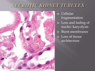
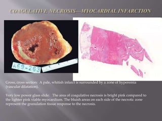
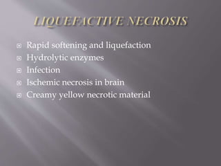
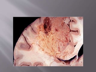
 Necrosis refers to a spectrum of Morphological
changes that follows cell death
 Enzymatic digestion of cell
 Denaturation of intracellular protein
 Membrane damage
 Increased esinophilia
 Glassy homogenous appearance
 Vacuolated cytoplasm – moth eaten
appearance
 Intra cytoplasmic myelin figures
 Karyolysis
 Pyknosis
 Karyorhexsis
 Epithelial cells stain
evenly pink
(eosinophilic) in
cytoplasm, with
purple, basophilic,
nucleic acids
confined to the
nuclei
 Apical surfaces are
ciliated
 Interstitia not
infiltrated with
immune cells nor
congested with
proteins
 Increased
eosinophilic staining
 Decreased
basophilic staining
(RNA)
 Plasma membrane
rounding, blebbing,
loss of cilia, due to
loss of connections
with cytoskeleton
 Integrity of tubules
degrading, but
basement
membranes intact
 Nuclei largely intact,
slightly narrowed,
pyknotic
 Cellular
fragmentation
 Loss and fading of
nuclei--karyolysis
 Burst membranes
 Loss of tissue
architecture
 Coagulative necrosis
 Liquefactive necrosis
 Caseous necrosis
 Gangrenous necrosis
 Fat necrosis
 Fibrinoid necrosis
 Most common
 Caused by ischemia
 Heart , kidney , spleen
 May also in viral hepatitis , thermal burns
 Architecture of dead tissue is preserved
 Esinophilic anucleate cells persist for weeks
 Phagocytosed
Denaturation vs enzymatic digestion
When there is marked cellular injury, there
is cell death and necrosis. This microscopic
appearance of myocardium shown here is
a mess because so many cells have died
that the tissue is not recognizable. Many
nuclei have become pyknotic (shrunken
and dark) and have then undergone
karyorrhexis (fragmentation) and
karyolysis (dissolution). The cytoplasm
and cell borders are no longer
recognizable. In this case, loss of the blood
supply from a major coronary artery led to
ischemia and cell death.
Here is myocardium in which the cells are
dying as a result of ischemic injury from
coronary artery occlusion. This is early in
the process of necrosis. The nuclei of the
myocardial fibers are being lost. The
cytoplasm is losing its structure, because
no well-defined cross-striations are seen.
Gross, cross section: A pale, whitish infarct is surrounded by a zone of hyperemia
(vascular dilatation).
Very low power glass slide: The area of coagulative necrosis is bright pink compared to
the lighter pink viable myocardium. The bluish areas on each side of the necrotic zone
represent the granulation tissue response to the necrosis.
 Rapid softening and liquefaction
 Hydrolytic enzymes
 Infection
 Ischemic necrosis in brain
 Creamy yellow necrotic material
 Variant of coagulative necrosis
 Mc in tuberculosis
 Friable white infracted tissue
 Architecture ?
 Collection Fragmented lysed cells
 Amorphous granular debris
 Distinctive inflammatory border
 Granuloma
 Common in clinical practice
 Usually lower limb
 Form of coagulative necrosis
 Types – 1.dry
2.wet
 Dry gangrene – variant of coagulative necrosis
 Wet gangrene - liiquefactive superimposed on
coagulative
 Enzymatic fat necrosis
 Traumatic fat necrosis
Cellular injury to the pancreatic acini leads to
release of powerful enzymes which damage fat by
the production of soaps, the chalky white areas seen
here on the cut surfaces. Microscopically, the
necrotic fat cells at the right have vague cellular
outlines, have lost their peripheral nuclei, and their
cytoplasm has become a pink amorphous mass of
necrotic material. There are some remaining
steatocytes at the left which are not necrotic.
 Special form of necrosis
 Immune reactions in blood vessles
 Immune complex with fibrin deposition
 Bright pink amorphous appearance in H.E
 Causes
malignant hypertension
SLE ,RA,HSP,PAN,HBV
acute rheumatic fever
Necrosis
Necrosis
Necrosis
Necrosis

Necrosis

  • 3.
     Necrosis refersto a spectrum of Morphological changes that follows cell death  Enzymatic digestion of cell  Denaturation of intracellular protein
  • 4.
     Membrane damage Increased esinophilia  Glassy homogenous appearance  Vacuolated cytoplasm – moth eaten appearance  Intra cytoplasmic myelin figures
  • 5.
  • 6.
     Epithelial cellsstain evenly pink (eosinophilic) in cytoplasm, with purple, basophilic, nucleic acids confined to the nuclei  Apical surfaces are ciliated  Interstitia not infiltrated with immune cells nor congested with proteins
  • 7.
     Increased eosinophilic staining Decreased basophilic staining (RNA)  Plasma membrane rounding, blebbing, loss of cilia, due to loss of connections with cytoskeleton  Integrity of tubules degrading, but basement membranes intact  Nuclei largely intact, slightly narrowed, pyknotic
  • 8.
     Cellular fragmentation  Lossand fading of nuclei--karyolysis  Burst membranes  Loss of tissue architecture
  • 10.
     Coagulative necrosis Liquefactive necrosis  Caseous necrosis  Gangrenous necrosis  Fat necrosis  Fibrinoid necrosis
  • 11.
     Most common Caused by ischemia  Heart , kidney , spleen  May also in viral hepatitis , thermal burns  Architecture of dead tissue is preserved  Esinophilic anucleate cells persist for weeks  Phagocytosed
  • 12.
  • 14.
    When there ismarked cellular injury, there is cell death and necrosis. This microscopic appearance of myocardium shown here is a mess because so many cells have died that the tissue is not recognizable. Many nuclei have become pyknotic (shrunken and dark) and have then undergone karyorrhexis (fragmentation) and karyolysis (dissolution). The cytoplasm and cell borders are no longer recognizable. In this case, loss of the blood supply from a major coronary artery led to ischemia and cell death.
  • 15.
    Here is myocardiumin which the cells are dying as a result of ischemic injury from coronary artery occlusion. This is early in the process of necrosis. The nuclei of the myocardial fibers are being lost. The cytoplasm is losing its structure, because no well-defined cross-striations are seen.
  • 16.
    Gross, cross section:A pale, whitish infarct is surrounded by a zone of hyperemia (vascular dilatation). Very low power glass slide: The area of coagulative necrosis is bright pink compared to the lighter pink viable myocardium. The bluish areas on each side of the necrotic zone represent the granulation tissue response to the necrosis.
  • 17.
     Rapid softeningand liquefaction  Hydrolytic enzymes  Infection  Ischemic necrosis in brain  Creamy yellow necrotic material
  • 19.
     Variant ofcoagulative necrosis  Mc in tuberculosis  Friable white infracted tissue  Architecture ?  Collection Fragmented lysed cells  Amorphous granular debris  Distinctive inflammatory border  Granuloma
  • 21.
     Common inclinical practice  Usually lower limb  Form of coagulative necrosis  Types – 1.dry 2.wet
  • 22.
     Dry gangrene– variant of coagulative necrosis  Wet gangrene - liiquefactive superimposed on coagulative
  • 25.
     Enzymatic fatnecrosis  Traumatic fat necrosis
  • 26.
    Cellular injury tothe pancreatic acini leads to release of powerful enzymes which damage fat by the production of soaps, the chalky white areas seen here on the cut surfaces. Microscopically, the necrotic fat cells at the right have vague cellular outlines, have lost their peripheral nuclei, and their cytoplasm has become a pink amorphous mass of necrotic material. There are some remaining steatocytes at the left which are not necrotic.
  • 28.
     Special formof necrosis  Immune reactions in blood vessles  Immune complex with fibrin deposition  Bright pink amorphous appearance in H.E  Causes malignant hypertension SLE ,RA,HSP,PAN,HBV acute rheumatic fever