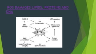
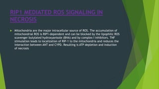
NECROSIS BY
Ekbal ali molla
m.pharm(pharmacology)
CIPT & AHS
1st year ,1st semester
INTRODUCTION
Necrosis is irreversible damage leads cell death, it is molecularly
regulated event that is associated with pathologies such as
ischemia-reperfusion injury, neurodegeneration and pathogen
infection. The serine/threonine kinase receptor-interacting Protein
1(RIP1) plays a crucial role during the Initiation of necrosis by
ligand-receptor Interactions. ATP depletion is an initiating Factor in
ischemia-induced necrotic cell death. It is always pathological.
NECROSIS
 Necrosis is the morphological changes that follow cell
death in living tissue or organ. Those morphological
changes are called necrosis.
 Necrotic cell death is characterized by cellular swelling,
plasma membrane rapture and the subsequent loss of the
intercellular contents. Necrotic cell death may be carried
out by a set of signal transduction pathway and execution
mechanism.
 [RIPK1-receptor interacting protein kinase 1, TRAF2- TNF
receptor associated factor 2, PARP- Polly ADP ribose
polymerase, ROS- reactive oxygen species.]
TYPES OF NECROSIS
 Coagulative necrosis
 Liquefactive necrosis
 Caseous necrosis
 Fat necrosis
 Fibrinoid necrosis
 Gangrenous necrosis
COAGULATIVE NECROSIS
 “In this type necrosis, the necrotic cell retains its
cellular outline for several days”
 Coagulative necrosis typically occurs in solid organs
such as kidney, heart and adrenal gland usually as a
result of deficient blood supply and anoxia.
 Denaturation of protein is basic mechanism of
coagulative necrosis. Denatures not only the structural
proteins but also the enzymic proteins, thus blocking
the cellular proteolysis.
LIQUEFACTIVE NECROSIS
 It is that type of necrosis that occurs due to autolytic and
heterolytic actions of enzymes that convert the proteins of
cell in liquid.
 Softening and liquefaction of tissue .(e.g. ischemic necrosis
of brain. In this necrosis complete loss of cellular detail and
cellular outline also destroyed.
CASEOUS NECROSIS
 Combination of coagulative and liquefactive necrosis
characterized by the presence of soft, dry, cheesy
homogenous necrotic material but it is not liquified.
Formulation of granuloma cell takes place.
 EXAMPLE- in tuberculous granuloma.
FAT NECROSIS
 In adipose tissue which are full of triglycerides, mostly seen
in pancreas, breast
 In acute pancreatitis necrosis in adipose tissue induced by
the action of pancreatic enzymes which are lead due
trauma to the pancreas.
 Chalky white opaque spots surrounded by inflammatory
margins are seen .
FIBRINOID NECROSIS
 Deposition of fibrin like material seen in
immunologic cell injury, hypertension, peptic ulcer.
 Mostly seen in autoimmune disease (e. g-rheumatic
fever) and malignant hypertension
GANGRENOUS NECROSIS
 Gangrene is the necrosis of tissue with enzymatic
decomposition (putrefaction)
 In which extensive tissue necrosis is complicated
to a variable degree by secondary bacterial
infection.
CAUSES OF GANGRENE
 burns
 Chemicals
 Embolus
 Thrombosis of atherosclerotic artery.
DRY GANGRENE
 It is usually secondary to slow occlusive vascular
disease.
 Gradual loss of arterial supply to an organ or tissue
as happen in arteriosclerosis, trauma, ergot
poisoning.
 Common sites are limbs and foot.
WET GANGRENE
 In this type of gangrene tissue appears moist
because it shows the severe infection and
putrefaction of tissue with edema.
 Blackening of the tissue is due to formation of iron
sulphide.
 Common sites are intestine, appendix, limbs.
METABOLIC CHANGES DURING
NECROSIS
 Metabolic changes that lead to necrotic cell death in IR injury. When oxygen
supply is limited, oxidative phosphorylation is inhibited and mitochondrial ATP
production is blocked.to maintain ATP production, cells resort to anaerobic
glycolysis using glycogen store and remaining glucose in the surrounding tissue
fluid. Which can lead to accumulation of lactic acid and drop in intercellular
PH. ATP production is much less because of limited supplies of oxygen and
glucose. The cell counteracts the PH drop by activation of Na+/H+ antiport,
causing increased levels of intercellular NA+. this excess Na+ can not be
pumped out by the Na+/K+ ATPase due to the reduced level of ATP. Then, the
cell loads with extracellular CA2+ through the Na+/Ca2+ antiport. The cytosolic
Ca2+ concentration can also be increase by release of Ca2+ from ER. Now Ca2+
enter the mitochondria via a uniporter then also Na+/Ca2+ antiport becomes
started and in mitochondrial matrix Ca2+ level increase. The mitochondria can
no longer regulate their matrix Ca2+ concentration, and mitochondrial Ca2+
overload occurs.
METABOLIC CHANGES DURING
NECROSIS
NECROTIC CELL DEATH INDUCED BY
DEATH DOMAIN RECEPTOR
 Mechanism of tumour necrosis factor-α (TNF-α)
induced necroptosis. Binding of TNF-α to its receptor
results in formation of Complex I. Activation of cIAP
and tartrate resistant acid phosphatase activates
downstream NF-κB signalling and subsequently
promote cell survival. Complex II acts as a switch
between apoptosis and necroptosis. Activation of
caspase-8 guides the cells to apoptosis. Inhibition of
caspase-8 leads to formation of a necrosome.
Membrane translocation of phosphorylated MLKL
disrupts cell membrane.
 [DAMPs: damage associated molecular patterns; MLKL:
mixed lineage kinase domain-like protein, cIAP: calf
intestinal alkaline phosphatise; TRADD: tumor necrosis
factor receptor associated death domain; TRAF: TNFR-
associated factors; RIP1: receptor-interacting protein 1;
CYLD: cylindromatosis, NF-kB: nuclear factor kappa.]
ROS DAMAGES LIPIDS, PROTEINS AND
DNA
RIP1 MEDIATED ROS SIGNALING IN
NECROSIS
 Mitochondria are the major intracellular source of ROS. The accumulation of
mitochondrial ROS is RIP1-dependent and can be blocked by the lipophilic ROS
scavenger butylated hydroxyanisole (BHA) and by complex I inhibitors. TNF
stimulation leads to localization of RIP-1 to the mitochondria and reduces the
interaction between ANT and CYPD. Resulting is ATP depletion and induction
of necrosis
ROS MEDIATED DNA DAMAGE IN
NECROSIS
Mitochondrial DNA is particularly susceptible to oxidative damage because of the
absence of protective histones, limited base excision repair mechanisms, and
close proximity to the electron transport chain, all of which lead to
mitochondrial genomic instability and consequent respiratory dysfunction.
Genomic DNA damage by ROS causes the DNA damage response, including
activation of p53 and PARP-1. It has been shown that necrotic cell death induced
by alkylating DNA damage does not depend on p53.
ROS MEDIATED LIPID PEROXIDATION IN
NECROSIS
 Important targets susceptible to ROS activity are the polyunsaturated fatty
acids in the cellular membranes. Reactive aldehydes derived from lipid
peroxidation can compromise the membrane function by interacting with both
the protein and the lipid moieties in the membrane. In mitochondria, the
lipid peroxidation products negatively affect oxidative phosphorylation, inner
membrane barrier properties, maintenance of mitochondrial membrane
potential, and the mitochondrial Ca2+ buffering capacity introduce to
necrosis.
REFERENCES
 [1] Clarke, P.G. (1990) Anatomy and Embryology, 181, 195-213.
 [2] Hengartner, M.O. (2000) Nature, 407, 770-776.
 [3] Levine, B. and Klionsky, D.J. (2004) Dev. Cell, 6, 463-477.
 [4] Festjens, N., Vanden Berghe, T. and Vandenabeele, P.
 [5] Yoshida, H., Kong, Y.Y., Yoshida, R., Elia, A.J., Hakem, A.,
Hakem, R., Penninger, J.M. and Mak, T.W. (1998) Cell, 94,
739-750.
THANK YOU

Necrosis

  • 1.
    NECROSIS BY Ekbal alimolla m.pharm(pharmacology) CIPT & AHS 1st year ,1st semester
  • 2.
    INTRODUCTION Necrosis is irreversibledamage leads cell death, it is molecularly regulated event that is associated with pathologies such as ischemia-reperfusion injury, neurodegeneration and pathogen infection. The serine/threonine kinase receptor-interacting Protein 1(RIP1) plays a crucial role during the Initiation of necrosis by ligand-receptor Interactions. ATP depletion is an initiating Factor in ischemia-induced necrotic cell death. It is always pathological.
  • 3.
    NECROSIS  Necrosis isthe morphological changes that follow cell death in living tissue or organ. Those morphological changes are called necrosis.  Necrotic cell death is characterized by cellular swelling, plasma membrane rapture and the subsequent loss of the intercellular contents. Necrotic cell death may be carried out by a set of signal transduction pathway and execution mechanism.  [RIPK1-receptor interacting protein kinase 1, TRAF2- TNF receptor associated factor 2, PARP- Polly ADP ribose polymerase, ROS- reactive oxygen species.]
  • 4.
    TYPES OF NECROSIS Coagulative necrosis  Liquefactive necrosis  Caseous necrosis  Fat necrosis  Fibrinoid necrosis  Gangrenous necrosis
  • 5.
    COAGULATIVE NECROSIS  “Inthis type necrosis, the necrotic cell retains its cellular outline for several days”  Coagulative necrosis typically occurs in solid organs such as kidney, heart and adrenal gland usually as a result of deficient blood supply and anoxia.  Denaturation of protein is basic mechanism of coagulative necrosis. Denatures not only the structural proteins but also the enzymic proteins, thus blocking the cellular proteolysis.
  • 6.
    LIQUEFACTIVE NECROSIS  Itis that type of necrosis that occurs due to autolytic and heterolytic actions of enzymes that convert the proteins of cell in liquid.  Softening and liquefaction of tissue .(e.g. ischemic necrosis of brain. In this necrosis complete loss of cellular detail and cellular outline also destroyed.
  • 7.
    CASEOUS NECROSIS  Combinationof coagulative and liquefactive necrosis characterized by the presence of soft, dry, cheesy homogenous necrotic material but it is not liquified. Formulation of granuloma cell takes place.  EXAMPLE- in tuberculous granuloma.
  • 8.
    FAT NECROSIS  Inadipose tissue which are full of triglycerides, mostly seen in pancreas, breast  In acute pancreatitis necrosis in adipose tissue induced by the action of pancreatic enzymes which are lead due trauma to the pancreas.  Chalky white opaque spots surrounded by inflammatory margins are seen .
  • 9.
    FIBRINOID NECROSIS  Depositionof fibrin like material seen in immunologic cell injury, hypertension, peptic ulcer.  Mostly seen in autoimmune disease (e. g-rheumatic fever) and malignant hypertension
  • 10.
    GANGRENOUS NECROSIS  Gangreneis the necrosis of tissue with enzymatic decomposition (putrefaction)  In which extensive tissue necrosis is complicated to a variable degree by secondary bacterial infection.
  • 11.
    CAUSES OF GANGRENE burns  Chemicals  Embolus  Thrombosis of atherosclerotic artery.
  • 12.
    DRY GANGRENE  Itis usually secondary to slow occlusive vascular disease.  Gradual loss of arterial supply to an organ or tissue as happen in arteriosclerosis, trauma, ergot poisoning.  Common sites are limbs and foot.
  • 13.
    WET GANGRENE  Inthis type of gangrene tissue appears moist because it shows the severe infection and putrefaction of tissue with edema.  Blackening of the tissue is due to formation of iron sulphide.  Common sites are intestine, appendix, limbs.
  • 14.
    METABOLIC CHANGES DURING NECROSIS Metabolic changes that lead to necrotic cell death in IR injury. When oxygen supply is limited, oxidative phosphorylation is inhibited and mitochondrial ATP production is blocked.to maintain ATP production, cells resort to anaerobic glycolysis using glycogen store and remaining glucose in the surrounding tissue fluid. Which can lead to accumulation of lactic acid and drop in intercellular PH. ATP production is much less because of limited supplies of oxygen and glucose. The cell counteracts the PH drop by activation of Na+/H+ antiport, causing increased levels of intercellular NA+. this excess Na+ can not be pumped out by the Na+/K+ ATPase due to the reduced level of ATP. Then, the cell loads with extracellular CA2+ through the Na+/Ca2+ antiport. The cytosolic Ca2+ concentration can also be increase by release of Ca2+ from ER. Now Ca2+ enter the mitochondria via a uniporter then also Na+/Ca2+ antiport becomes started and in mitochondrial matrix Ca2+ level increase. The mitochondria can no longer regulate their matrix Ca2+ concentration, and mitochondrial Ca2+ overload occurs.
  • 15.
  • 16.
    NECROTIC CELL DEATHINDUCED BY DEATH DOMAIN RECEPTOR  Mechanism of tumour necrosis factor-α (TNF-α) induced necroptosis. Binding of TNF-α to its receptor results in formation of Complex I. Activation of cIAP and tartrate resistant acid phosphatase activates downstream NF-κB signalling and subsequently promote cell survival. Complex II acts as a switch between apoptosis and necroptosis. Activation of caspase-8 guides the cells to apoptosis. Inhibition of caspase-8 leads to formation of a necrosome. Membrane translocation of phosphorylated MLKL disrupts cell membrane.  [DAMPs: damage associated molecular patterns; MLKL: mixed lineage kinase domain-like protein, cIAP: calf intestinal alkaline phosphatise; TRADD: tumor necrosis factor receptor associated death domain; TRAF: TNFR- associated factors; RIP1: receptor-interacting protein 1; CYLD: cylindromatosis, NF-kB: nuclear factor kappa.]
  • 17.
    ROS DAMAGES LIPIDS,PROTEINS AND DNA
  • 18.
    RIP1 MEDIATED ROSSIGNALING IN NECROSIS  Mitochondria are the major intracellular source of ROS. The accumulation of mitochondrial ROS is RIP1-dependent and can be blocked by the lipophilic ROS scavenger butylated hydroxyanisole (BHA) and by complex I inhibitors. TNF stimulation leads to localization of RIP-1 to the mitochondria and reduces the interaction between ANT and CYPD. Resulting is ATP depletion and induction of necrosis
  • 19.
    ROS MEDIATED DNADAMAGE IN NECROSIS Mitochondrial DNA is particularly susceptible to oxidative damage because of the absence of protective histones, limited base excision repair mechanisms, and close proximity to the electron transport chain, all of which lead to mitochondrial genomic instability and consequent respiratory dysfunction. Genomic DNA damage by ROS causes the DNA damage response, including activation of p53 and PARP-1. It has been shown that necrotic cell death induced by alkylating DNA damage does not depend on p53.
  • 20.
    ROS MEDIATED LIPIDPEROXIDATION IN NECROSIS  Important targets susceptible to ROS activity are the polyunsaturated fatty acids in the cellular membranes. Reactive aldehydes derived from lipid peroxidation can compromise the membrane function by interacting with both the protein and the lipid moieties in the membrane. In mitochondria, the lipid peroxidation products negatively affect oxidative phosphorylation, inner membrane barrier properties, maintenance of mitochondrial membrane potential, and the mitochondrial Ca2+ buffering capacity introduce to necrosis.
  • 21.
    REFERENCES  [1] Clarke,P.G. (1990) Anatomy and Embryology, 181, 195-213.  [2] Hengartner, M.O. (2000) Nature, 407, 770-776.  [3] Levine, B. and Klionsky, D.J. (2004) Dev. Cell, 6, 463-477.  [4] Festjens, N., Vanden Berghe, T. and Vandenabeele, P.  [5] Yoshida, H., Kong, Y.Y., Yoshida, R., Elia, A.J., Hakem, A., Hakem, R., Penninger, J.M. and Mak, T.W. (1998) Cell, 94, 739-750.
  • 22.