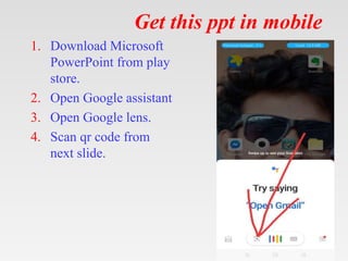
Tips on using my ppt.
1. You can freely download, edit, modify and put your
name etc.
2. Don’t be concerned about number of slides. Half the
slides are blanks except for the title.
3. First show the blank slides (eg. Aetiology ) > Ask
students what they already know about ethology of
today's topic. > Then show next slide which enumerates
aetiologies.
4. At the end rerun the show – show blank> ask questions >
show next slide.
5. This will be an ACTIVE LEARNING SESSION x
three revisions.
6. Good for self study also.
7. See notes for bibliography.
Lymphangioma & Cystic
hygroma
Lymphangioma & Cystic
hygroma
• Congenital malformation
Epidemiology
Epidemiology
• The incidence of CH is estimated to be 1
case per 6,000-16,000 live births.
• Mortality  as high as 2-6% in some series
 secondary to pneumonia, bronchiectasis,
and airway compromise  in the larger-
sized lesions.
• morbidity depends on the anatomic location
of the CH  related to cosmetic
disfigurement and impingement on other
critical structures such as nerves, vessels,
Cont’d
• no racial predominance
• The sex distribution is equal.
• 50-65% are evident at birth, with 80-90% of CHs
presenting by age 2 years.
• CH can be visualized using abdominal
ultrasonography by 10 weeks gestation
• Fast-spin MRI can also be used to determine the
extent of fetal CH.
• Elevated alpha fetoprotein levels in amniocentesis
fluid has been reported in pregnancies with CH.
Pathophysiology
Pathophysiology
• Lymphangiomas are thought to arise from a combination of the
following:
– a failure of lymphatics to connect to the venous system,
– abnormal budding of lymphatic tissue,
– sequestered lymphatic rests that retain their embryonic growth
potential.
• These lymphatic rests can penetrate adjacent structures or
dissect along fascial planes and eventually become canalized.
• These spaces retain their secretions and develop cystic
components because of the lack of a venous outflow tract.
• The nature of the surrounding tissue determines whether the
lymphangioma is capillary, cavernous, or cystic.
Presentation
Presentation
• History
– signs and symptoms vary depending on the lesion's location.
– The microcystic form tends to predominate over CH in the oral
cavity and oropharynx.
– Microcystic lymphangiomas commonly appear as clusters of
clear, black, or red vesicles on the buccal mucosa or tongue.
– CHs tend to predominate below the mylohyoid muscle and can
involve both the anterior and posterior triangles of the neck.
– typically large and thick walled cysts and have little involvement
of surrounding tissue.
Cont’d
• History:
– The overlying skin can take on a bluish hue or may appear
normal.
– CHs often present after a sudden increase in size
secondary to infection or intralesional bleeding.
– Spontaneous decompression or shrinkage is uncommon
– Rarely, children with CH display symptoms of newly
onset obstructive sleep apnea syndrome (OSAS).
– This situation may involve children with CH or other
space-occupying lesions of the supraglottis or paraglottic
region.
Cont’d
• History
– Suprahyoid lymphangiomas tend to cause more
breathing difficulties than infrahyoid lesions.
– Potentially life-threatening airway compromise that
manifests as noisy breathing (stridor) and cyanosis is a
possible symptom of lymphangiomas.
– Feeding difficulties, as well as failure to thrive, 
potential lymphangioma.
– This is especially true when the lesion affects structures
of the upper aerodigestive tract
Cont’d
• Physical findings may include the following:
– CHs are typically soft, painless, compressible
(doughy) masses.
– A CH typically transilluminates.
– closely evaluate for tracheal deviation or other
evidence of impending airway obstruction.
– Closely inspect the tongue, oral cavity, hypopharynx,
and larynx because any involvement may lead to
airway obstruction.
Cont’d
• Imaging Studies
– MRI, CT scanning, and ultrasonography are all helpful in delineating the
nature of a cystic neck mass.
– CT scanning and MRI reveal ring-like margin enhancement with sharp
demarcation of cystic areas.
– The cystic areas tend to appear circumscribed and discrete.
– A poorly defined isodense mass that obscures muscle and fatty planes is
more consistent with a microcystic lymphatic malformation than a CH.
Cont’d
• Imaging Study
– MRI:
• MRI is the consensus study of choice. It provides the best soft tissue detail and can delineate the
relationship of the lesion to underlying structures.
• Contrast can be used to differentiate hemangiomas from lymphangiomas. On MRI, CHs appear
hyperintense on T2-weighted images and hypointense on T1-weighted images.
• CT scanning:
– CT scanning is faster and may be more readily available than MRI.
– CT scanning carries the risk of radiation exposure, and detail is lost if the CH is surrounded by
tissue of similar attenuation.
– Contrast helps to enhance cyst wall visualization and the relationship to surrounding blood
vessels. On CT scans, CHs appear isodense to cerebrospinal fluid (CSF).
Cont’d
• Imaging Study
– Ultrasonography: This is the least invasive study.
– It is very useful in demonstrating the relationship of CH
to the surrounding structures.
– Ultrasonography has limited ability in assessing
mediastinal and retropharyngeal structures.
– It can be used to detect CH in utero.
– Echographic visualization of multiple septae in fetal
CH has been postulated to be a poor prognostic
indicator.
Cont’d
• Imaging Study
– Plain radiography:
• With any large mass of the head and neck, airway
radiography can be helpful in delineating possible
airway compromise.
• Plain radiography is a reasonable initial imaging
modality in the evaluation of a neck mass with a
potential airway manifestation.
• Central in a discussion of CH is the
understanding that it is synonymous with
macrocystic lymphatic malformation and
cystic lymphangioma.
• proposed categorization of lymphangiomas
based on the size of the cystic component,
as follows:
– Macrocystic - Cystic spaces at least 2 cm
– Microcystic - Spaces less than 2 cm
– Mixed lesions
Treatment
Treatment
• Medical Care
– Although some authors have reported watchful waiting of
cystic hygroma (CH), it should be considered only in
patients who are asymptomatic.
– The medical treatment of CH consists of the administration
of sclerosing agents.
– Sclerosing agents include:
• OK-432 (an inactive strain of group A Streptococcus pyogenes),
• bleomycin,
• pure ethanol,
• sodium tetradecyl sulfate,
• doxycycline.
• Medical Treatment
– An infected CH should be treated with intravenous
antibiotics, and definitive surgery should be performed once
the infection has resolved.
– Incision and drainage or aspiration results in only temporary
shrinkage, and subsequent fibrosis can further complicate the
resection.
– Radiotherapy has not been demonstrated to be effective.
– The preferred treatment of all CH is surgical resection. Only
resection can truly offer the potential for cure.
• Surgical Care
– The mainstay of treatment is surgical excision
– both the operating team and the family of the patient should go forward with
the knowledge that CH is a benign lesion.
– If acute infection occurs prior to resection, surgery should be delayed at least
3 months.
– The surgical team should attempt to completely remove the lymphangioma
or to remove as much as possible, sparing all vital neurovascular structures.
– Complete excision has been estimated to be possible in roughly 40% of
cases.
– CHs are ideally removed in one procedure because secondary excisions are
complicated by fibrosis and distorted anatomical landmarks.
• Microcystic lesions are much more difficult to remove
because of their intimate association with nearby tissues.
• Laser therapy is a recent advancement in the treatment of
microcystic lesions.
• The exceptions to excision at the time of diagnosis are:
– premature infants
– small in size
– involvement of crucial neurovascular structures that are small
and difficult to identify (eg, facial nerve).
• If no airway obstruction is present, surgery
can be delayed until the child is aged 2
years or older, especially when operating
around the facial nerve in the parotid area.
• Signs of airway obstruction require surgical
evaluation at the time of diagnosis.
• In emergency situations, aspiration with an
18-gauge or 20-gauge needle may obviate
the need for an emergency tracheostomy.
• Although traditional wisdom has dictated not aspirating lymphatic
malformations, a study by Burezq et al documented success with serial
aspiration of CH.
• In their series, 14 patients were treated with aspiration alone (3 needed
multiple aspirations), with a mean follow-up of 5.75 years.
• No failures were reported. This technique may hold promise for the
future management of CH.
• Other authors contend aspiration has no role and believe that aspiration
is often followed by recurrence, hemorrhage, or infection.
• Radiofrequency ablation has been advocated for use with intraoral
lymphatic malformations, especially microcystic lesions.
• Magnetic resonance–controlled laser-induced
interstitial thermotherapy is a novel therapy that has
been proposed for treatment of lymphangiomas.
• CH can present on routine prenatal ultrasonography
as a large obstructing airway mass, as can other
pathologic conditions such as a teratoma or
rhabdomyosarcoma.
• If such a mass is visible on ultrasonography, MRI
should be performed to further delineate the mass.
Cont’d
• In these cases, a multispecialty team
including a high-risk obstetrician, pediatric
otolaryngologist, pediatric surgeon, and
neonatologist should be present at the ex
utero intrapartum treatment (EXIT)
procedure.
• A planned cesarean delivery is performed,
and intubation or tracheostomy is used to
establish an airway.
Complications
Complications
• Complications include airway obstruction, hemorrhage, infection, and
deformation of surrounding bony structures or teeth if left untreated.
• Complications from the surgical excision of a cystic hygroma (CH) are
myriad and are related to the location and structures adjacent to the mass:
– damage to a neurovascular structure (including cranial nerves),
– chylous fistula,
– chylothorax,
– hemorrhage,
– recurrence.
• Most recurrences occur within the first year but have been reported to
occur as long as 10 years after excision.
Prognosis
Prognosis
• Unlike in hemangiomas, spontaneous
resolution of CH is uncommon.
• Recurrence is rare when all gross disease is
removed.
• If residual tissue is left behind, the expected
recurrence rate is approximately 15%.
• In prenatal CH, diagnosis after 30 weeks'
gestation is considered a positive
prognosticator.
Get this ppt in mobile
1. Download Microsoft
PowerPoint from play
store.
2. Open Google assistant
3. Open Google lens.
4. Scan qr code from
next slide.
Get this ppt in mobile
Get my ppt collection
• https://www.slideshare.net/drpradeeppande/
edit_my_uploads
• https://www.dropbox.com/sh/x600md3cvj8
5woy/AACVMHuQtvHvl_K8ehc3ltkEa?dl
=0
• https://www.facebook.com/doctorpradeeppa
nde/?ref=pages_you_manage

Cystic hygroma.pptx

  • 1.
    Tips on usingmy ppt. 1. You can freely download, edit, modify and put your name etc. 2. Don’t be concerned about number of slides. Half the slides are blanks except for the title. 3. First show the blank slides (eg. Aetiology ) > Ask students what they already know about ethology of today's topic. > Then show next slide which enumerates aetiologies. 4. At the end rerun the show – show blank> ask questions > show next slide. 5. This will be an ACTIVE LEARNING SESSION x three revisions. 6. Good for self study also. 7. See notes for bibliography.
  • 2.
  • 3.
    Lymphangioma & Cystic hygroma •Congenital malformation
  • 4.
  • 5.
    Epidemiology • The incidenceof CH is estimated to be 1 case per 6,000-16,000 live births. • Mortality  as high as 2-6% in some series  secondary to pneumonia, bronchiectasis, and airway compromise  in the larger- sized lesions. • morbidity depends on the anatomic location of the CH  related to cosmetic disfigurement and impingement on other critical structures such as nerves, vessels,
  • 6.
    Cont’d • no racialpredominance • The sex distribution is equal. • 50-65% are evident at birth, with 80-90% of CHs presenting by age 2 years. • CH can be visualized using abdominal ultrasonography by 10 weeks gestation • Fast-spin MRI can also be used to determine the extent of fetal CH. • Elevated alpha fetoprotein levels in amniocentesis fluid has been reported in pregnancies with CH.
  • 8.
  • 9.
    Pathophysiology • Lymphangiomas arethought to arise from a combination of the following: – a failure of lymphatics to connect to the venous system, – abnormal budding of lymphatic tissue, – sequestered lymphatic rests that retain their embryonic growth potential. • These lymphatic rests can penetrate adjacent structures or dissect along fascial planes and eventually become canalized. • These spaces retain their secretions and develop cystic components because of the lack of a venous outflow tract. • The nature of the surrounding tissue determines whether the lymphangioma is capillary, cavernous, or cystic.
  • 10.
  • 11.
    Presentation • History – signsand symptoms vary depending on the lesion's location. – The microcystic form tends to predominate over CH in the oral cavity and oropharynx. – Microcystic lymphangiomas commonly appear as clusters of clear, black, or red vesicles on the buccal mucosa or tongue. – CHs tend to predominate below the mylohyoid muscle and can involve both the anterior and posterior triangles of the neck. – typically large and thick walled cysts and have little involvement of surrounding tissue.
  • 12.
    Cont’d • History: – Theoverlying skin can take on a bluish hue or may appear normal. – CHs often present after a sudden increase in size secondary to infection or intralesional bleeding. – Spontaneous decompression or shrinkage is uncommon – Rarely, children with CH display symptoms of newly onset obstructive sleep apnea syndrome (OSAS). – This situation may involve children with CH or other space-occupying lesions of the supraglottis or paraglottic region.
  • 13.
    Cont’d • History – Suprahyoidlymphangiomas tend to cause more breathing difficulties than infrahyoid lesions. – Potentially life-threatening airway compromise that manifests as noisy breathing (stridor) and cyanosis is a possible symptom of lymphangiomas. – Feeding difficulties, as well as failure to thrive,  potential lymphangioma. – This is especially true when the lesion affects structures of the upper aerodigestive tract
  • 14.
    Cont’d • Physical findingsmay include the following: – CHs are typically soft, painless, compressible (doughy) masses. – A CH typically transilluminates. – closely evaluate for tracheal deviation or other evidence of impending airway obstruction. – Closely inspect the tongue, oral cavity, hypopharynx, and larynx because any involvement may lead to airway obstruction.
  • 16.
    Cont’d • Imaging Studies –MRI, CT scanning, and ultrasonography are all helpful in delineating the nature of a cystic neck mass. – CT scanning and MRI reveal ring-like margin enhancement with sharp demarcation of cystic areas. – The cystic areas tend to appear circumscribed and discrete. – A poorly defined isodense mass that obscures muscle and fatty planes is more consistent with a microcystic lymphatic malformation than a CH.
  • 17.
    Cont’d • Imaging Study –MRI: • MRI is the consensus study of choice. It provides the best soft tissue detail and can delineate the relationship of the lesion to underlying structures. • Contrast can be used to differentiate hemangiomas from lymphangiomas. On MRI, CHs appear hyperintense on T2-weighted images and hypointense on T1-weighted images. • CT scanning: – CT scanning is faster and may be more readily available than MRI. – CT scanning carries the risk of radiation exposure, and detail is lost if the CH is surrounded by tissue of similar attenuation. – Contrast helps to enhance cyst wall visualization and the relationship to surrounding blood vessels. On CT scans, CHs appear isodense to cerebrospinal fluid (CSF).
  • 18.
    Cont’d • Imaging Study –Ultrasonography: This is the least invasive study. – It is very useful in demonstrating the relationship of CH to the surrounding structures. – Ultrasonography has limited ability in assessing mediastinal and retropharyngeal structures. – It can be used to detect CH in utero. – Echographic visualization of multiple septae in fetal CH has been postulated to be a poor prognostic indicator.
  • 19.
    Cont’d • Imaging Study –Plain radiography: • With any large mass of the head and neck, airway radiography can be helpful in delineating possible airway compromise. • Plain radiography is a reasonable initial imaging modality in the evaluation of a neck mass with a potential airway manifestation.
  • 20.
    • Central ina discussion of CH is the understanding that it is synonymous with macrocystic lymphatic malformation and cystic lymphangioma. • proposed categorization of lymphangiomas based on the size of the cystic component, as follows: – Macrocystic - Cystic spaces at least 2 cm – Microcystic - Spaces less than 2 cm – Mixed lesions
  • 21.
  • 22.
    Treatment • Medical Care –Although some authors have reported watchful waiting of cystic hygroma (CH), it should be considered only in patients who are asymptomatic. – The medical treatment of CH consists of the administration of sclerosing agents. – Sclerosing agents include: • OK-432 (an inactive strain of group A Streptococcus pyogenes), • bleomycin, • pure ethanol, • sodium tetradecyl sulfate, • doxycycline.
  • 23.
    • Medical Treatment –An infected CH should be treated with intravenous antibiotics, and definitive surgery should be performed once the infection has resolved. – Incision and drainage or aspiration results in only temporary shrinkage, and subsequent fibrosis can further complicate the resection. – Radiotherapy has not been demonstrated to be effective. – The preferred treatment of all CH is surgical resection. Only resection can truly offer the potential for cure.
  • 24.
    • Surgical Care –The mainstay of treatment is surgical excision – both the operating team and the family of the patient should go forward with the knowledge that CH is a benign lesion. – If acute infection occurs prior to resection, surgery should be delayed at least 3 months. – The surgical team should attempt to completely remove the lymphangioma or to remove as much as possible, sparing all vital neurovascular structures. – Complete excision has been estimated to be possible in roughly 40% of cases. – CHs are ideally removed in one procedure because secondary excisions are complicated by fibrosis and distorted anatomical landmarks.
  • 25.
    • Microcystic lesionsare much more difficult to remove because of their intimate association with nearby tissues. • Laser therapy is a recent advancement in the treatment of microcystic lesions. • The exceptions to excision at the time of diagnosis are: – premature infants – small in size – involvement of crucial neurovascular structures that are small and difficult to identify (eg, facial nerve).
  • 26.
    • If noairway obstruction is present, surgery can be delayed until the child is aged 2 years or older, especially when operating around the facial nerve in the parotid area. • Signs of airway obstruction require surgical evaluation at the time of diagnosis. • In emergency situations, aspiration with an 18-gauge or 20-gauge needle may obviate the need for an emergency tracheostomy.
  • 27.
    • Although traditionalwisdom has dictated not aspirating lymphatic malformations, a study by Burezq et al documented success with serial aspiration of CH. • In their series, 14 patients were treated with aspiration alone (3 needed multiple aspirations), with a mean follow-up of 5.75 years. • No failures were reported. This technique may hold promise for the future management of CH. • Other authors contend aspiration has no role and believe that aspiration is often followed by recurrence, hemorrhage, or infection. • Radiofrequency ablation has been advocated for use with intraoral lymphatic malformations, especially microcystic lesions.
  • 28.
    • Magnetic resonance–controlledlaser-induced interstitial thermotherapy is a novel therapy that has been proposed for treatment of lymphangiomas. • CH can present on routine prenatal ultrasonography as a large obstructing airway mass, as can other pathologic conditions such as a teratoma or rhabdomyosarcoma. • If such a mass is visible on ultrasonography, MRI should be performed to further delineate the mass.
  • 29.
    Cont’d • In thesecases, a multispecialty team including a high-risk obstetrician, pediatric otolaryngologist, pediatric surgeon, and neonatologist should be present at the ex utero intrapartum treatment (EXIT) procedure. • A planned cesarean delivery is performed, and intubation or tracheostomy is used to establish an airway.
  • 30.
  • 31.
    Complications • Complications includeairway obstruction, hemorrhage, infection, and deformation of surrounding bony structures or teeth if left untreated. • Complications from the surgical excision of a cystic hygroma (CH) are myriad and are related to the location and structures adjacent to the mass: – damage to a neurovascular structure (including cranial nerves), – chylous fistula, – chylothorax, – hemorrhage, – recurrence. • Most recurrences occur within the first year but have been reported to occur as long as 10 years after excision.
  • 32.
  • 33.
    Prognosis • Unlike inhemangiomas, spontaneous resolution of CH is uncommon. • Recurrence is rare when all gross disease is removed. • If residual tissue is left behind, the expected recurrence rate is approximately 15%. • In prenatal CH, diagnosis after 30 weeks' gestation is considered a positive prognosticator.
  • 34.
    Get this pptin mobile 1. Download Microsoft PowerPoint from play store. 2. Open Google assistant 3. Open Google lens. 4. Scan qr code from next slide.
  • 35.
    Get this pptin mobile
  • 36.
    Get my pptcollection • https://www.slideshare.net/drpradeeppande/ edit_my_uploads • https://www.dropbox.com/sh/x600md3cvj8 5woy/AACVMHuQtvHvl_K8ehc3ltkEa?dl =0 • https://www.facebook.com/doctorpradeeppa nde/?ref=pages_you_manage

Editor's Notes

  • #2 drpradeeppande@gmail.com 7697305442