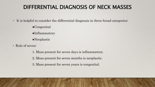
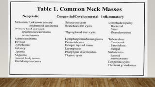
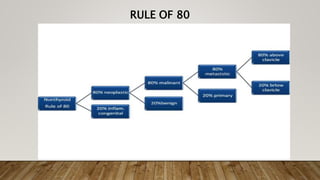
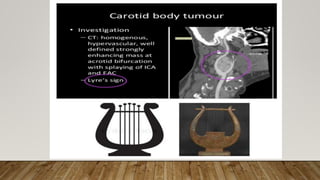
UNIVERSITY OF RWANDA
CHUK/ENT Department
Topic: Neck Masses
Prepared and presented by IRAKIZA Jacques Desire
during ENT Clinical Rotation
Aug 13, 2019
INTRODUCTION
• A neck mass is any abnormal enlargement, swelling, or growth from the level of the
base of skull to the clavicles
• The location of the mass can focus the differential
MAJOR STRUCTURES
• The major structures are located largely in the anterior triangle.
• The borders of the anterior triangle are the inferior border of the mandible, the
sternocleidomastoid muscle and the midline.
• The borders of the posterior triangle are the sternocleidomastoid muscle, the trapezius
muscle and the clavicle.
• The major structures that can be palpated in the midline, within the anterior triangles
and from superior to inferior, are the hyoid bone, the thyroid cartilage with its notch (the
‘Adam’s apple’), the cricothyroid membrane, the cricoid cartilage and the trachea
MAJOR STRUCTURES CONT’D
• The isthmus of the thyroid gland may be palpated over the first 2 tracheal rings and its right and left
lobes lie over the cricoid and thyroid cartilages laterally. A normal thyroid gland is not easily palpable.
• The carotid bulb can be palpated near the anterior border of the sternocleidomastoid muscle at the level
of the hyoid bone.
• The parotid gland lies over the angle of the mandible, in front of and below the ear. It extends medially
between the mastoid process and the posterior border of the mandible. Its borders are indistinct and
difficult to delineate on palpation(as thyroid gland!)
• The submandibular salivary glands are located just below the body of the mandible. Normal glands are
often palpable in thin individuals. The glands may be distinguished from submandibular lymph nodes in
that the salivary glands are palpable bimanually via the floor of the mouth and the neck.
• For more anatomy check my Tracheostomy slides…
LYMPH NODES
• The location of cervical lymph nodes can be divided
into six levels and level of the lymph nodes can be
predictive as to the source of the problem.
• Level I includes submandibular and submental
nodes.
• Levels II, III and IV encompass lymph nodes along
the internal jugular vein, deep to the
sternocleidomastoid muscle in the upper, middle and
lower thirds of the neck respectively.
• Level V contains the nodes in the posterior triangle.
These are commonly enlarged in viral infections, e.g.
mononucleosis.
• Level VI lies between the carotid sheaths in the
anterior triangle and contains the prelaryngeal and
pretracheal nodes.
CONT’D
• Note that lymphadenopathy due to inflammatory diseases usually resolves within 4
- 6 weeks.
• Therefore, any node which persists beyond 2 weeks requires further evaluation.
Other suspicious features include lymph nodes more than 1.5 cm in diameter, firm,
rubbery lymph nodes, matted lymph nodes and nodes that are fixed or have
decreased mobility.
HISTORY
• A careful history can provide important clues to the diagnosis of a neck mass. Duration of symptoms is one of the
most important points in the history.
• Inflammatory neck masses are usually acute in onset and resolve within several weeks. Cervical lymphadenitis,
the most common cause of neck masses, is often associated with upper respiratory tract infections.
• Congenital neck masses are often present for an extended duration sometimes, but not always, since birth. For
example branchial cysts usually present in young adults in their twenties. Furthermore, rapid enlargement of a
small congenital mass may occur following an upper respiratory tract infection.
• Malignant neck masses, as in metastatic carcinoma to cervical lymph nodes, tend to have a history of progressive
enlargement. The most common origin of these metastases is squamous cell carcinoma of the upper aerodigestive
tract.
• More than 80% of these tumours are associated with tobacco and alcohol use in persons over 40 years of age.
Further features of malignancy include voice change, odynophagia, dysphagia, haemoptysis and previous radiation,
especially with thyroid tumours.
• Additional important features are: oral lesions, referred ear pain, muffled or decreased hearing and constitutional
symptoms (e.g. night sweats, anorexia, weight loss), unilateral nasal discharge or epistaxis, family history of cancer
and previous tumours.
EXAMINATION
• Examination should include the mass itself, the rest of the neck, the skin of the head
and neck and the ENT system (ears, oral cavity, nasal cavity, nasopharynx,
oropharynx, hypopharynx and the larynx). In cases where pathology is suspected in
an area that is difficult to examine without specialised equipment, for example the
nasopharynx, hypopharynx and larynx, patients should be referred to an
otolaryngologist.
• The size, consistency, tenderness and mobility of the mass provide diagnostic clues.
• Acute inflammatory masses tend to be soft, tender and mobile. Chronic
inflammatory masses are often non-tender and rubbery and either mobile or matted.
Congenital masses are usually soft, mobile and non-tender unless infected. Vascular
masses may be pulsatile or have a bruit. Malignant masses may be hard, nontender
and fixed.
P.E. CONT’D
• The scalp and skin of the head and neck should be examined for primary cutaneous
tumours. The ear may reveal serous otitis media associated with a nasopharyngeal
carcinoma or a fistula in the external auditory canal associated with some branchial
cleft abnormalities. Cranial nerve examination is also necessary.
• Nasal examination may reveal a unilateral nasal mass or discharge suspicious of a
neoplasm. The mucosa of the oral cavity/oropharynx may reveal a primary
malignancy
• A unilateral, asymmetrically enlarged tonsil may suggest a neoplasm
• Assessment of the mass with swallowing is important as movement from swallowing
suggests a lesion in the thyroid gland or a thyroglossal cyst
DIFFERENTIAL DIAGNOSIS OF NECK MASSES
• It is helpful to consider the differential diagnosis in three broad categories:
●Congenital
●Inflammatory
●Neoplastic
• Rule of seven:
1. Mass present for seven days is inflammatory.
2. Mass present for seven months is neoplastic.
3. Mass present for seven years is congenital.
CONGENITAL NECK MASSES
• Usually present at birth, but may present at any age.
• Most common non-inflammatory neck mass in children.
• Malignancy in adult until proven otherwise
• Cystic lesions such as branchial cleft cysts can present in adulthood,
and should be investigated to ensure malignancy is not present.
• Carcinomas of the tonsil, tongue base, and thyroid may all present as
cystic neck masses.
THYROGLOSSAL DISC CYSTS (TGDC)
• Midline anterior neck.
• Rounded with a diameter of 2-4cm
• Diagnosed in childhood, but up to 40 percent may present
after age 20
• Asymptomatic except when infected by URTI
• Move with tongue protrusion
• Rarely carcinoma develop in cyst.
MANAGEMENT OF TGDC
• Excision to confirm the diagnosis and to prevent future infections.
• The Sistrunk operation is the procedure of choice.
• Thyroid carcinoma can be present in (1 to 2%) of thyroglossal duct
cysts,
• All thyroglossal duct cysts and tracts should undergo a careful
histologic examination
CYSTIC HYGROMAS (LYMPHANGIOMAS)
• Cystic hygromas are present as soft, fluctuant and transilluminable
masses just under the skin.
• Nearly all present by the age of 2 to 3 years, with 60% occurring in the
head and neck region (usually in the posterior triangle) and most
presenting at birth.
• They are multiloculated and painless.
• Ultrasound is useful to confirm the diagnosis and CT scanning is
essential if surgery is contemplated.
ECTOPIC THYROID
• 90% are lingual
• Symptoms are of base of tongue obstruction,
dysphagia
• Surgical Excision
PLUNGING RANULA
• A ranula is a mucocele or retention cyst arising from an
obstruction in the sublingual glands in the floor of mouth.
• Simple ranula- unilateral oral cavity cystic lesion
• Painless and slow-growing.
• They are most often located in the submentum.
• When they extend through the mylohyoid muscle into the
neck they are referred to as "plunging ranula".
MANAGEMENT
• Plunging ranula- pierce the mylohyoid to present as a
paramedian or lateral neck mass.
• CT scan/MRI
• Treatment is intraoral excision to include the sublingual
gland of origin
BRANCHIAL CLEFT CYST
• Almost 20 percent of pediatric neck masses.
• Present in late childhood or early adulthood when a previously unrecognized cyst
becomes infected.
• Only a very small percentage first present in adulthood.
• Relatively consistent in their location in the neck, anterior to the SCM.
• Painless swelling
• Young adults
• M= F ratio
• Unilateral, 75% on left side
• The 2nd is the most Common (90%) branchial anomaly and the 3rd is rare!
MANAGEMENT
• Management of branchial cleft cysts begins with controlling
infection, if present.
• Once the infection has resolved, the mass is usually excised
to prevent future problems
THYMIC CYST
• Thymus develop from 3rd pharyngeal pouch and descend to neck to
the mediastinum.
• Thymic remnants may persist anywhere in its path from angle
mandible to midline of neck.
• Swelling either cystic or solid.
• Can occur in children or adults by presents of anterior neck mass and
deep to middle SCM.
• Rare condition
• Tx: Surgical excision + sternotomy if extend into mediastinum.
SUBLINGUAL DERMOID CYST
• Midline submental swelling but does NOT move on
protrusion of tongue.
• Can be arises from floor of mouth and need to be
diffrentiated with ranula.
• Tx: Surgical excision
TERATOID CYSTS AND TERATOMAS
• All three germ cell layers- Endoderm, mesoderm and ectoderm.
Larger midline masses, present earlier in life.
• 20% associated maternal polyhydramnios
• Unlike adult teratomas, they rarely demonstrate malignant
degeneration.
• Surgical excision.
INFECTIVE AND INFLAMMATORY MASSES
• The most common cause of a neck lump is
inflammatory/infective reactive lymphadenopathy,
• They’re commonly caused by a self-limiting bacterial,
parasitic or viral infection that resolves within weeks
• There can be non-infectious inflammatory disorders (eg:
sarcoidosis)
LYMPHADENITIS ETIOLOGIES
• Bacterial: streptococcal and staph infections (group A beta-haemolytic streptococcus
is the most common cause); mycobacterial infections (TB and atypical mycobacteria);
lymphadenitis secondary to dental infection and tonsillitis; unusual disorders (cat-
scratch disease, actinomyces, tularaemia)
• Viral: Epstein-Barr virus (EBV), cytomegalovirus (CMV), herpes simplex virus
(HSV), other viruses causing URTIs, HIV
• Parasitic: toxoplasmosis
• Fungal: coccidiomycosis
MYCOBACTERIAL LYMPHADENITIS
• Mycobacterial lymphadenitis should be suspected in an
acute lymphadenitis, with only mild tenderness and a
partial response to antibiotics.
• Other rare granulomatous causes of adenopathy include cat
scratch disease and actinomycosis.
HIV INFECTION
• Cervical lymphadenopathy is very common in patients with HIV
infection.
• Lymphadenopathy syndrome is a mild form of HIV disease that
represents one of the initial stages of the infection.
• Patients can remain stable for months to years, with little in the way
of symptoms.
• This diagnosis should be considered in any adult with persistent
generalised lymphadenopathy and the relevant risk factors.
ACUTE SIALADENITIS
• Acute infection of the salivary glands can be bacterial or viral in
origin.
• Bacterial sialadenitis occurs more frequently in the parotid glands, is
more common in the elderly and is associated with reduced salivary
flow from dehydration.
• Treatment is with broad-spectrum antibiotics covering S. aureus, the
most common pathogen causing infection of the salivary glands, in
addition to supportive measures (rehydration, analgesics and gland
massage to encourage salivary flow).
• Appropriate antibiotics include flucloxacillin, cephalexin and
clindamycin.
• Surgical drainage may be required if an abscess develops.
VIRAL SIALADENITIS
• Most commonly due to the mumps virus, which typically
affects the parotid glands bilaterally.
• The mumps virus most often affects children, with peak
incidence at ages 4 to 6 years.
• Other causes include coxsackie virus, cytomegalovirus and
HIV
THYROIDITIS
• The most common inflammatory goitre is Hashimoto’s thyroiditis.
• Autoantibodies against thyroid peroxidase are produced, resulting in
lymphocytic infiltration of the thyroid and eventually a goiter, which
is typically firm and rubbery.
• Management by an endocrinologist is usually necessary because of the
initial hyperthyroidism and subsequent hypothyroidism.
• Occasionally surgery is required for obstructive symptoms.
NEOPLASTIC MASSES
• Benign ones include lipoma (most common benign tomor of soft tissue in
neck), fibroma, neuroma and schwannoma
• Malignant neoplastic masses includes:
• primary neck tumours — sarcoma, salivary gland tumours, thyroid
gland tumours, parathyroid gland tumours
• lymphoma
• metastases from supraclavicular primary tumours, e.g. upper
aerodigestive tract squamous cell carcinoma (SCC), skin SCC, melanoma,
thyroid or salivary gland metastases
• metastases from infraclavicular primary tumour — lung,
oesophagus, stomach.
RULE OF 80
SQUAMOUS CELL CARCINOMA
• SCC is the most common cause of a malignant neck lump.
• Metastatic SCC most commonly arises from the mucosa of the upper
aerodigestive tract (oral cavity, nasopharynx, oropharynx and
laryngopharynx).
• Cutaneous malignancies (SCC and melanoma) may also metastasise
to the parotid gland or lateral cervical lymph nodes, sometimes years
after the primary tumour was excised.
LYMPHOMA
• The nodes are typically ‘rubbery’ in consistency. Associated symptoms
include night sweats, lethargy and weight loss.
• Lymphoma is the most common cause of a malignant neck lump in
children and should therefore, despite being rare, be considered in the
differential diagnosis of any progressive or persistent childhood
lymphadenopathy.
• FNAB and CT scan are indicated, with referral to a haematologist if
cytology is suggestive of lymphoma
ADENOCARCINOMA
• Adenocarcinoma is a type of cancer that forms in mucus secreting
glands throughout the body.
• Metastatic adenocarcinoma to the upper cervical lymph nodes may
originate from the salivary glands or sinonasal cavity.
• Metastatic adenocarcinoma in the lower neck may arise from a site
below the clavicles (e.g. lung, oesophagus or stomach).
• Virchow’s node (also referred to as Troisier’s sign) refers to metastatic
adenocarcinoma occurring in the left supraclavicular fossa and
usually arising from the stomach
THYROID CANCER
• Most thyroid cancers present clinically with a palpable thyroid nodule,
which is often asymptomatic. About half of thyroid cancers are initially
noticed by the patient, whereas the remainder are detected during routine
physical examination, by chance on imaging studies often for unrelated
medical conditions or during surgery for benign thyroid disease.
• Occasionally thyroid cancer can present with a metastatic neck node, and
the diagnosis is confirmed on FNAB.
• Management:
• When thyroid cancer is suspected or demonstrated on FNAB, prompt referral to a ENT
surgeon is warranted.
• Total thyroidectomy and adjuvant iodine ablation therapy is indicated for most patients
diagnosed with thyroid cancer.
SALIVARY GLAND MALIGNANCY
• Salivary gland cancers include adenocarcinoma and metastatic
cutaneous SCC.
• Symptoms and signs that suggest malignancy include pain, rapid
growth, a hard mass, fixity to the skin or mandible and facial nerve
palsy.
• FNAB and CT/MRI scanning are essential to assess the extent of
disease and to plan surgery.
• High-grade salivary gland malignancy often requires neck dissection
and postoperative radiotherapy.
ANOTHER RULE OF 80…
CAROTID BODY TUMOURS / PARAGANGLIOMAS
• Carotid body tumors are rare benign tumors of the carotid body neural
plexus.
• They usually present as a painless pulsatile mass at the level of the
carotid bifurcation, and typically can be moved side to side but not
vertically.
• The tumours are extremely vascular, and are diagnosed using a
combination of CT scan, MRI, magnetic resonance angiography and
carotid doppler scanning. (Findings on the next slide)
• Following comprehensive assessment, surgery is usually performed by
a head and neck surgeon and a vascular surgeon.
OTHER DIFFERENTIAL DIAGNOSISES
• Vascular masses include paragangliomas and vascular malformations, such as
haemangioma, AV malformation, aneurysm.
• Traumatic masses: haematoma, false aneurysm, AV fistula.
• Metabolic, idiopathic and auto-immune conditions are rare, e.g. inflammatory
pseudotumours.
• Thyroid gland masses include multinodular goitre, colloid goitre, thyroiditis, etc.
• Salivary gland masses e.g. prominence with ageing, sialadenitis, sialolithiasis
(calculus obstructing the duct can result in a tender, inflamed, swollen gland. This
is most common in the submandibular gland), salivary cysts (HIV), Sjögren’s
syndrome and acute parotitis due to mumps.
• Parapharyngeal masses should be considered, especially with a high neck mass and
a medially displaced tonsil.
ANY QUESTION OR ADDITION IS WELCOMED !!

Neck masses

  • 1.
    UNIVERSITY OF RWANDA CHUK/ENTDepartment Topic: Neck Masses Prepared and presented by IRAKIZA Jacques Desire during ENT Clinical Rotation Aug 13, 2019
  • 2.
    INTRODUCTION • A neckmass is any abnormal enlargement, swelling, or growth from the level of the base of skull to the clavicles • The location of the mass can focus the differential
  • 3.
    MAJOR STRUCTURES • Themajor structures are located largely in the anterior triangle. • The borders of the anterior triangle are the inferior border of the mandible, the sternocleidomastoid muscle and the midline. • The borders of the posterior triangle are the sternocleidomastoid muscle, the trapezius muscle and the clavicle. • The major structures that can be palpated in the midline, within the anterior triangles and from superior to inferior, are the hyoid bone, the thyroid cartilage with its notch (the ‘Adam’s apple’), the cricothyroid membrane, the cricoid cartilage and the trachea
  • 4.
    MAJOR STRUCTURES CONT’D •The isthmus of the thyroid gland may be palpated over the first 2 tracheal rings and its right and left lobes lie over the cricoid and thyroid cartilages laterally. A normal thyroid gland is not easily palpable. • The carotid bulb can be palpated near the anterior border of the sternocleidomastoid muscle at the level of the hyoid bone. • The parotid gland lies over the angle of the mandible, in front of and below the ear. It extends medially between the mastoid process and the posterior border of the mandible. Its borders are indistinct and difficult to delineate on palpation(as thyroid gland!) • The submandibular salivary glands are located just below the body of the mandible. Normal glands are often palpable in thin individuals. The glands may be distinguished from submandibular lymph nodes in that the salivary glands are palpable bimanually via the floor of the mouth and the neck.
  • 5.
    • For moreanatomy check my Tracheostomy slides…
  • 6.
    LYMPH NODES • Thelocation of cervical lymph nodes can be divided into six levels and level of the lymph nodes can be predictive as to the source of the problem. • Level I includes submandibular and submental nodes. • Levels II, III and IV encompass lymph nodes along the internal jugular vein, deep to the sternocleidomastoid muscle in the upper, middle and lower thirds of the neck respectively. • Level V contains the nodes in the posterior triangle. These are commonly enlarged in viral infections, e.g. mononucleosis. • Level VI lies between the carotid sheaths in the anterior triangle and contains the prelaryngeal and pretracheal nodes.
  • 7.
    CONT’D • Note thatlymphadenopathy due to inflammatory diseases usually resolves within 4 - 6 weeks. • Therefore, any node which persists beyond 2 weeks requires further evaluation. Other suspicious features include lymph nodes more than 1.5 cm in diameter, firm, rubbery lymph nodes, matted lymph nodes and nodes that are fixed or have decreased mobility.
  • 8.
    HISTORY • A carefulhistory can provide important clues to the diagnosis of a neck mass. Duration of symptoms is one of the most important points in the history. • Inflammatory neck masses are usually acute in onset and resolve within several weeks. Cervical lymphadenitis, the most common cause of neck masses, is often associated with upper respiratory tract infections. • Congenital neck masses are often present for an extended duration sometimes, but not always, since birth. For example branchial cysts usually present in young adults in their twenties. Furthermore, rapid enlargement of a small congenital mass may occur following an upper respiratory tract infection. • Malignant neck masses, as in metastatic carcinoma to cervical lymph nodes, tend to have a history of progressive enlargement. The most common origin of these metastases is squamous cell carcinoma of the upper aerodigestive tract. • More than 80% of these tumours are associated with tobacco and alcohol use in persons over 40 years of age. Further features of malignancy include voice change, odynophagia, dysphagia, haemoptysis and previous radiation, especially with thyroid tumours. • Additional important features are: oral lesions, referred ear pain, muffled or decreased hearing and constitutional symptoms (e.g. night sweats, anorexia, weight loss), unilateral nasal discharge or epistaxis, family history of cancer and previous tumours.
  • 9.
    EXAMINATION • Examination shouldinclude the mass itself, the rest of the neck, the skin of the head and neck and the ENT system (ears, oral cavity, nasal cavity, nasopharynx, oropharynx, hypopharynx and the larynx). In cases where pathology is suspected in an area that is difficult to examine without specialised equipment, for example the nasopharynx, hypopharynx and larynx, patients should be referred to an otolaryngologist. • The size, consistency, tenderness and mobility of the mass provide diagnostic clues. • Acute inflammatory masses tend to be soft, tender and mobile. Chronic inflammatory masses are often non-tender and rubbery and either mobile or matted. Congenital masses are usually soft, mobile and non-tender unless infected. Vascular masses may be pulsatile or have a bruit. Malignant masses may be hard, nontender and fixed.
  • 11.
    P.E. CONT’D • Thescalp and skin of the head and neck should be examined for primary cutaneous tumours. The ear may reveal serous otitis media associated with a nasopharyngeal carcinoma or a fistula in the external auditory canal associated with some branchial cleft abnormalities. Cranial nerve examination is also necessary. • Nasal examination may reveal a unilateral nasal mass or discharge suspicious of a neoplasm. The mucosa of the oral cavity/oropharynx may reveal a primary malignancy • A unilateral, asymmetrically enlarged tonsil may suggest a neoplasm • Assessment of the mass with swallowing is important as movement from swallowing suggests a lesion in the thyroid gland or a thyroglossal cyst
  • 12.
    DIFFERENTIAL DIAGNOSIS OFNECK MASSES • It is helpful to consider the differential diagnosis in three broad categories: ●Congenital ●Inflammatory ●Neoplastic • Rule of seven: 1. Mass present for seven days is inflammatory. 2. Mass present for seven months is neoplastic. 3. Mass present for seven years is congenital.
  • 14.
    CONGENITAL NECK MASSES •Usually present at birth, but may present at any age. • Most common non-inflammatory neck mass in children. • Malignancy in adult until proven otherwise • Cystic lesions such as branchial cleft cysts can present in adulthood, and should be investigated to ensure malignancy is not present. • Carcinomas of the tonsil, tongue base, and thyroid may all present as cystic neck masses.
  • 15.
    THYROGLOSSAL DISC CYSTS(TGDC) • Midline anterior neck. • Rounded with a diameter of 2-4cm • Diagnosed in childhood, but up to 40 percent may present after age 20 • Asymptomatic except when infected by URTI • Move with tongue protrusion • Rarely carcinoma develop in cyst.
  • 16.
    MANAGEMENT OF TGDC •Excision to confirm the diagnosis and to prevent future infections. • The Sistrunk operation is the procedure of choice. • Thyroid carcinoma can be present in (1 to 2%) of thyroglossal duct cysts, • All thyroglossal duct cysts and tracts should undergo a careful histologic examination
  • 17.
    CYSTIC HYGROMAS (LYMPHANGIOMAS) •Cystic hygromas are present as soft, fluctuant and transilluminable masses just under the skin. • Nearly all present by the age of 2 to 3 years, with 60% occurring in the head and neck region (usually in the posterior triangle) and most presenting at birth. • They are multiloculated and painless. • Ultrasound is useful to confirm the diagnosis and CT scanning is essential if surgery is contemplated.
  • 18.
    ECTOPIC THYROID • 90%are lingual • Symptoms are of base of tongue obstruction, dysphagia • Surgical Excision
  • 19.
    PLUNGING RANULA • Aranula is a mucocele or retention cyst arising from an obstruction in the sublingual glands in the floor of mouth. • Simple ranula- unilateral oral cavity cystic lesion • Painless and slow-growing. • They are most often located in the submentum. • When they extend through the mylohyoid muscle into the neck they are referred to as "plunging ranula".
  • 20.
    MANAGEMENT • Plunging ranula-pierce the mylohyoid to present as a paramedian or lateral neck mass. • CT scan/MRI • Treatment is intraoral excision to include the sublingual gland of origin
  • 21.
    BRANCHIAL CLEFT CYST •Almost 20 percent of pediatric neck masses. • Present in late childhood or early adulthood when a previously unrecognized cyst becomes infected. • Only a very small percentage first present in adulthood. • Relatively consistent in their location in the neck, anterior to the SCM. • Painless swelling • Young adults • M= F ratio • Unilateral, 75% on left side • The 2nd is the most Common (90%) branchial anomaly and the 3rd is rare!
  • 22.
    MANAGEMENT • Management ofbranchial cleft cysts begins with controlling infection, if present. • Once the infection has resolved, the mass is usually excised to prevent future problems
  • 23.
    THYMIC CYST • Thymusdevelop from 3rd pharyngeal pouch and descend to neck to the mediastinum. • Thymic remnants may persist anywhere in its path from angle mandible to midline of neck. • Swelling either cystic or solid. • Can occur in children or adults by presents of anterior neck mass and deep to middle SCM. • Rare condition • Tx: Surgical excision + sternotomy if extend into mediastinum.
  • 24.
    SUBLINGUAL DERMOID CYST •Midline submental swelling but does NOT move on protrusion of tongue. • Can be arises from floor of mouth and need to be diffrentiated with ranula. • Tx: Surgical excision
  • 25.
    TERATOID CYSTS ANDTERATOMAS • All three germ cell layers- Endoderm, mesoderm and ectoderm. Larger midline masses, present earlier in life. • 20% associated maternal polyhydramnios • Unlike adult teratomas, they rarely demonstrate malignant degeneration. • Surgical excision.
  • 26.
    INFECTIVE AND INFLAMMATORYMASSES • The most common cause of a neck lump is inflammatory/infective reactive lymphadenopathy, • They’re commonly caused by a self-limiting bacterial, parasitic or viral infection that resolves within weeks • There can be non-infectious inflammatory disorders (eg: sarcoidosis)
  • 27.
    LYMPHADENITIS ETIOLOGIES • Bacterial:streptococcal and staph infections (group A beta-haemolytic streptococcus is the most common cause); mycobacterial infections (TB and atypical mycobacteria); lymphadenitis secondary to dental infection and tonsillitis; unusual disorders (cat- scratch disease, actinomyces, tularaemia) • Viral: Epstein-Barr virus (EBV), cytomegalovirus (CMV), herpes simplex virus (HSV), other viruses causing URTIs, HIV • Parasitic: toxoplasmosis • Fungal: coccidiomycosis
  • 28.
    MYCOBACTERIAL LYMPHADENITIS • Mycobacteriallymphadenitis should be suspected in an acute lymphadenitis, with only mild tenderness and a partial response to antibiotics. • Other rare granulomatous causes of adenopathy include cat scratch disease and actinomycosis.
  • 29.
    HIV INFECTION • Cervicallymphadenopathy is very common in patients with HIV infection. • Lymphadenopathy syndrome is a mild form of HIV disease that represents one of the initial stages of the infection. • Patients can remain stable for months to years, with little in the way of symptoms. • This diagnosis should be considered in any adult with persistent generalised lymphadenopathy and the relevant risk factors.
  • 30.
    ACUTE SIALADENITIS • Acuteinfection of the salivary glands can be bacterial or viral in origin. • Bacterial sialadenitis occurs more frequently in the parotid glands, is more common in the elderly and is associated with reduced salivary flow from dehydration. • Treatment is with broad-spectrum antibiotics covering S. aureus, the most common pathogen causing infection of the salivary glands, in addition to supportive measures (rehydration, analgesics and gland massage to encourage salivary flow). • Appropriate antibiotics include flucloxacillin, cephalexin and clindamycin. • Surgical drainage may be required if an abscess develops.
  • 31.
    VIRAL SIALADENITIS • Mostcommonly due to the mumps virus, which typically affects the parotid glands bilaterally. • The mumps virus most often affects children, with peak incidence at ages 4 to 6 years. • Other causes include coxsackie virus, cytomegalovirus and HIV
  • 32.
    THYROIDITIS • The mostcommon inflammatory goitre is Hashimoto’s thyroiditis. • Autoantibodies against thyroid peroxidase are produced, resulting in lymphocytic infiltration of the thyroid and eventually a goiter, which is typically firm and rubbery. • Management by an endocrinologist is usually necessary because of the initial hyperthyroidism and subsequent hypothyroidism. • Occasionally surgery is required for obstructive symptoms.
  • 33.
    NEOPLASTIC MASSES • Benignones include lipoma (most common benign tomor of soft tissue in neck), fibroma, neuroma and schwannoma • Malignant neoplastic masses includes: • primary neck tumours — sarcoma, salivary gland tumours, thyroid gland tumours, parathyroid gland tumours • lymphoma • metastases from supraclavicular primary tumours, e.g. upper aerodigestive tract squamous cell carcinoma (SCC), skin SCC, melanoma, thyroid or salivary gland metastases • metastases from infraclavicular primary tumour — lung, oesophagus, stomach.
  • 34.
  • 35.
    SQUAMOUS CELL CARCINOMA •SCC is the most common cause of a malignant neck lump. • Metastatic SCC most commonly arises from the mucosa of the upper aerodigestive tract (oral cavity, nasopharynx, oropharynx and laryngopharynx). • Cutaneous malignancies (SCC and melanoma) may also metastasise to the parotid gland or lateral cervical lymph nodes, sometimes years after the primary tumour was excised.
  • 36.
    LYMPHOMA • The nodesare typically ‘rubbery’ in consistency. Associated symptoms include night sweats, lethargy and weight loss. • Lymphoma is the most common cause of a malignant neck lump in children and should therefore, despite being rare, be considered in the differential diagnosis of any progressive or persistent childhood lymphadenopathy. • FNAB and CT scan are indicated, with referral to a haematologist if cytology is suggestive of lymphoma
  • 37.
    ADENOCARCINOMA • Adenocarcinoma isa type of cancer that forms in mucus secreting glands throughout the body. • Metastatic adenocarcinoma to the upper cervical lymph nodes may originate from the salivary glands or sinonasal cavity. • Metastatic adenocarcinoma in the lower neck may arise from a site below the clavicles (e.g. lung, oesophagus or stomach). • Virchow’s node (also referred to as Troisier’s sign) refers to metastatic adenocarcinoma occurring in the left supraclavicular fossa and usually arising from the stomach
  • 38.
    THYROID CANCER • Mostthyroid cancers present clinically with a palpable thyroid nodule, which is often asymptomatic. About half of thyroid cancers are initially noticed by the patient, whereas the remainder are detected during routine physical examination, by chance on imaging studies often for unrelated medical conditions or during surgery for benign thyroid disease. • Occasionally thyroid cancer can present with a metastatic neck node, and the diagnosis is confirmed on FNAB. • Management: • When thyroid cancer is suspected or demonstrated on FNAB, prompt referral to a ENT surgeon is warranted. • Total thyroidectomy and adjuvant iodine ablation therapy is indicated for most patients diagnosed with thyroid cancer.
  • 39.
    SALIVARY GLAND MALIGNANCY •Salivary gland cancers include adenocarcinoma and metastatic cutaneous SCC. • Symptoms and signs that suggest malignancy include pain, rapid growth, a hard mass, fixity to the skin or mandible and facial nerve palsy. • FNAB and CT/MRI scanning are essential to assess the extent of disease and to plan surgery. • High-grade salivary gland malignancy often requires neck dissection and postoperative radiotherapy.
  • 40.
  • 41.
    CAROTID BODY TUMOURS/ PARAGANGLIOMAS • Carotid body tumors are rare benign tumors of the carotid body neural plexus. • They usually present as a painless pulsatile mass at the level of the carotid bifurcation, and typically can be moved side to side but not vertically. • The tumours are extremely vascular, and are diagnosed using a combination of CT scan, MRI, magnetic resonance angiography and carotid doppler scanning. (Findings on the next slide) • Following comprehensive assessment, surgery is usually performed by a head and neck surgeon and a vascular surgeon.
  • 43.
    OTHER DIFFERENTIAL DIAGNOSISES •Vascular masses include paragangliomas and vascular malformations, such as haemangioma, AV malformation, aneurysm. • Traumatic masses: haematoma, false aneurysm, AV fistula. • Metabolic, idiopathic and auto-immune conditions are rare, e.g. inflammatory pseudotumours. • Thyroid gland masses include multinodular goitre, colloid goitre, thyroiditis, etc. • Salivary gland masses e.g. prominence with ageing, sialadenitis, sialolithiasis (calculus obstructing the duct can result in a tender, inflamed, swollen gland. This is most common in the submandibular gland), salivary cysts (HIV), Sjögren’s syndrome and acute parotitis due to mumps. • Parapharyngeal masses should be considered, especially with a high neck mass and a medially displaced tonsil.
  • 44.
    ANY QUESTION ORADDITION IS WELCOMED !!

Editor's Notes

  • #7 The location of cervical lymph nodes can be divided into six levels, as shown in Fig. 1. The level of the lymph nodes can be predictive as to the source of the problem. Level I includes submandibular and submental nodes. Levels II, III and IV encompass lymph nodes along the internal jugular vein, deep to the sternocleidomastoid muscle in the upper, middle and lower thirds of the neck respectively. Level V contains the nodes in the posterior triangle. These are commonly enlarged in viral infections, e.g. mononucleosis. Level VI lies between the carotid sheaths in the anterior triangle and contains the prelaryngeal and pretracheal nodes.
  • #35 80 of metastatic are SCC