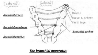
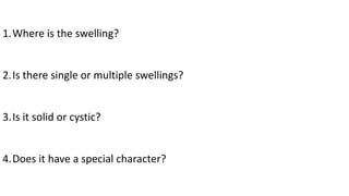
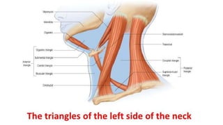
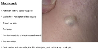
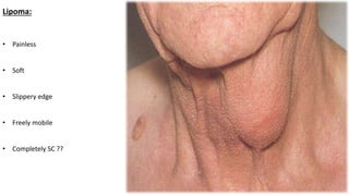
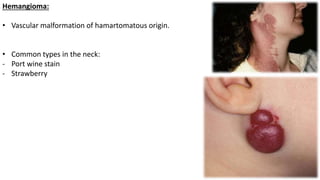
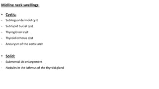
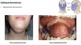
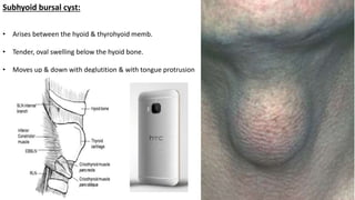
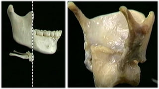
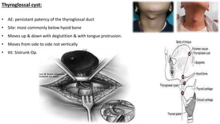
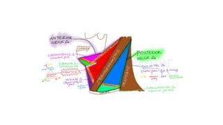
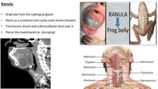
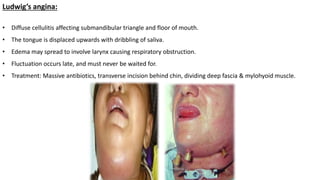
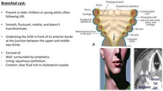
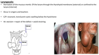
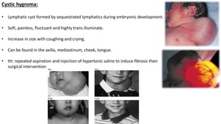
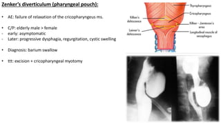
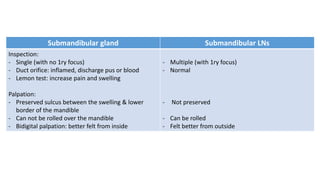
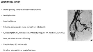
Neck swellings
By
Ahmed Zaki
Lecturer of General Surgery
Mansoura University
The branchial apparatus
1.Where is the swelling?
2.Is there single or multiple swellings?
3.Is it solid or cystic?
4.Does it have a special character?
• Swellings that might appear anywhere:
1- Skin: sebaceous cyst
2- S.C.: Lipoma
3- Vessels: hemangioma
4- Lymphatics: lymphangioma, cold abscess
Sebaceous cyst:
• Retention cyst of a sebaceous gland.
• Well defined hemispherical tense cystic.
• Smooth surface.
• Not tender
• Not fixed to deeper structures unless infected.
• Not translucent.
• Duct: blocked and attached to the skin at one point, punctum looks as a black spot.
• Swellings that might appear anywhere:
1- Skin: sebaceous cyst
2- S.C.: Lipoma
3- Vessels: hemangioma
4- Lymphatics: lymphangioma
Lipoma:
• Painless
• Soft
• Slippery edge
• Freely mobile
• Completely SC ??
• Swellings that might appear anywhere:
1- Skin: sebaceous cyst
2- S.C.: Lipoma
3- Vessels: hemangioma
4- Lymphatics: lymphangioma
Hemangioma:
• Vascular malformation of hamartomatous origin.
• Common types in the neck:
- Port wine stain
- Strawberry
Midline neck swellings:
• Cystic:
- Sublingual dermoid cyst
- Subhyoid bursal cyst
- Thyroglossal cyst
- Thyroid isthmus cyst
- Aneurysm of the aortic arch
• Solid:
- Submental LN enlargement
- Nodules in the isthmus of the thyroid gland
Sublingual dermoid cyst:
• Sequestration dermoid cyst
Infra myelohyoid dermoid Supra myelohyoid dermoid
Subhyoid bursal cyst:
• Arises between the hyoid & thyrohyoid memb.
• Tender, oval swelling below the hyoid bone.
• Moves up & down with deglutition & with tongue protrusion
Thyroglossal cyst:
• AE: persistant patency of the thyroglossal duct
• Site: most commonly below hyoid bone
• Moves up & down with deglutition & with tongue protrusion.
• Moves from side to side not vertically
• ttt: Sistrunk Op.
Lateral neck swellings:
• Cystic swellings:
- Submandibular : plunging ranula, Ludwig’s angina
- Carotid : Branchial cyst, neurofibroma or schwannoma of the vagus, caroid artery aneurysm, laryngocele
- Posterior : Cystic hygroma, pharyngeal pouch
- Thyroid cystic swelling
• Solid swellings:
- Submandibular : enlarged submandibular gland or LN
- Carotid : LNs, Enlarged thyroid gland, carotid body tumor
- Posterior : LNs, neurofibroma or schwannoma of brachial plexus, cervical rib, pancost tumor
Ranula:
• Originate from the sublingual gland
• Starts as a unilateral oral cavity cystic lesion (simple)
• Translucent, bluish and submandibular duct over it.
• Pierce the myelohyoid ms. (plunging)
Ludwig’s angina:
• Diffuse cellulitis affecting submandibular triangle and floor of mouth.
• The tongue is displaced upwards with dribbling of saliva.
• Edema may spread to involve larynx causing respiratory obstruction.
• Fluctuation occurs late, and must never be waited for.
• Treatment: Massive antibiotics, transverse incision behind chin, dividing deep fascia & mylohyoid muscle.
Lateral neck swellings:
• Cystic swellings:
- Submandibular : plunging ranula, Ludwig’s angina
- Carotid : Branchial cyst, laryngocele, neurofibroma or schwannoma of the vagus, caroid artery aneurysm.
- Posterior : Cystic hygroma, pharyngeal pouch
- Thyroid cystic swelling
• Solid swellings:
- Submandibular : enlarged submandibular gland or LN
- Carotid : LNs, Enlarged thyroid gland, carotid body tumor
- Posterior : LNs, neurofibroma or schwannoma of brachial plexus, cervical rib, pancost tumor
Branchial cyst:
• Present in older children or young adults often
following URI.
• Smooth, fluctuant, mobile, and doesn’t
transilluminate.
• Underlying the SCM in front of its anterior border
at the junction between the upper and middle
two thirds.
• Formed of:
- Wall: surrounded by lymphatics
- Lining: squamous epithelium
- Content: clear fluid rich in cholesterol crystals
Laryngocele:
• Herniation of the mucous memb. Of the larynx through the thyrohyoid membrane (external) or confined to the
larynx (internal)
• Occur in singers and teachers
• C/P: resonant, translucent cystic swelling below the hyoid bone
• ttt: excision + repair of the defect + avoid straining
Lateral neck swellings:
• Cystic swellings:
- Submandibular : plunging ranula, Ludwig’s angina
- Carotid : Branchial cyst, laryngocele, neurofibroma or schwannoma of the vagus, caroid artery aneurysm.
- Posterior : Cystic hygroma, pharyngeal pouch
- Thyroid cystic swelling
• Solid swellings:
- Submandibular : enlarged submandibular gland or LN
- Carotid : LNs, Enlarged thyroid gland, carotid body tumor
- Posterior : LNs, neurofibroma or schwannoma of brachial plexus, cervical rib, pancost tumor
Cystic hygroma:
• Lymphatic cyst formed by sequestrated lymphatics during embryonic development.
• Soft, painless, fluctuant and highly trans illuminate.
• Increase in size with coughing and crying.
• Can be found in the axilla, mediastinum, cheek, tongue.
• ttt: repeated aspiration and injection of hypertonic saline to induce fibrosis then
surgical intervention
Zenker’s diverticulum (pharyngeal pouch):
• AE: failure of relaxation of the cricopharyngeus ms.
• C/P: elderly male > female
- early: asymptomatic
- Later: progressive dysphagia, regurgitation, cystic swelling
• Diagnosis: barium swallow
• ttt: excision + cricopharyngeal myotomy
Lateral neck swellings:
• Cystic swellings:
- Submandibular : plunging ranula, Ludwig’s angina
- Carotid : Branchial cyst, laryngocele, neurofibroma or schwannoma of the vagus, caroid artery aneurysm.
- Posterior : Cystic hygroma, pharyngeal pouch
- Thyroid cystic swelling
• Solid swellings:
- Submandibular : enlarged submandibular gland or LN
- Carotid : LNs, Enlarged thyroid gland, carotid body tumor
- Posterior : LNs, neurofibroma or schwannoma of brachial plexus, cervical rib, pancost tumor
Submandibular gland Submandibular LNs
Inspection:
- Single (with no 1ry focus)
- Duct orifice: inflamed, discharge pus or blood
- Lemon test: increase pain and swelling
Palpation:
- Preserved sulcus between the swelling & lower
border of the mandible
- Can not be rolled over the mandible
- Bidigital palpation: better felt from inside
- Multiple (with 1ry focus)
- Normal
- Not preserved
- Can be rolled
- Felt better from outside
Lateral neck swellings:
• Cystic swellings:
- Submandibular : plunging ranula, Ludwig’s angina
- Carotid : Branchial cyst, laryngocele, neurofibroma or schwannoma of the vagus, caroid artery aneurysm.
- Posterior : Cystic hygroma, pharyngeal pouch
- Thyroid cystic swelling
• Solid swellings:
- Submandibular : enlarged submandibular gland or LN
- Carotid : LNs, Enlarged thyroid gland, carotid body tumor
- Posterior : LNs, neurofibroma or schwannoma of brachial plexus, cervical rib, pancost tumor
Carotid body tumor:
• Slowly growing tumor at the carotid bifurcation
• Locally invasive
• Rare in children
• Pulsatile, compressible mass, moves from side to side
• C/P: asymptomatic, nervousness, irritability, irregular HR, headache, sweating
fever, recurrent attacks of fainting
• Investigations: CT angiography
• ttt: close observation or surgical excision.
Lymph drainage of the head & neck
a) Circular chain of LNs
I) Inner ring (Waldeyer ring): Nasopharyngeal, palatine and lingual tonsils.
II) Outer ring:
• Submental, submandibular, buccal, parotid, pre and post auricular and occipital
b) Vertical chains of LNs
• Ext. jugular
• Ant. Jugular
• Pre-laryngeal
• Pre-tracheal
• Para-tracheal
• Retropharyngeal
• Upper & lower deep cx. LNs
And finally the upper & lower deep cx. LNs collect to form the jugular lymph trunk
then to the thoracic duct (Lt. side) or Rt. Lymphatic duct (Rt. Side)
Block Neck Dissection
a) Radical block neck dissection:
Structures removed:
• Lymph nodes of anterior and posterior triangles of neck.
• Sternomastoid→ to expose internal jugular vein.
• Internal jugular vein → is removed from base of skull to its root in the neck.
• Spinal accessory nerve.
• Cervical fascia from jaw to clavicle.
• Submandibular salivary gland → easier removal of submandibular L.N.
• Lower part of parotid salivary gland → contains lymph glands.
b) Modified radical neck dissection:
Preservation of one or more of the following: CN XI, IJV, sternomastoid ms.
c) Suprahyoid block dissection (for carcinoma of the lower lip): level I, II
d) Supra omohyoid block dissection: level I, II, III
e) Lateral block dissection: level II, III, IV
f) Postero-lateral block dissection: level II, III, IV, V
g) Central neck dissection: level VI
Neck swellings.pptx

Neck swellings.pptx

  • 1.
    Neck swellings By Ahmed Zaki Lecturerof General Surgery Mansoura University
  • 2.
  • 3.
    1.Where is theswelling? 2.Is there single or multiple swellings? 3.Is it solid or cystic? 4.Does it have a special character?
  • 5.
    • Swellings thatmight appear anywhere: 1- Skin: sebaceous cyst 2- S.C.: Lipoma 3- Vessels: hemangioma 4- Lymphatics: lymphangioma, cold abscess
  • 6.
    Sebaceous cyst: • Retentioncyst of a sebaceous gland. • Well defined hemispherical tense cystic. • Smooth surface. • Not tender • Not fixed to deeper structures unless infected. • Not translucent. • Duct: blocked and attached to the skin at one point, punctum looks as a black spot.
  • 7.
    • Swellings thatmight appear anywhere: 1- Skin: sebaceous cyst 2- S.C.: Lipoma 3- Vessels: hemangioma 4- Lymphatics: lymphangioma
  • 8.
    Lipoma: • Painless • Soft •Slippery edge • Freely mobile • Completely SC ??
  • 9.
    • Swellings thatmight appear anywhere: 1- Skin: sebaceous cyst 2- S.C.: Lipoma 3- Vessels: hemangioma 4- Lymphatics: lymphangioma
  • 10.
    Hemangioma: • Vascular malformationof hamartomatous origin. • Common types in the neck: - Port wine stain - Strawberry
  • 11.
    Midline neck swellings: •Cystic: - Sublingual dermoid cyst - Subhyoid bursal cyst - Thyroglossal cyst - Thyroid isthmus cyst - Aneurysm of the aortic arch • Solid: - Submental LN enlargement - Nodules in the isthmus of the thyroid gland
  • 12.
    Sublingual dermoid cyst: •Sequestration dermoid cyst Infra myelohyoid dermoid Supra myelohyoid dermoid
  • 13.
    Subhyoid bursal cyst: •Arises between the hyoid & thyrohyoid memb. • Tender, oval swelling below the hyoid bone. • Moves up & down with deglutition & with tongue protrusion
  • 15.
    Thyroglossal cyst: • AE:persistant patency of the thyroglossal duct • Site: most commonly below hyoid bone • Moves up & down with deglutition & with tongue protrusion. • Moves from side to side not vertically • ttt: Sistrunk Op.
  • 17.
    Lateral neck swellings: •Cystic swellings: - Submandibular : plunging ranula, Ludwig’s angina - Carotid : Branchial cyst, neurofibroma or schwannoma of the vagus, caroid artery aneurysm, laryngocele - Posterior : Cystic hygroma, pharyngeal pouch - Thyroid cystic swelling • Solid swellings: - Submandibular : enlarged submandibular gland or LN - Carotid : LNs, Enlarged thyroid gland, carotid body tumor - Posterior : LNs, neurofibroma or schwannoma of brachial plexus, cervical rib, pancost tumor
  • 18.
    Ranula: • Originate fromthe sublingual gland • Starts as a unilateral oral cavity cystic lesion (simple) • Translucent, bluish and submandibular duct over it. • Pierce the myelohyoid ms. (plunging)
  • 19.
    Ludwig’s angina: • Diffusecellulitis affecting submandibular triangle and floor of mouth. • The tongue is displaced upwards with dribbling of saliva. • Edema may spread to involve larynx causing respiratory obstruction. • Fluctuation occurs late, and must never be waited for. • Treatment: Massive antibiotics, transverse incision behind chin, dividing deep fascia & mylohyoid muscle.
  • 20.
    Lateral neck swellings: •Cystic swellings: - Submandibular : plunging ranula, Ludwig’s angina - Carotid : Branchial cyst, laryngocele, neurofibroma or schwannoma of the vagus, caroid artery aneurysm. - Posterior : Cystic hygroma, pharyngeal pouch - Thyroid cystic swelling • Solid swellings: - Submandibular : enlarged submandibular gland or LN - Carotid : LNs, Enlarged thyroid gland, carotid body tumor - Posterior : LNs, neurofibroma or schwannoma of brachial plexus, cervical rib, pancost tumor
  • 21.
    Branchial cyst: • Presentin older children or young adults often following URI. • Smooth, fluctuant, mobile, and doesn’t transilluminate. • Underlying the SCM in front of its anterior border at the junction between the upper and middle two thirds. • Formed of: - Wall: surrounded by lymphatics - Lining: squamous epithelium - Content: clear fluid rich in cholesterol crystals
  • 22.
    Laryngocele: • Herniation ofthe mucous memb. Of the larynx through the thyrohyoid membrane (external) or confined to the larynx (internal) • Occur in singers and teachers • C/P: resonant, translucent cystic swelling below the hyoid bone • ttt: excision + repair of the defect + avoid straining
  • 23.
    Lateral neck swellings: •Cystic swellings: - Submandibular : plunging ranula, Ludwig’s angina - Carotid : Branchial cyst, laryngocele, neurofibroma or schwannoma of the vagus, caroid artery aneurysm. - Posterior : Cystic hygroma, pharyngeal pouch - Thyroid cystic swelling • Solid swellings: - Submandibular : enlarged submandibular gland or LN - Carotid : LNs, Enlarged thyroid gland, carotid body tumor - Posterior : LNs, neurofibroma or schwannoma of brachial plexus, cervical rib, pancost tumor
  • 24.
    Cystic hygroma: • Lymphaticcyst formed by sequestrated lymphatics during embryonic development. • Soft, painless, fluctuant and highly trans illuminate. • Increase in size with coughing and crying. • Can be found in the axilla, mediastinum, cheek, tongue. • ttt: repeated aspiration and injection of hypertonic saline to induce fibrosis then surgical intervention
  • 25.
    Zenker’s diverticulum (pharyngealpouch): • AE: failure of relaxation of the cricopharyngeus ms. • C/P: elderly male > female - early: asymptomatic - Later: progressive dysphagia, regurgitation, cystic swelling • Diagnosis: barium swallow • ttt: excision + cricopharyngeal myotomy
  • 26.
    Lateral neck swellings: •Cystic swellings: - Submandibular : plunging ranula, Ludwig’s angina - Carotid : Branchial cyst, laryngocele, neurofibroma or schwannoma of the vagus, caroid artery aneurysm. - Posterior : Cystic hygroma, pharyngeal pouch - Thyroid cystic swelling • Solid swellings: - Submandibular : enlarged submandibular gland or LN - Carotid : LNs, Enlarged thyroid gland, carotid body tumor - Posterior : LNs, neurofibroma or schwannoma of brachial plexus, cervical rib, pancost tumor
  • 27.
    Submandibular gland SubmandibularLNs Inspection: - Single (with no 1ry focus) - Duct orifice: inflamed, discharge pus or blood - Lemon test: increase pain and swelling Palpation: - Preserved sulcus between the swelling & lower border of the mandible - Can not be rolled over the mandible - Bidigital palpation: better felt from inside - Multiple (with 1ry focus) - Normal - Not preserved - Can be rolled - Felt better from outside
  • 28.
    Lateral neck swellings: •Cystic swellings: - Submandibular : plunging ranula, Ludwig’s angina - Carotid : Branchial cyst, laryngocele, neurofibroma or schwannoma of the vagus, caroid artery aneurysm. - Posterior : Cystic hygroma, pharyngeal pouch - Thyroid cystic swelling • Solid swellings: - Submandibular : enlarged submandibular gland or LN - Carotid : LNs, Enlarged thyroid gland, carotid body tumor - Posterior : LNs, neurofibroma or schwannoma of brachial plexus, cervical rib, pancost tumor
  • 29.
    Carotid body tumor: •Slowly growing tumor at the carotid bifurcation • Locally invasive • Rare in children • Pulsatile, compressible mass, moves from side to side • C/P: asymptomatic, nervousness, irritability, irregular HR, headache, sweating fever, recurrent attacks of fainting • Investigations: CT angiography • ttt: close observation or surgical excision.
  • 30.
    Lymph drainage ofthe head & neck a) Circular chain of LNs I) Inner ring (Waldeyer ring): Nasopharyngeal, palatine and lingual tonsils. II) Outer ring: • Submental, submandibular, buccal, parotid, pre and post auricular and occipital b) Vertical chains of LNs • Ext. jugular • Ant. Jugular • Pre-laryngeal • Pre-tracheal • Para-tracheal • Retropharyngeal • Upper & lower deep cx. LNs And finally the upper & lower deep cx. LNs collect to form the jugular lymph trunk then to the thoracic duct (Lt. side) or Rt. Lymphatic duct (Rt. Side)
  • 32.
  • 33.
    a) Radical blockneck dissection: Structures removed: • Lymph nodes of anterior and posterior triangles of neck. • Sternomastoid→ to expose internal jugular vein. • Internal jugular vein → is removed from base of skull to its root in the neck. • Spinal accessory nerve. • Cervical fascia from jaw to clavicle. • Submandibular salivary gland → easier removal of submandibular L.N. • Lower part of parotid salivary gland → contains lymph glands.
  • 34.
    b) Modified radicalneck dissection: Preservation of one or more of the following: CN XI, IJV, sternomastoid ms. c) Suprahyoid block dissection (for carcinoma of the lower lip): level I, II d) Supra omohyoid block dissection: level I, II, III e) Lateral block dissection: level II, III, IV f) Postero-lateral block dissection: level II, III, IV, V g) Central neck dissection: level VI

Editor's Notes

  • #3 These arches are cylindrical in shape, which give the neck a wavy appearance Branchial arches: gives cartilages, ms., nerves, arteries Branchial grooves (clefts): covered by ectoderm (the first gives external auditory canal and external tympanic membrane) and the rest obliterates Branchial pouches: 1st: eustachian tube, middle ear (mastoid air cells), inner tympanic membrane 2nd: supra tonsillar fossa 3rd: inferior parathyroid 4th: superior parathyroid, para-follicular C cells - Fusion of 2nd arch with 5th arch leads to: loss of wavy appearance …….. Cervical sinus of His
  • #5 - Contents of posterior triangle: Ms.: omohyoid Nerves: spinal accessory, trunks of brachial plexus Arteries: br. Of 1st subclivian: tra. Cx., suprascapular ……. 3rd subclavian Veins: subclavian v. and its tributary Contents of digastric triangle: Submandibular gland and LNs Facial artery and common facial vein Hypoglossal, lingual n. Contents of carotid triangle: Carotid arteries IJV with 3 1st tributaries: common facial, lingual, superior thyroid, middle thyroid, inferior petrosal, pharyngeal Last 3 cranial nerves: Others: cx. LNs, ansa cervicalis , sympathetic chain
  • #11 -Hamartoma: tumor like condition in which the tissue of a particular part of the body is haphazardly arranged with an excess of one or more of its components -Types: capillary (strawberry: resolve spontaneously at 7years …….. port wine: along distribution of trigeminal n. & never involute …… salmon patch: forehead in the midline ………. Spider nevi: with LCF) …………………………………………………………. Cavernous ………………mixed ………….. Cercoid aneurysm …………glomous body tumor
  • #13 -Sequestration dermoid: congenital inclusion of a piece of epithelium in the SC tissue at line of fusion e.g: angular dermoid, pre auricular, sublingual, submental, suprasternal -Implantation -Tubulo dermoid: distension of remnants of embryonic ducts e.g: thyroglossal cyst, branchial cyst -Teratomatous dermoid
  • #14 Bursa: fluid filled sac between the bones to act as a cushion
  • #16 Development of the thyroid gland: Thyroglossal duct: a median pub from the floor of the developing phrynx ….. Extends downwards from the foramen caecum to unite with the ultimobranchial body ….. Gives pyramidal lobe, ishmus and most of the 2 lobes Ultimobranchial body: from the 4th pharyngeal pouch ……… gives small part of the lobes Neural crest: gives parafollicular C cells which secretes thyrocalcitonine
  • #19 Either due to - Extravasation cyst: damage of sublingual gland and duct => escape of mucus under the mucosal lining of the mouth floor - Retention cyst: obstruction of the duct
  • #20 Don’t wait for fluctuation in : Parotid, prostate ……. Perinephric, perianal ……… Pulp space, palmer space ……… Breast, buttock, brain ………. Liver, lung, Ludwig’s angina
  • #22 The 2nd arch grows caudally and fuses with the 5th forming a cavity known as the cx sinus …… if persists => branchial cyst
  • #24 Schwannoma: arise from nerve sheath ……. C/P: medial tonsillar displacement, hoarsness, horner syndrome
  • #25 -Normally large accumulation of lymphatic vesicles is present lateral to the jugular vein (jugular lymph sac) -If some vesicles fail to join the jugular lymph sac they form the cystic hygroma
  • #26 Cricophryngeal myotomy: zenker Cardiomyotomy for achalasia Ramstedt pyloromyotomy: CHPS
  • #27 Schwannoma: arise from nerve sheath ……. C/P: medial tonsillar displacement, hoarsness, horner syndrome
  • #29 Schwannoma: arise from nerve sheath ……. C/P: medial tonsillar displacement, hoarsness, horner syndrome
  • #30 24 hrs Holter monitoring might reveals significant nocturnal cardiac pauses
  • #31 Upper deep cx.: along the upper half of the IJV (most important jugulo-digastric) Lower deep cx.: along the lower half of the IJV (most important jugulo-omohyoid & supraclavicular)