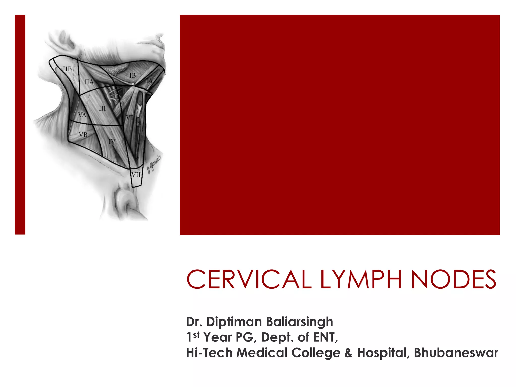
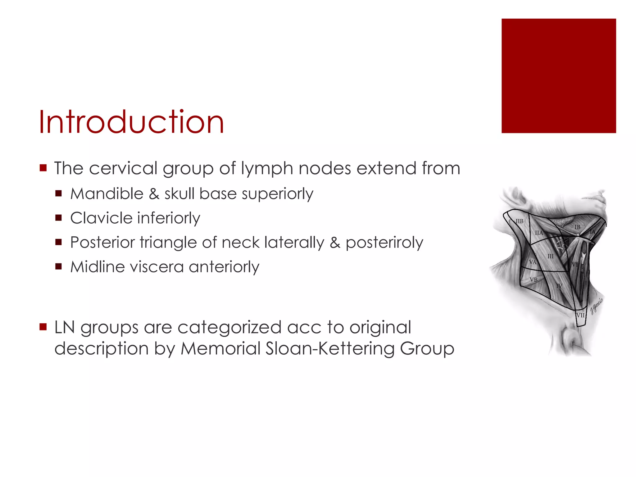
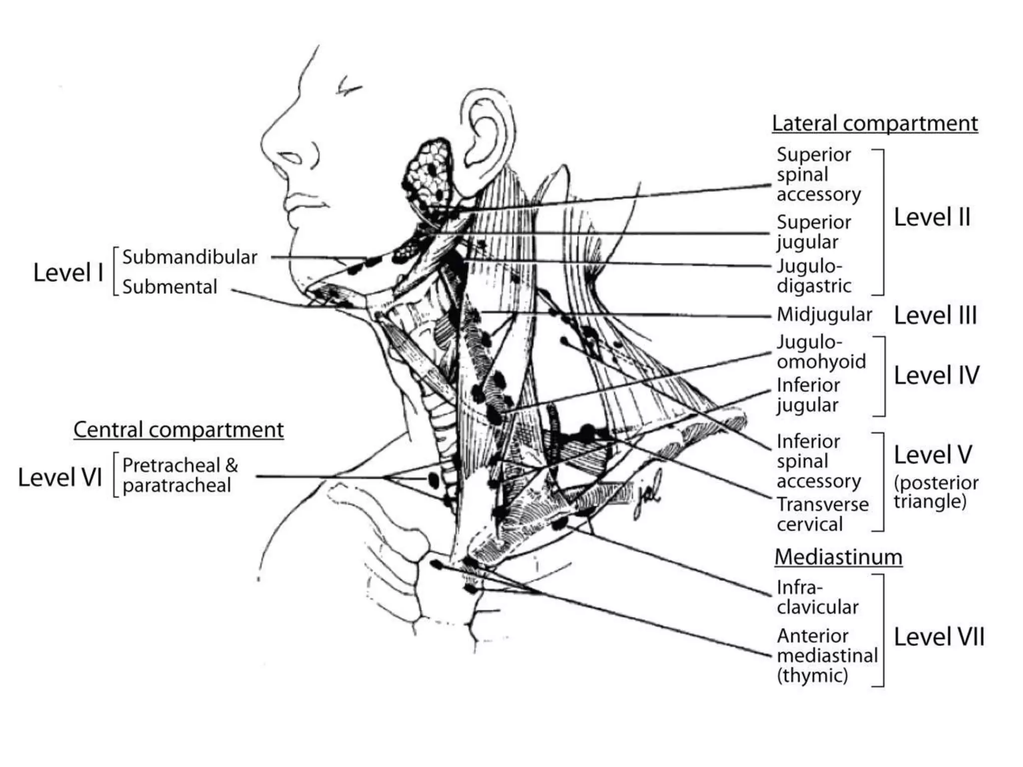
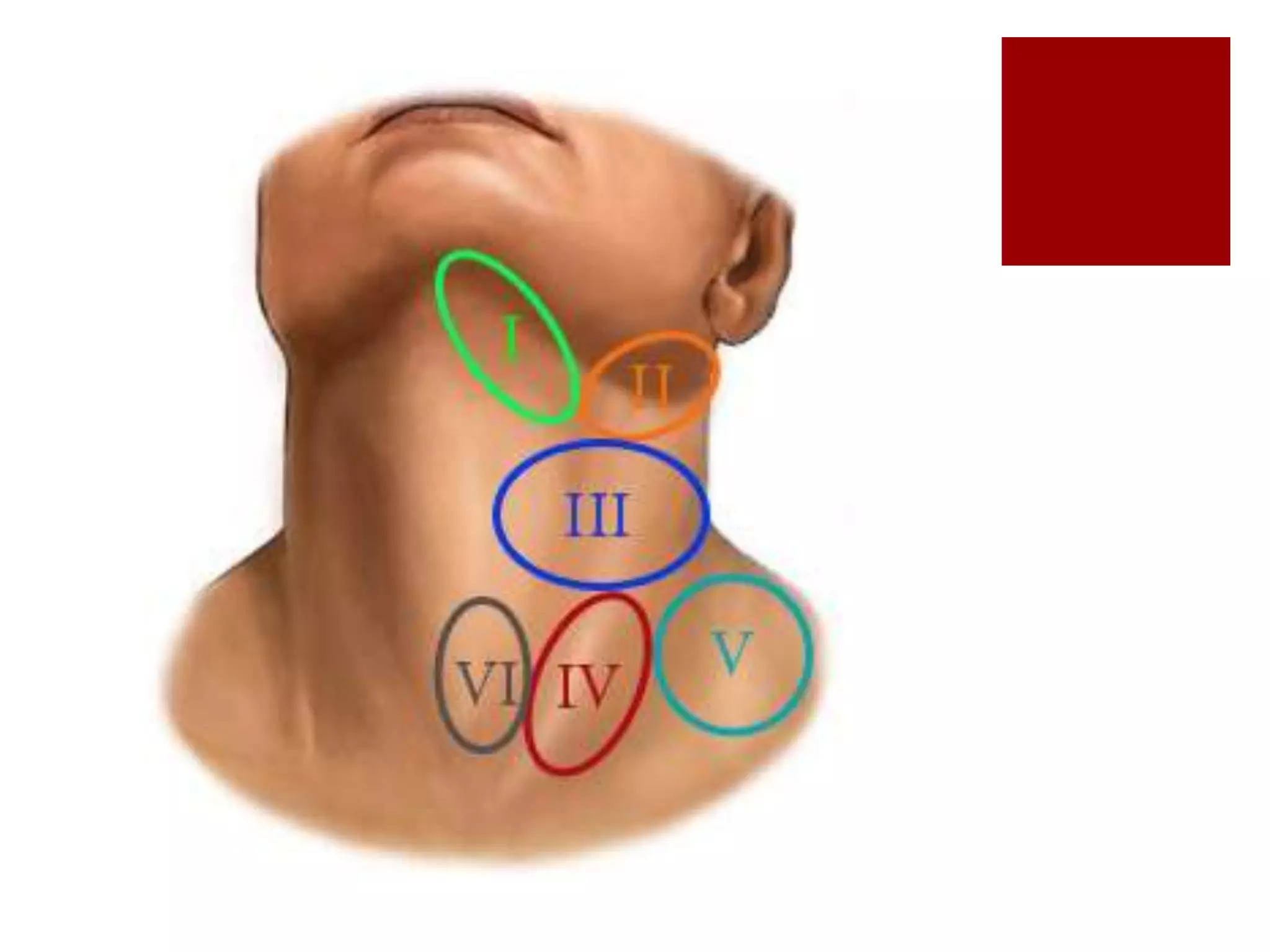
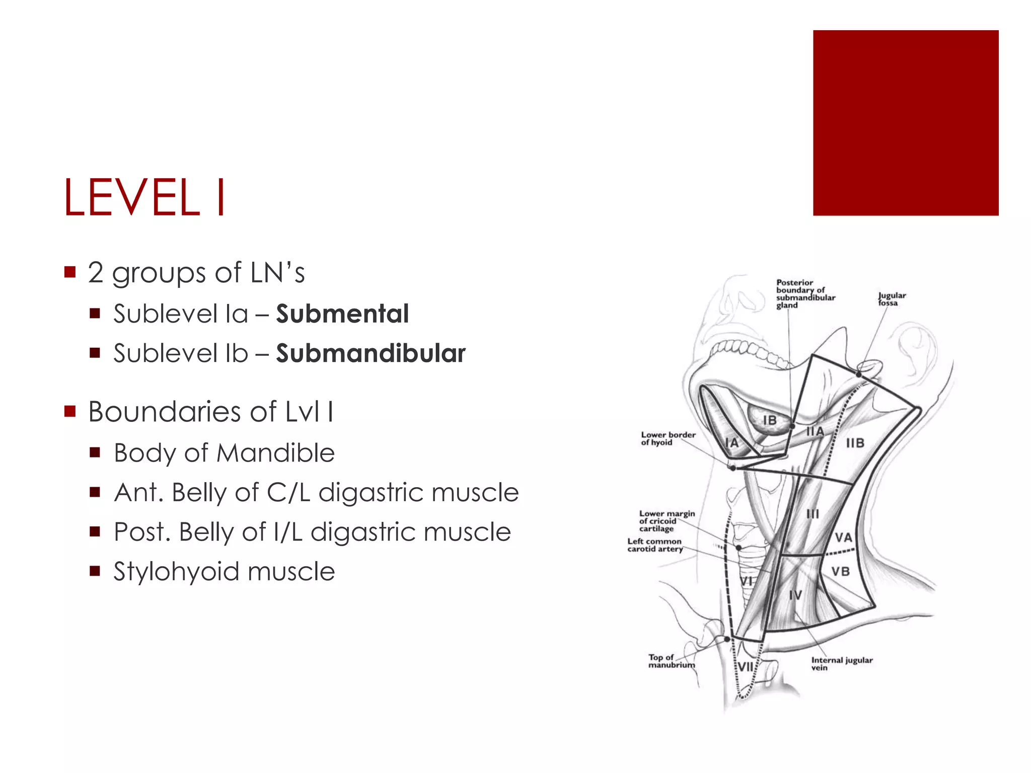
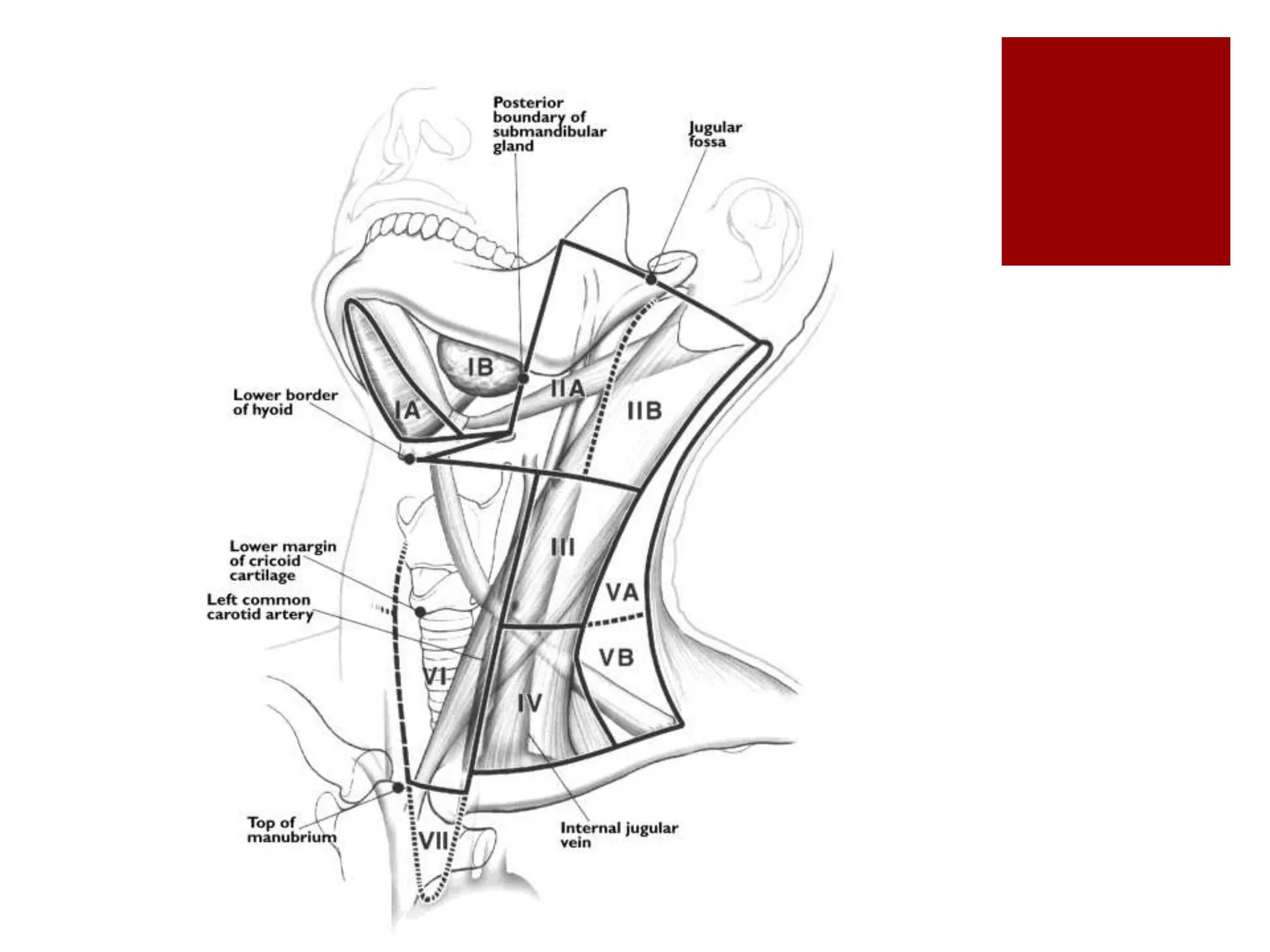
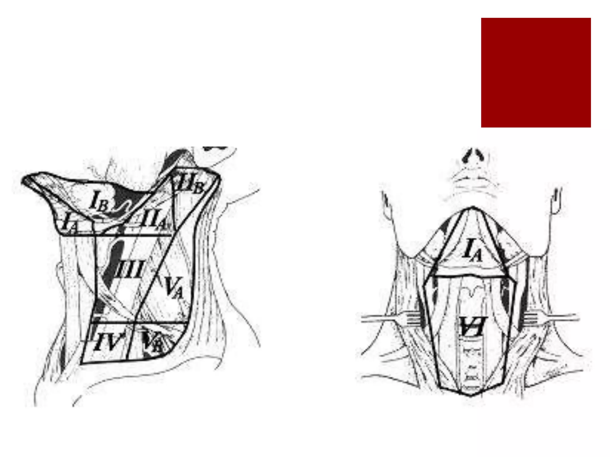
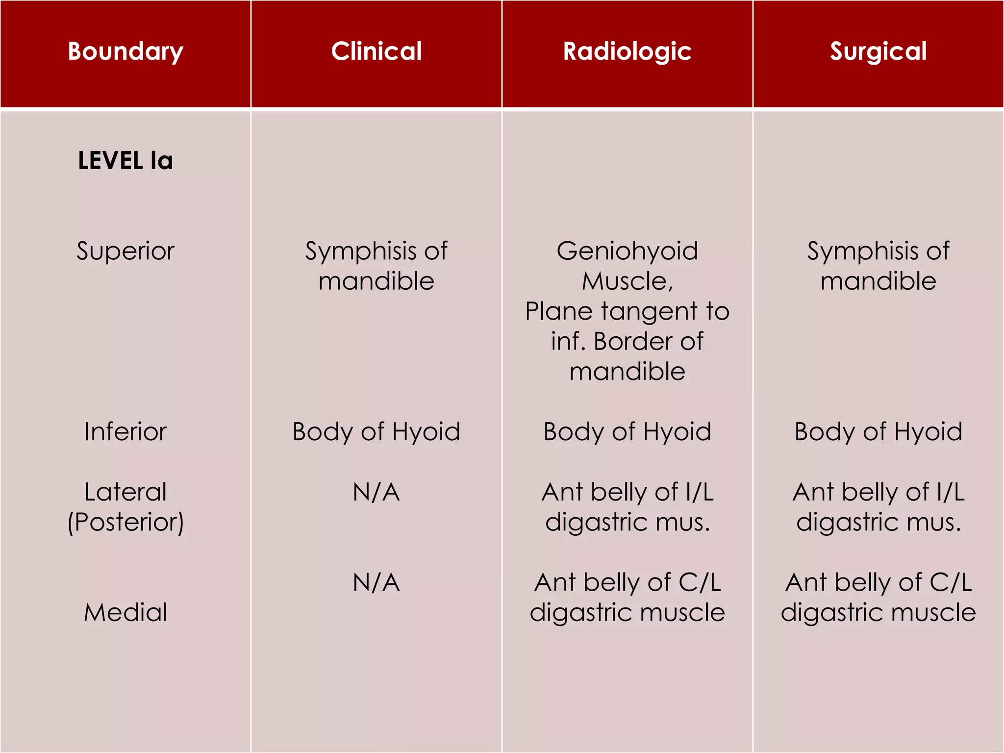
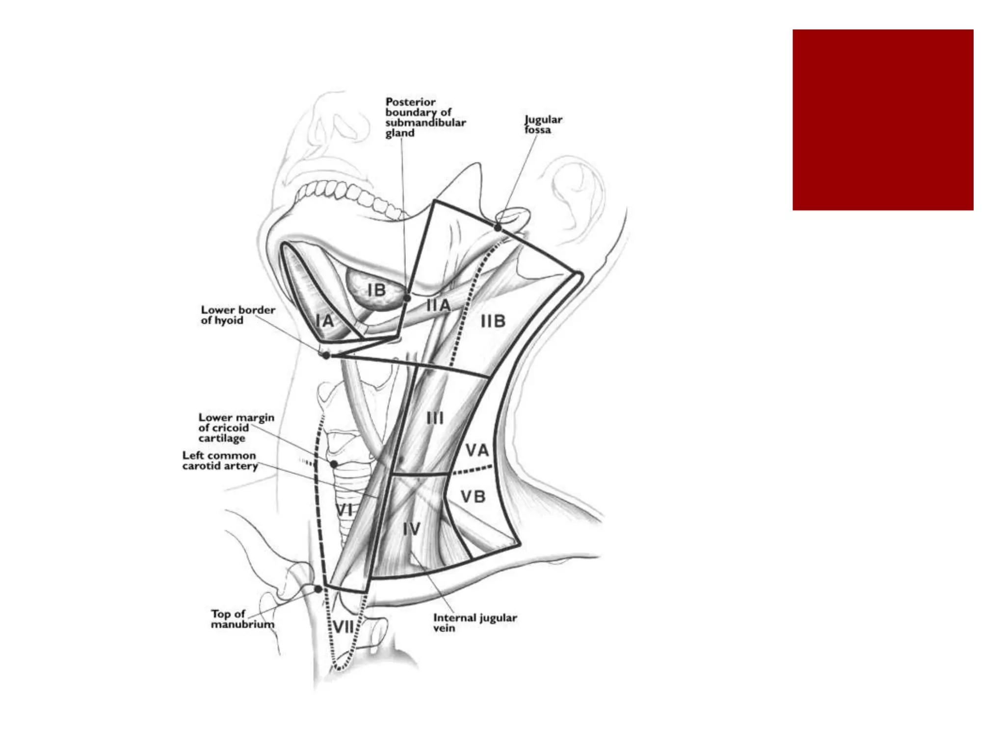
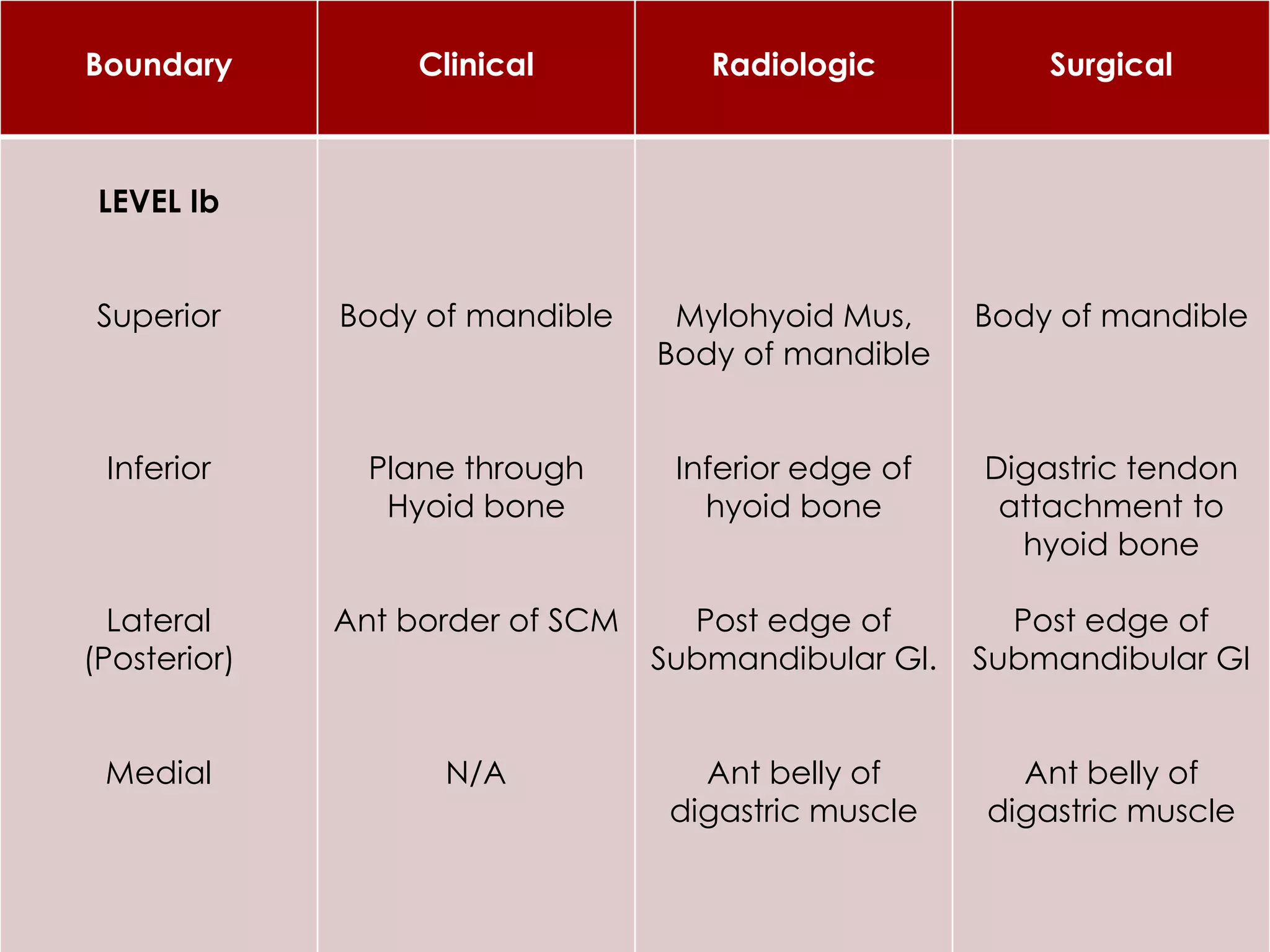
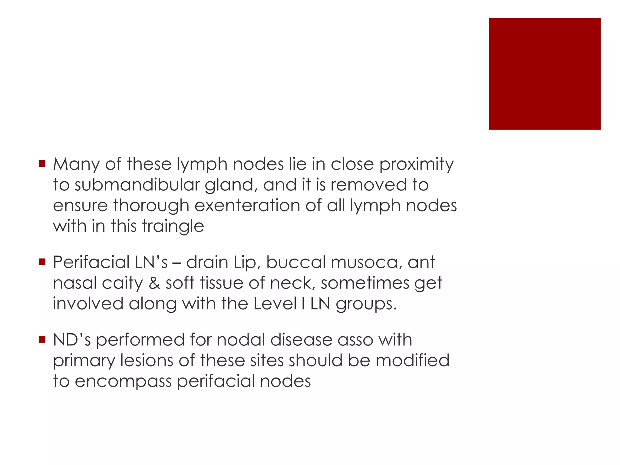
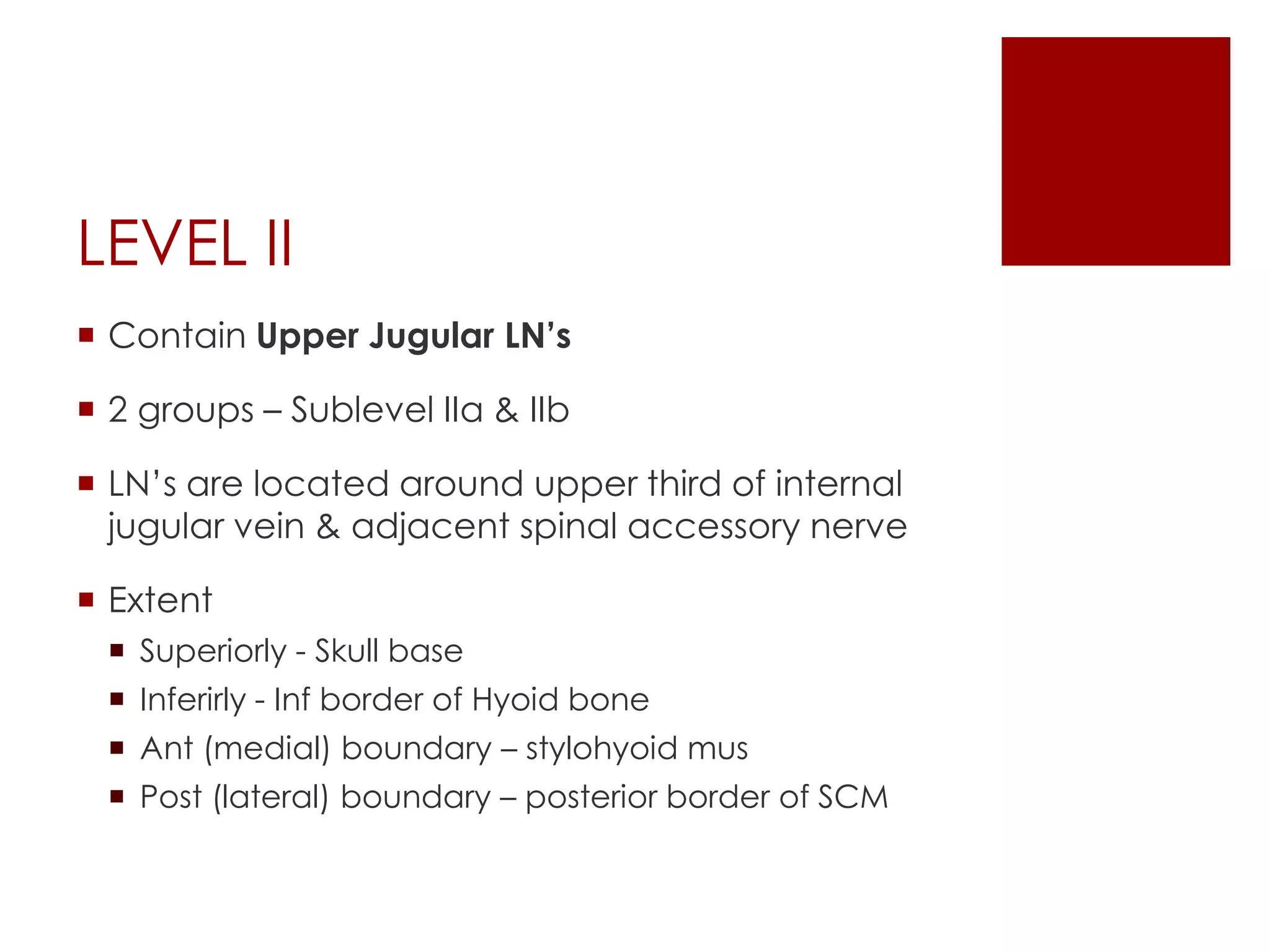
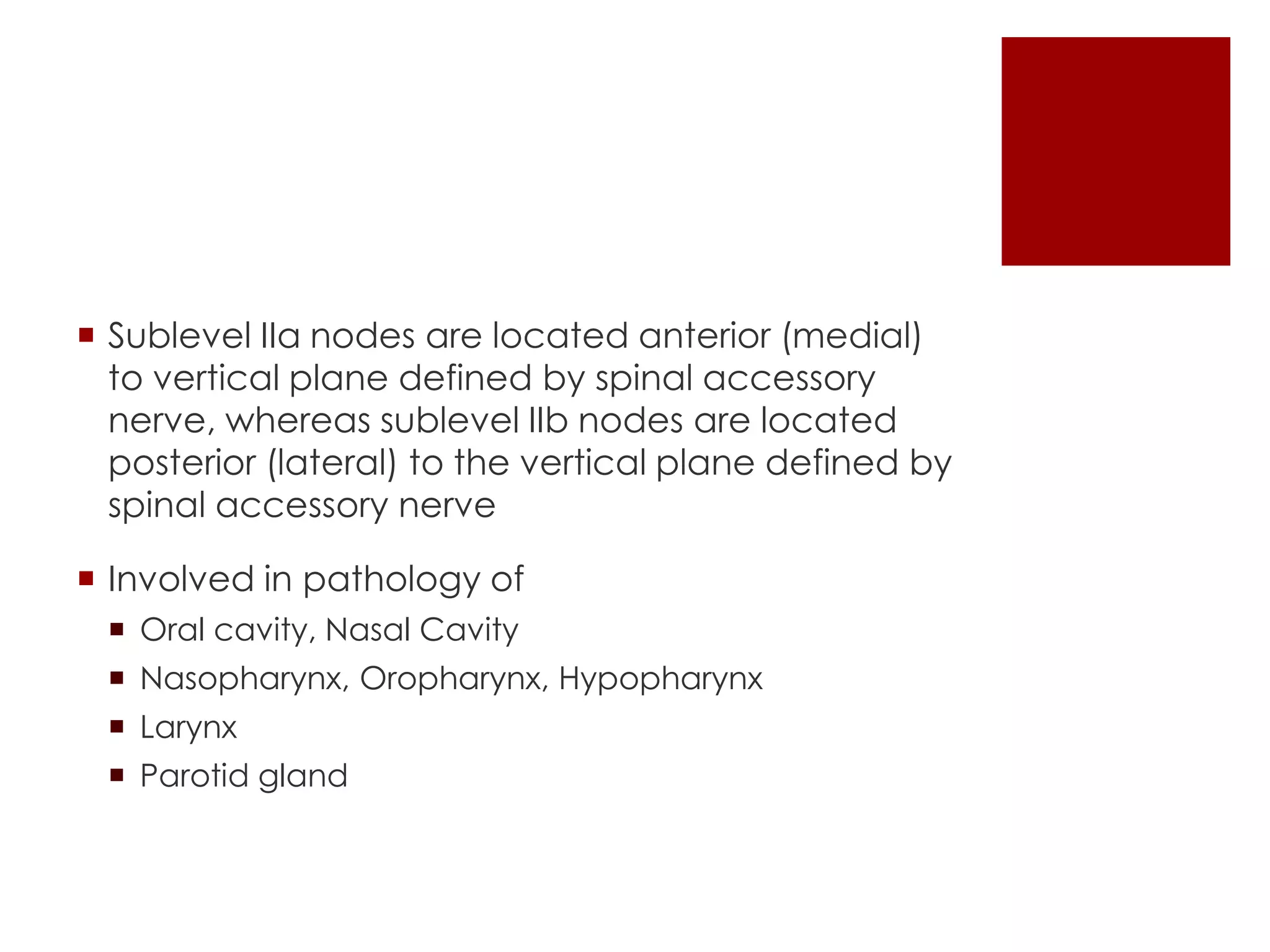
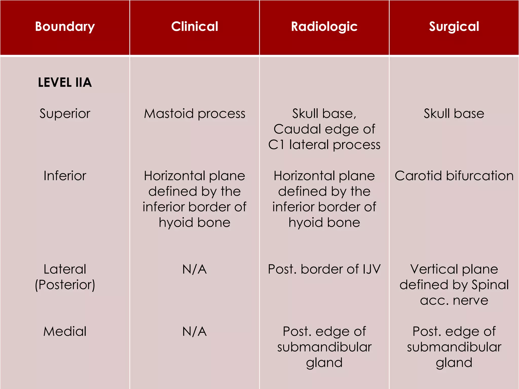
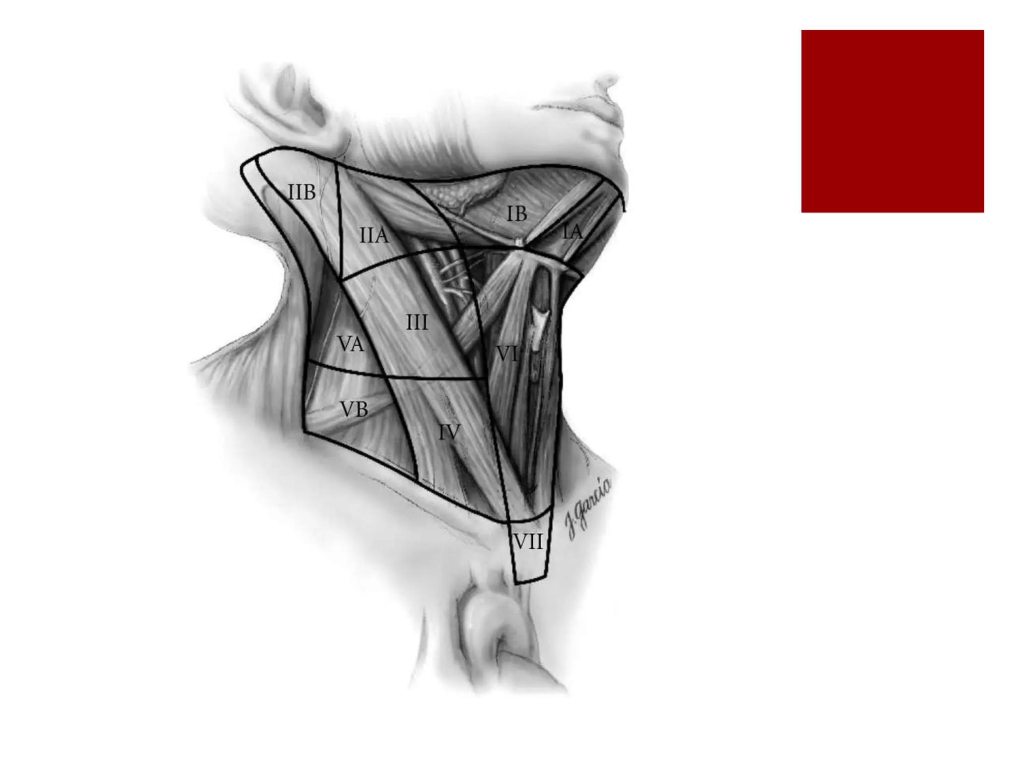
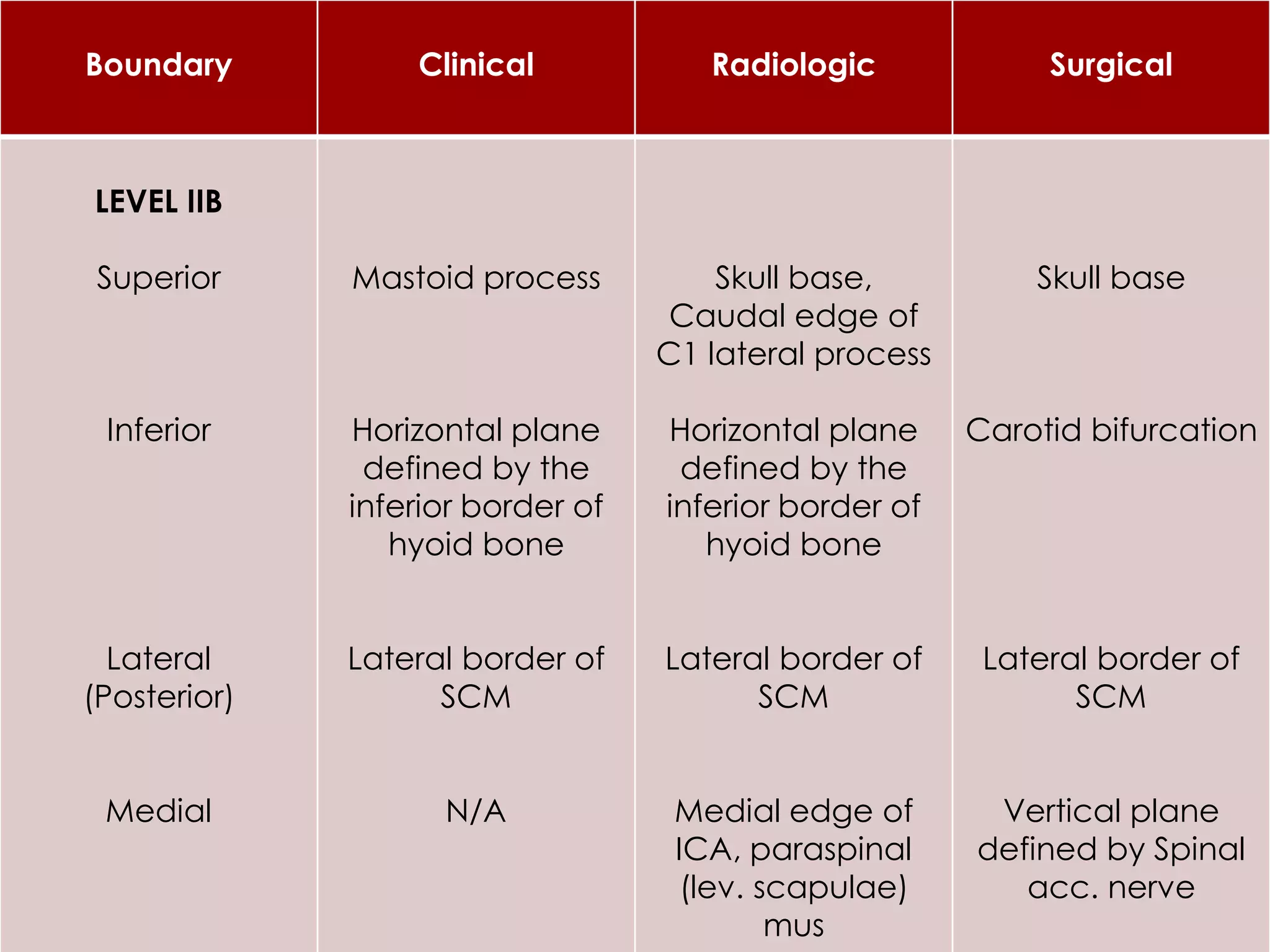
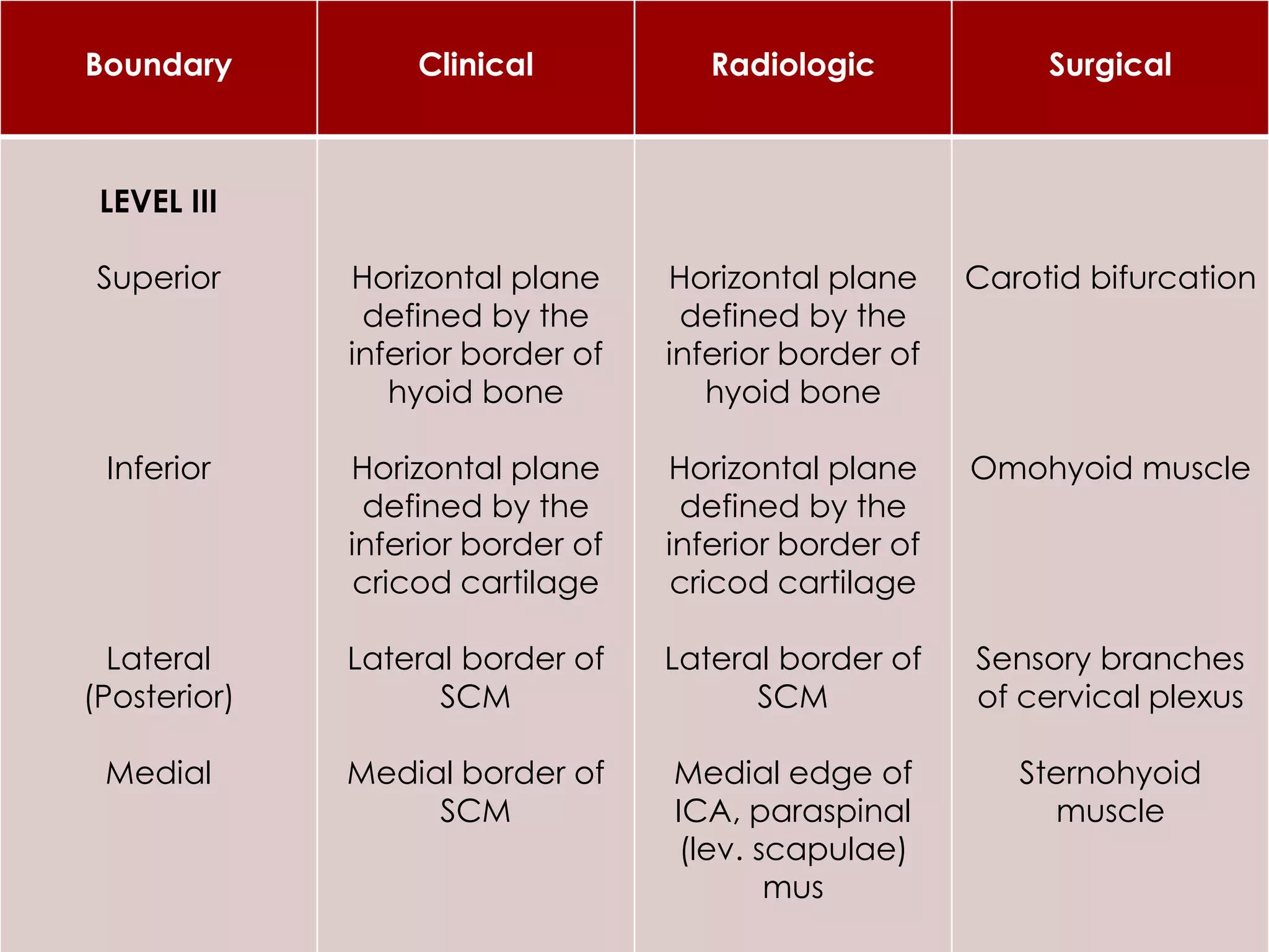
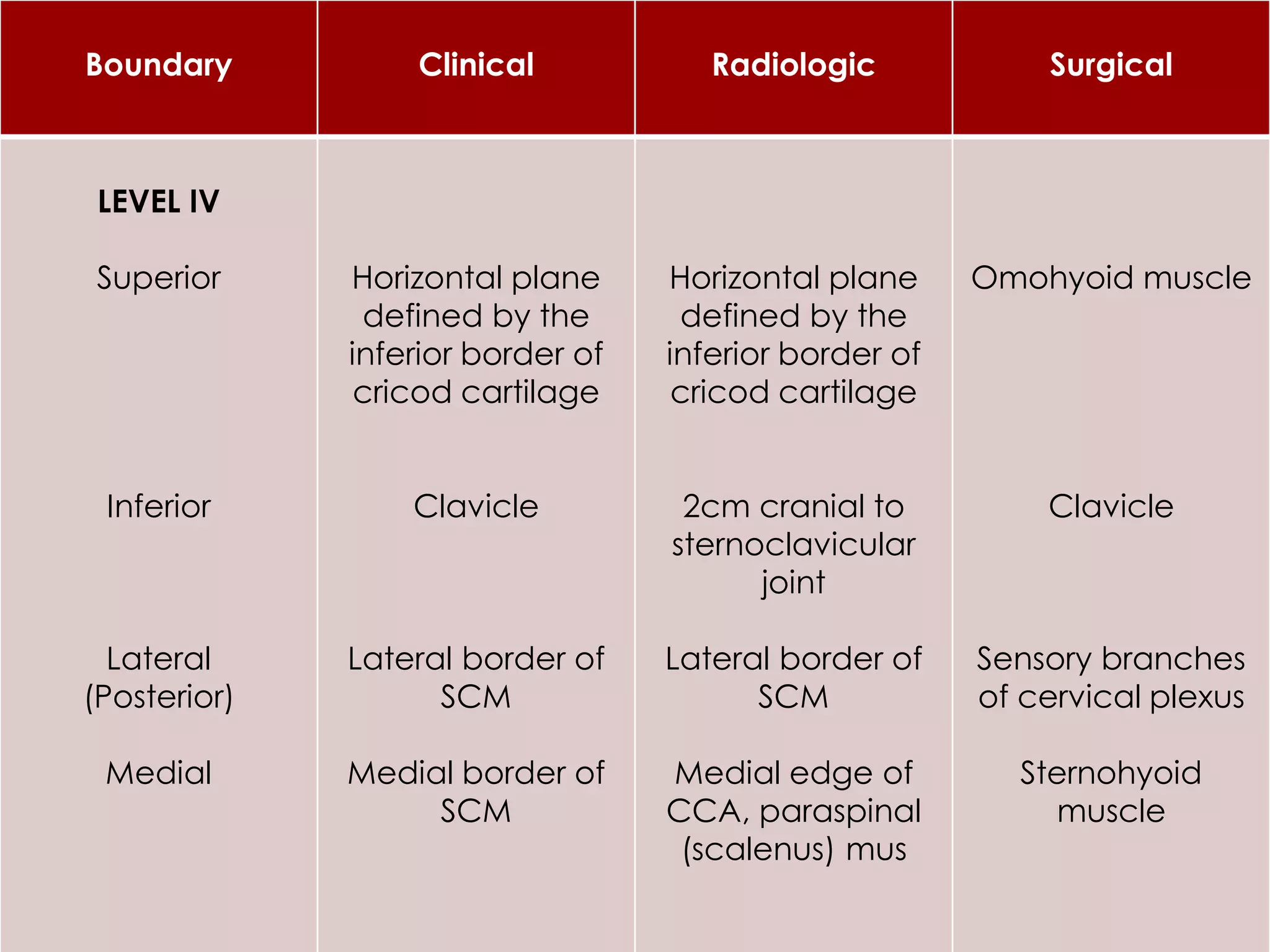
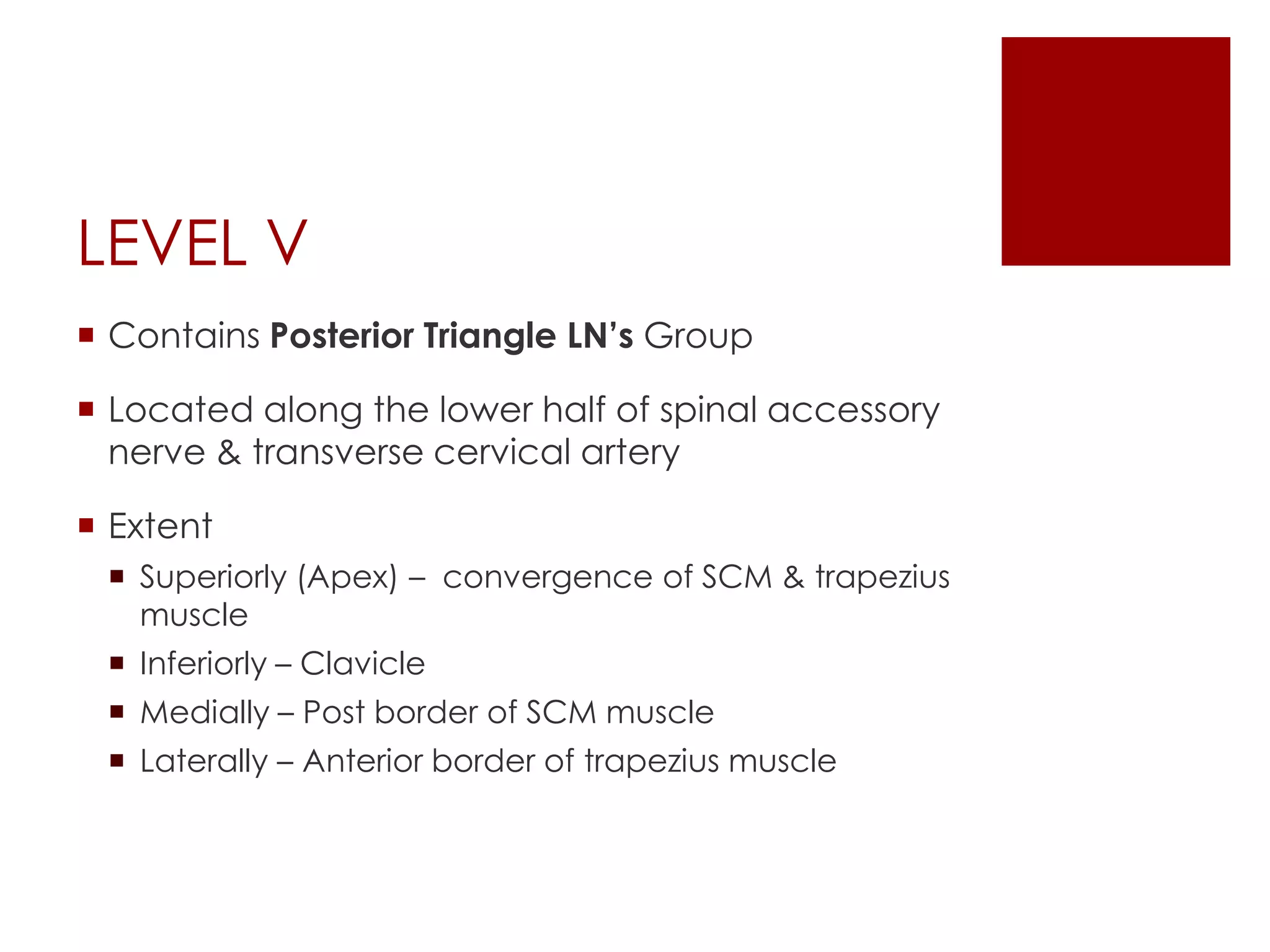
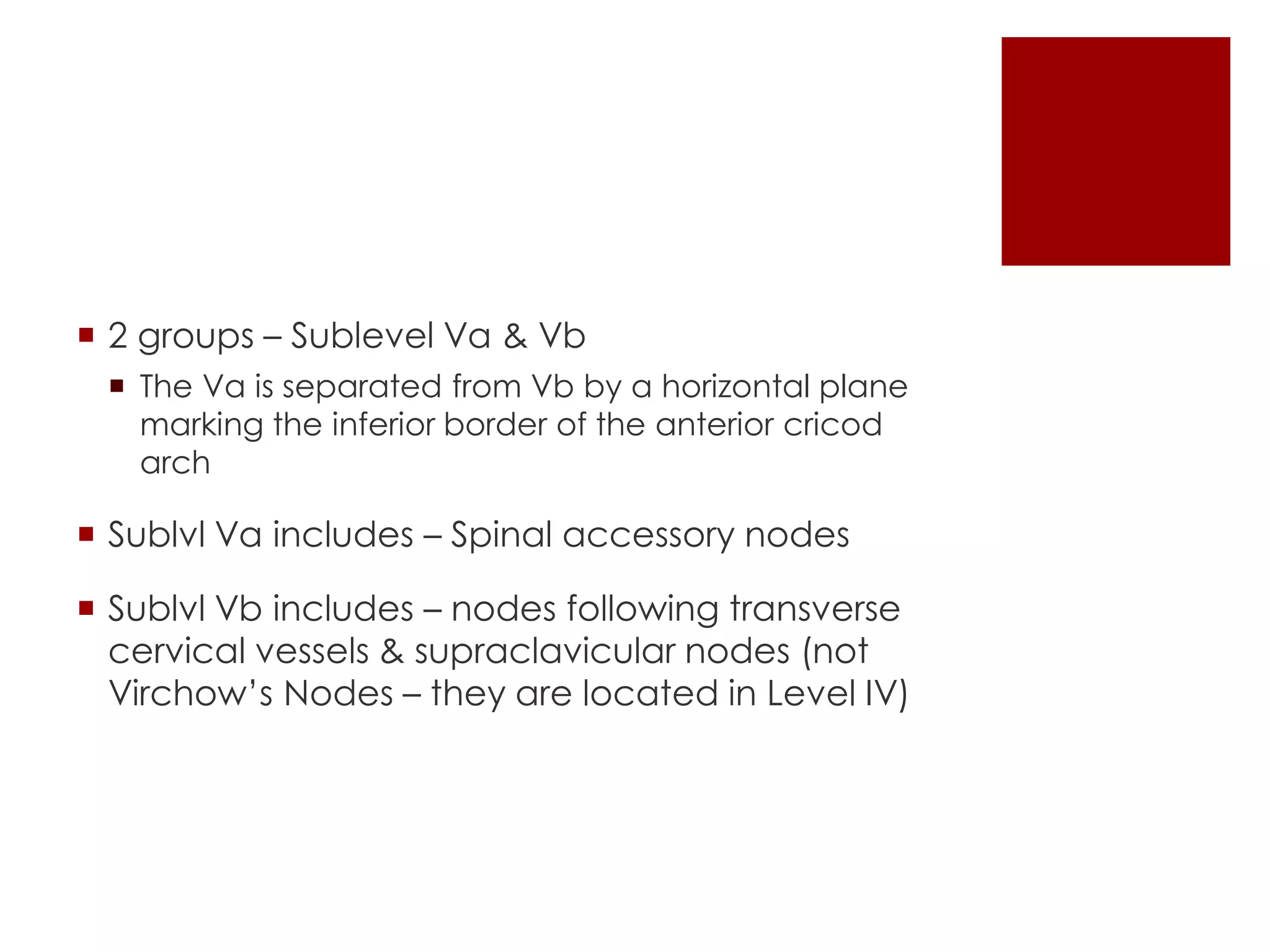
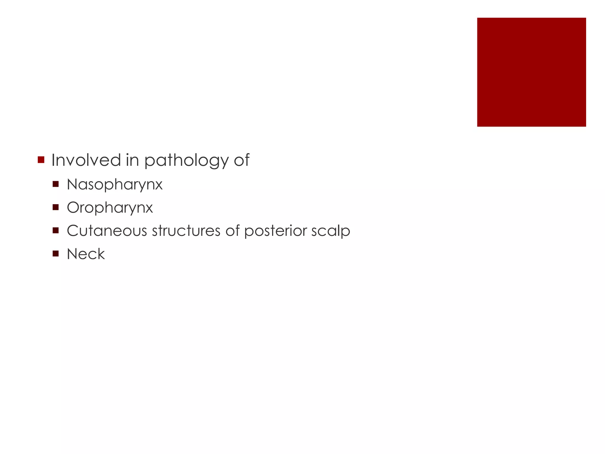
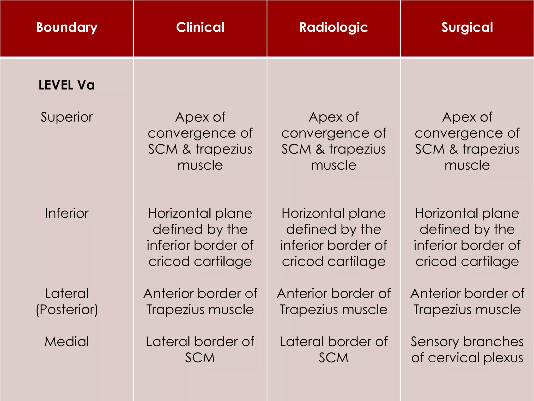
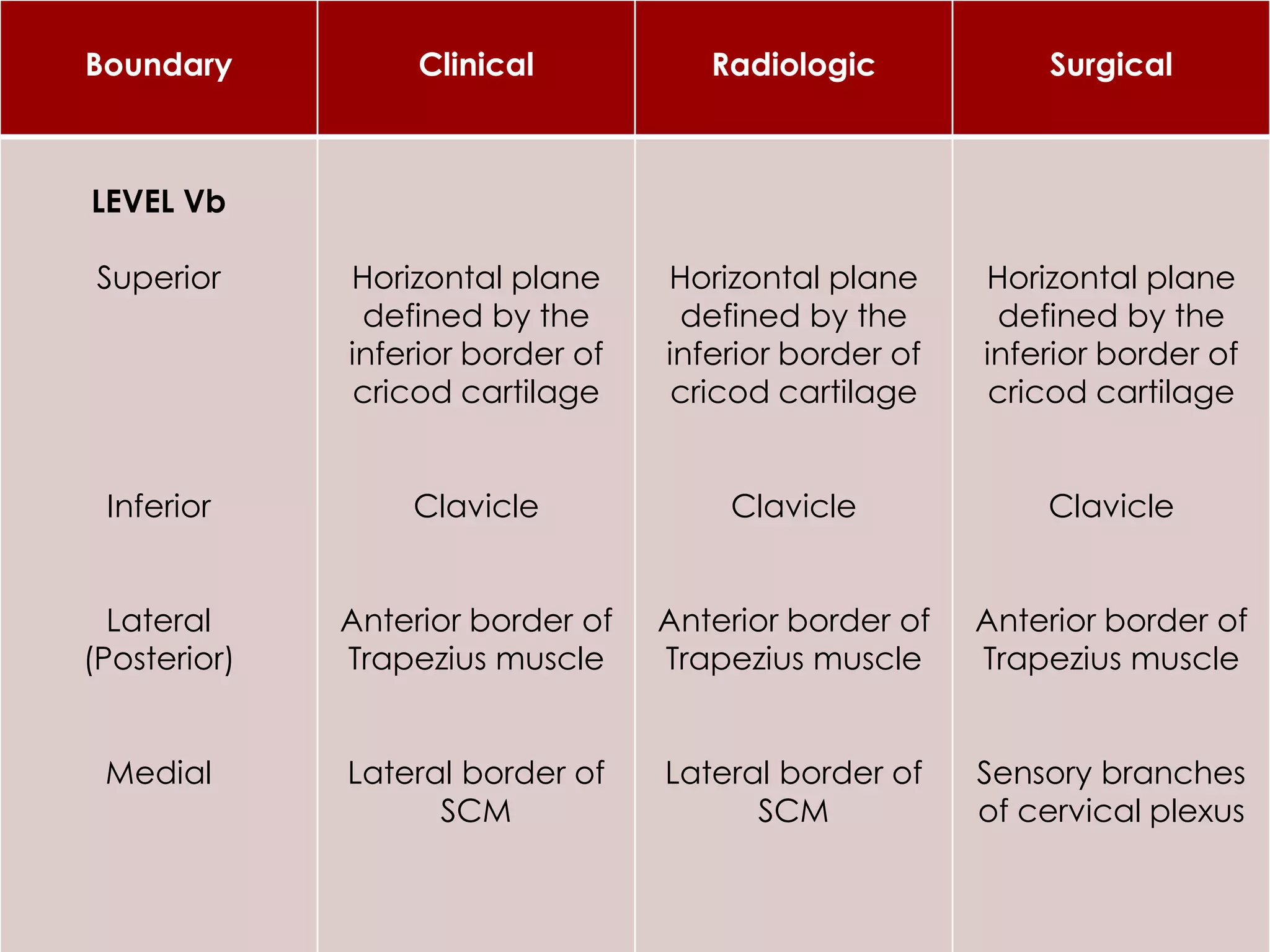
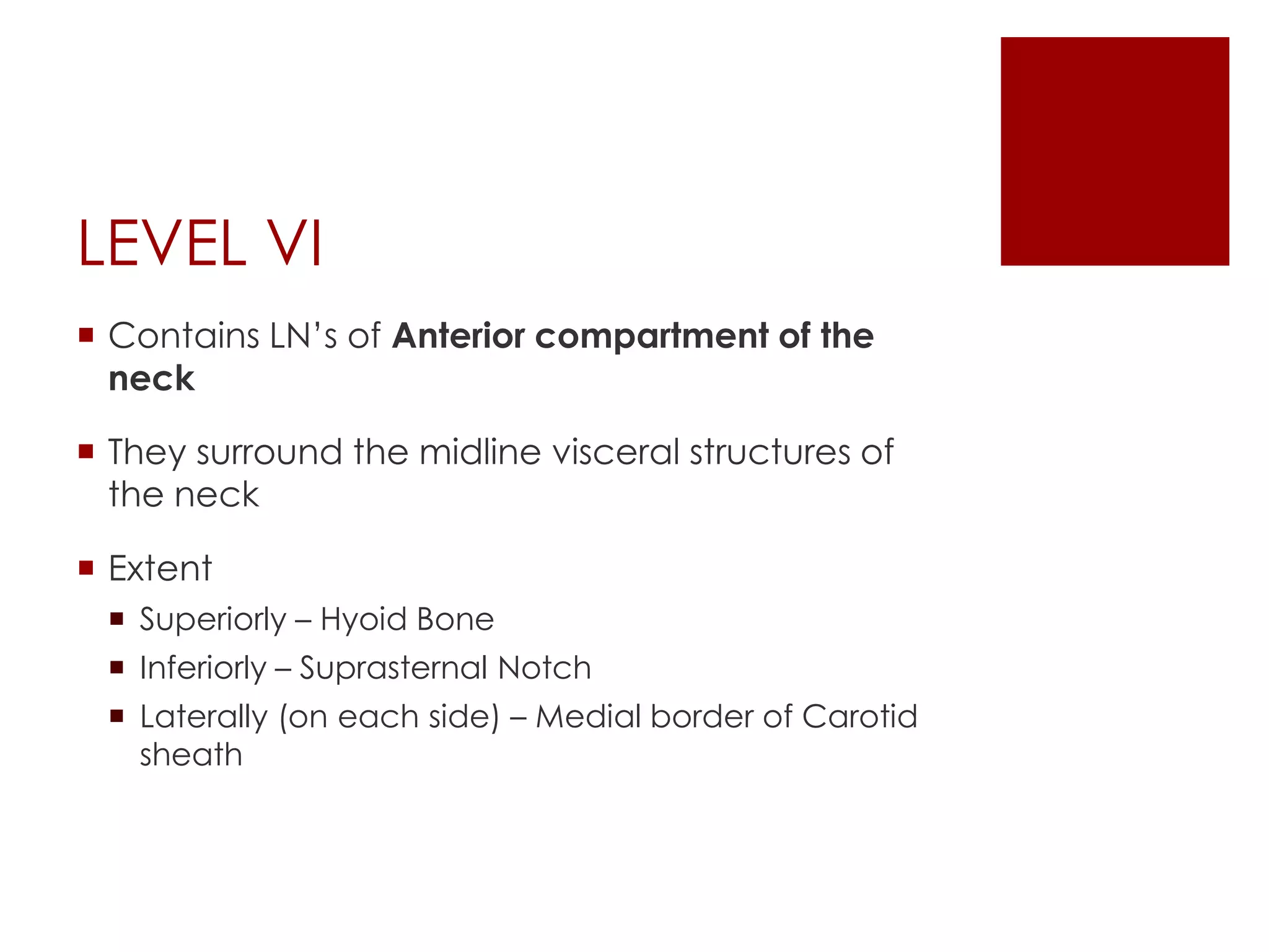
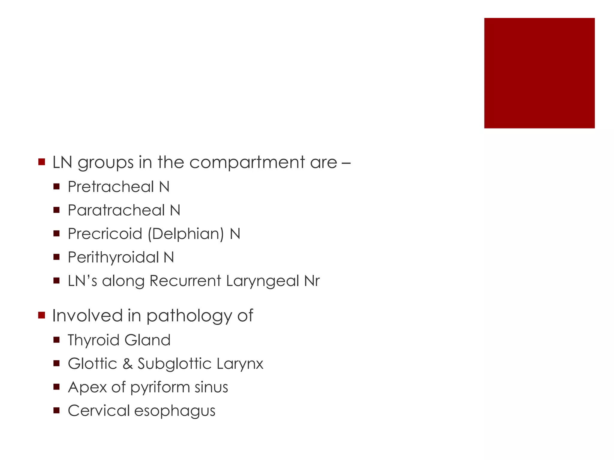
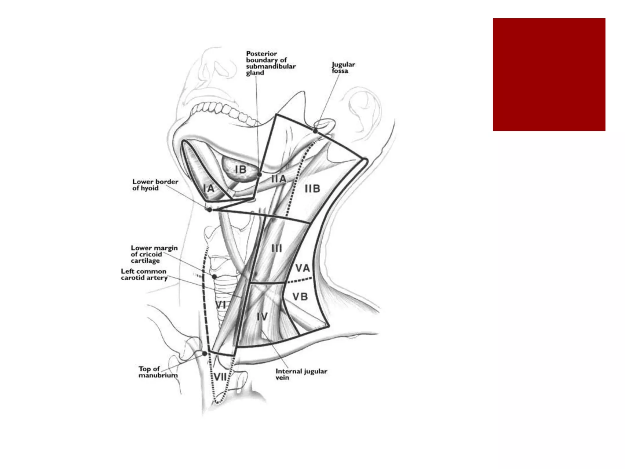
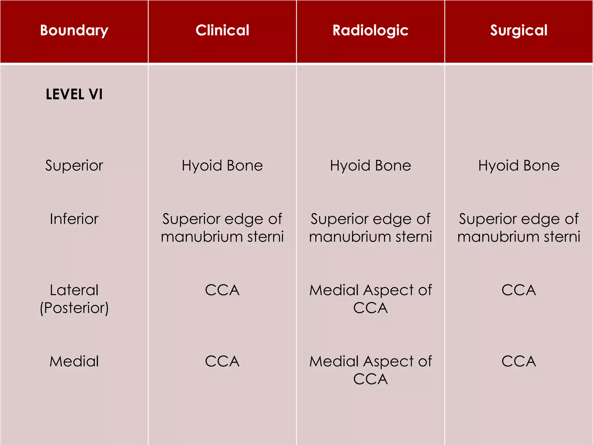
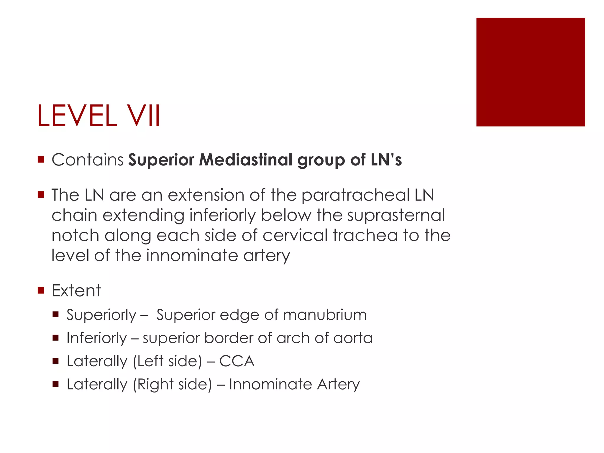
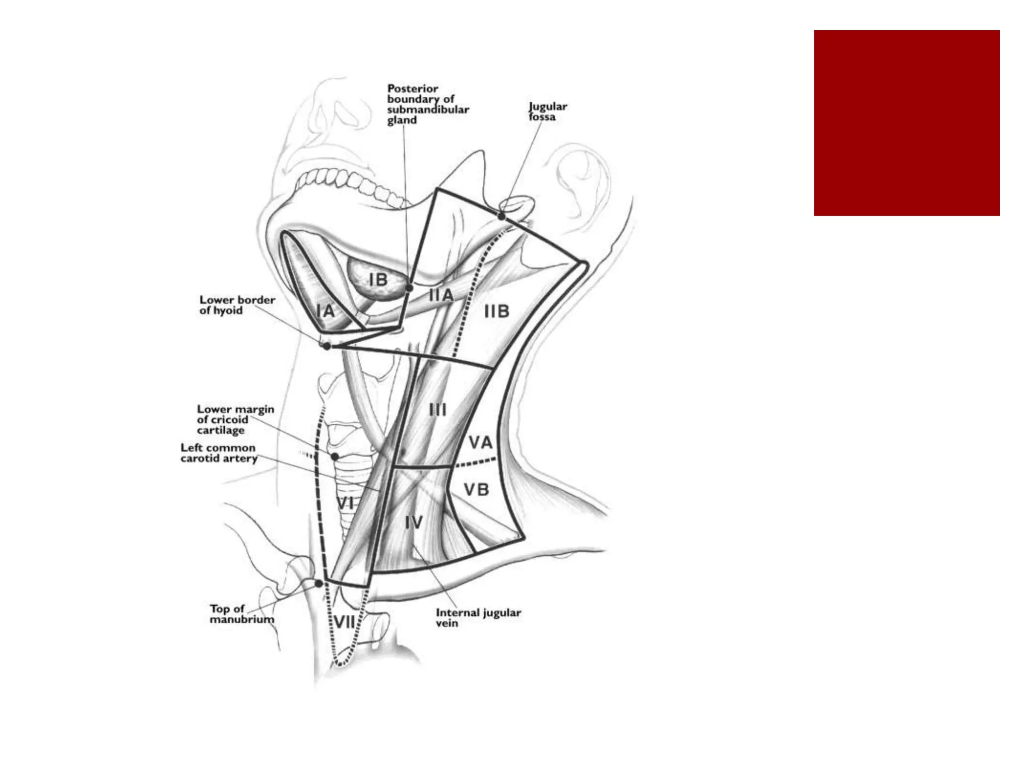
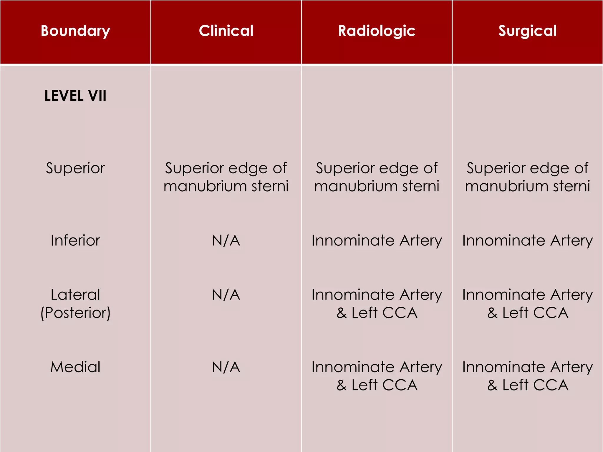
CERVICAL LYMPH NODES
Dr. Diptiman Baliarsingh
1st Year PG, Dept. of ENT,
Hi-Tech Medical College & Hospital, Bhubaneswar
Introduction
 The cervical group of lymph nodes extend from
 Mandible & skull base superiorly
 Clavicle inferiorly
 Posterior triangle of neck laterally & posteriroly
 Midline viscera anteriorly
 LN groups are categorized acc to original
description by Memorial Sloan-Kettering Group
Levels of Neck Nodes
 There are 7 levels of neck and most have
sublevels containing specific group of nodes
 Level I – Submental & Submandibular
 Level II – Upper Jugular
 Level III – Middle Jugular
 Level VI – Lower Jugular
 Level V – Posterior Triangle
 Level VI – Anterior/Central Compartment
 Level VII – Superior Mediastinal
LEVEL I
 2 groups of LN’s
 Sublevel Ia – Submental
 Sublevel Ib – Submandibular
 Boundaries of Lvl I
 Body of Mandible
 Ant. Belly of C/L digastric muscle
 Post. Belly of I/L digastric muscle
 Stylohyoid muscle
Submental (Sublevel Ia)
 LN’s in the triangular boundary formed by
 Ant belly of digastric muscles
 Hyoid Bone
 Involved in pathology of
 Floor of mouth
 Anterior oral tongue
 Anterior mandibular alveolar ridge
 Lower lip
Boundary Clinical Radiologic Surgical
LEVEL Ia
Superior
Inferior
Lateral
(Posterior)
Medial
Symphisis of
mandible
Body of Hyoid
N/A
N/A
Geniohyoid
Muscle,
Plane tangent to
inf. Border of
mandible
Body of Hyoid
Ant belly of I/L
digastric mus.
Ant belly of C/L
digastric muscle
Symphisis of
mandible
Body of Hyoid
Ant belly of I/L
digastric mus.
Ant belly of C/L
digastric muscle
Submandibular (Sublevel Ib)
 LN’s in the triangular boundary formed by
 Ant belly of digastric muscle
 Stylohyoid & Post belly of digastric muscle
 Body of Mandible
 Involved in pathology of
 Oral Cavity
 Anterior Nasal Cavity
 Soft tissue structures of midface
 Submandibular gland
Boundary Clinical Radiologic Surgical
LEVEL Ib
Superior
Inferior
Lateral
(Posterior)
Medial
Body of mandible
Plane through
Hyoid bone
Ant border of SCM
N/A
Mylohyoid Mus,
Body of mandible
Inferior edge of
hyoid bone
Post edge of
Submandibular Gl.
Ant belly of
digastric muscle
Body of mandible
Digastric tendon
attachment to
hyoid bone
Post edge of
Submandibular Gl
Ant belly of
digastric muscle
 Many of these lymph nodes lie in close proximity
to submandibular gland, and it is removed to
ensure thorough exenteration of all lymph nodes
with in this traingle
 Perifacial LN’s – drain Lip, buccal musoca, ant
nasal caity & soft tissue of neck, sometimes get
involved along with the Level I LN groups.
 ND’s performed for nodal disease asso with
primary lesions of these sites should be modified
to encompass perifacial nodes
LEVEL II
 Contain Upper Jugular LN’s
 2 groups – Sublevel IIa & IIb
 LN’s are located around upper third of internal
jugular vein & adjacent spinal accessory nerve
 Extent
 Superiorly - Skull base
 Inferirly - Inf border of Hyoid bone
 Ant (medial) boundary – stylohyoid mus
 Post (lateral) boundary – posterior border of SCM
 Sublevel IIa nodes are located anterior (medial)
to vertical plane defined by spinal accessory
nerve, whereas sublevel IIb nodes are located
posterior (lateral) to the vertical plane defined by
spinal accessory nerve
 Involved in pathology of
 Oral cavity, Nasal Cavity
 Nasopharynx, Oropharynx, Hypopharynx
 Larynx
 Parotid gland
Boundary Clinical Radiologic Surgical
LEVEL IIA
Superior
Inferior
Lateral
(Posterior)
Medial
Mastoid process
Horizontal plane
defined by the
inferior border of
hyoid bone
N/A
N/A
Skull base,
Caudal edge of
C1 lateral process
Horizontal plane
defined by the
inferior border of
hyoid bone
Post. border of IJV
Post. edge of
submandibular
gland
Skull base
Carotid bifurcation
Vertical plane
defined by Spinal
acc. nerve
Post. edge of
submandibular
gland
Boundary Clinical Radiologic Surgical
LEVEL IIB
Superior
Inferior
Lateral
(Posterior)
Medial
Mastoid process
Horizontal plane
defined by the
inferior border of
hyoid bone
Lateral border of
SCM
N/A
Skull base,
Caudal edge of
C1 lateral process
Horizontal plane
defined by the
inferior border of
hyoid bone
Lateral border of
SCM
Medial edge of
ICA, paraspinal
(lev. scapulae)
mus
Skull base
Carotid bifurcation
Lateral border of
SCM
Vertical plane
defined by Spinal
acc. nerve
 AHNS Committee recommends – perpendicular
plane formed by posterior aspect of
submandibular gland as radiologic marker of this
boundary
LEVEL III
 Contains Middle Jugular LN’s Group
 Located around middle third of IJV
 Extent
 Superiorly – Carotid bifurcation / Inferior aspect of
body of hyoid bone
 Inferiorly – Junction of Omohyoid mus with IJV / lower
border of cricoid arch(cartilage)
 Medially – Lateral border of sternohyiod muscle
 Laterally – Post border of SCM muscle
 Involved in pathology of
 Oral cavity
 Nasopharynx, Oropharynx, Hypopharynx
 Larynx
 AHNS Committee recommends – Lateral border
of CCA serves as Radiologic marker for medial
boundary
Boundary Clinical Radiologic Surgical
LEVEL III
Superior
Inferior
Lateral
(Posterior)
Medial
Horizontal plane
defined by the
inferior border of
hyoid bone
Horizontal plane
defined by the
inferior border of
cricod cartilage
Lateral border of
SCM
Medial border of
SCM
Horizontal plane
defined by the
inferior border of
hyoid bone
Horizontal plane
defined by the
inferior border of
cricod cartilage
Lateral border of
SCM
Medial edge of
ICA, paraspinal
(lev. scapulae)
mus
Carotid bifurcation
Omohyoid muscle
Sensory branches
of cervical plexus
Sternohyoid
muscle
LEVEL IV
 Contains Lower Jugular LN’s Group
 Located around lower third of IJV
 Extent
 Superiorly – Junction of Omohyoid mus with IJV /
lower border of cricoid arch(cartilage)
 Inferiorly – Clavicle
 Medially – Lateral border of sternohyiod muscle
 Laterally – Post border of SCM muscle
 Involved in pathology of
 Hypopharynx
 Thyroid
 Cervical Esophagus
 Larynx
 AHNS Committee recommends – Lateral border
of CCA serves as Radiologic marker for medial
boundary
Boundary Clinical Radiologic Surgical
LEVEL IV
Superior
Inferior
Lateral
(Posterior)
Medial
Horizontal plane
defined by the
inferior border of
cricod cartilage
Clavicle
Lateral border of
SCM
Medial border of
SCM
Horizontal plane
defined by the
inferior border of
cricod cartilage
2cm cranial to
sternoclavicular
joint
Lateral border of
SCM
Medial edge of
CCA, paraspinal
(scalenus) mus
Omohyoid muscle
Clavicle
Sensory branches
of cervical plexus
Sternohyoid
muscle
LEVEL V
 Contains Posterior Triangle LN’s Group
 Located along the lower half of spinal accessory
nerve & transverse cervical artery
 Extent
 Superiorly (Apex) – convergence of SCM & trapezius
muscle
 Inferiorly – Clavicle
 Medially – Post border of SCM muscle
 Laterally – Anterior border of trapezius muscle
 2 groups – Sublevel Va & Vb
 The Va is separated from Vb by a horizontal plane
marking the inferior border of the anterior cricod
arch
 Sublvl Va includes – Spinal accessory nodes
 Sublvl Vb includes – nodes following transverse
cervical vessels & supraclavicular nodes (not
Virchow’s Nodes – they are located in Level IV)
 Involved in pathology of
 Nasopharynx
 Oropharynx
 Cutaneous structures of posterior scalp
 Neck
Boundary Clinical Radiologic Surgical
LEVEL Va
Superior
Inferior
Lateral
(Posterior)
Medial
Apex of
convergence of
SCM & trapezius
muscle
Horizontal plane
defined by the
inferior border of
cricod cartilage
Anterior border of
Trapezius muscle
Lateral border of
SCM
Apex of
convergence of
SCM & trapezius
muscle
Horizontal plane
defined by the
inferior border of
cricod cartilage
Anterior border of
Trapezius muscle
Lateral border of
SCM
Apex of
convergence of
SCM & trapezius
muscle
Horizontal plane
defined by the
inferior border of
cricod cartilage
Anterior border of
Trapezius muscle
Sensory branches
of cervical plexus
Boundary Clinical Radiologic Surgical
LEVEL Vb
Superior
Inferior
Lateral
(Posterior)
Medial
Horizontal plane
defined by the
inferior border of
cricod cartilage
Clavicle
Anterior border of
Trapezius muscle
Lateral border of
SCM
Horizontal plane
defined by the
inferior border of
cricod cartilage
Clavicle
Anterior border of
Trapezius muscle
Lateral border of
SCM
Horizontal plane
defined by the
inferior border of
cricod cartilage
Clavicle
Anterior border of
Trapezius muscle
Sensory branches
of cervical plexus
LEVEL VI
 Contains LN’s of Anterior compartment of the
neck
 They surround the midline visceral structures of
the neck
 Extent
 Superiorly – Hyoid Bone
 Inferiorly – Suprasternal Notch
 Laterally (on each side) – Medial border of Carotid
sheath
 LN groups in the compartment are –
 Pretracheal N
 Paratracheal N
 Precricoid (Delphian) N
 Perithyroidal N
 LN’s along Recurrent Laryngeal Nr
 Involved in pathology of
 Thyroid Gland
 Glottic & Subglottic Larynx
 Apex of pyriform sinus
 Cervical esophagus
Boundary Clinical Radiologic Surgical
LEVEL VI
Superior
Inferior
Lateral
(Posterior)
Medial
Hyoid Bone
Superior edge of
manubrium sterni
CCA
CCA
Hyoid Bone
Superior edge of
manubrium sterni
Medial Aspect of
CCA
Medial Aspect of
CCA
Hyoid Bone
Superior edge of
manubrium sterni
CCA
CCA
LEVEL VII
 Contains Superior Mediastinal group of LN’s
 The LN are an extension of the paratracheal LN
chain extending inferiorly below the suprasternal
notch along each side of cervical trachea to the
level of the innominate artery
 Extent
 Superiorly – Superior edge of manubrium
 Inferiorly – superior border of arch of aorta
 Laterally (Left side) – CCA
 Laterally (Right side) – Innominate Artery
Boundary Clinical Radiologic Surgical
LEVEL VII
Superior
Inferior
Lateral
(Posterior)
Medial
Superior edge of
manubrium sterni
N/A
N/A
N/A
Superior edge of
manubrium sterni
Innominate Artery
Innominate Artery
& Left CCA
Innominate Artery
& Left CCA
Superior edge of
manubrium sterni
Innominate Artery
Innominate Artery
& Left CCA
Innominate Artery
& Left CCA
Thank You...
 REFERENCES
 CUMMING’S Otolaryngolagy Head & Neck Surgery -
5th Ed

Cervical lymph nodes

  • 1.
    CERVICAL LYMPH NODES Dr.Diptiman Baliarsingh 1st Year PG, Dept. of ENT, Hi-Tech Medical College & Hospital, Bhubaneswar
  • 2.
    Introduction  The cervicalgroup of lymph nodes extend from  Mandible & skull base superiorly  Clavicle inferiorly  Posterior triangle of neck laterally & posteriroly  Midline viscera anteriorly  LN groups are categorized acc to original description by Memorial Sloan-Kettering Group
  • 3.
    Levels of NeckNodes  There are 7 levels of neck and most have sublevels containing specific group of nodes  Level I – Submental & Submandibular  Level II – Upper Jugular  Level III – Middle Jugular  Level VI – Lower Jugular  Level V – Posterior Triangle  Level VI – Anterior/Central Compartment  Level VII – Superior Mediastinal
  • 6.
    LEVEL I  2groups of LN’s  Sublevel Ia – Submental  Sublevel Ib – Submandibular  Boundaries of Lvl I  Body of Mandible  Ant. Belly of C/L digastric muscle  Post. Belly of I/L digastric muscle  Stylohyoid muscle
  • 7.
    Submental (Sublevel Ia) LN’s in the triangular boundary formed by  Ant belly of digastric muscles  Hyoid Bone  Involved in pathology of  Floor of mouth  Anterior oral tongue  Anterior mandibular alveolar ridge  Lower lip
  • 10.
    Boundary Clinical RadiologicSurgical LEVEL Ia Superior Inferior Lateral (Posterior) Medial Symphisis of mandible Body of Hyoid N/A N/A Geniohyoid Muscle, Plane tangent to inf. Border of mandible Body of Hyoid Ant belly of I/L digastric mus. Ant belly of C/L digastric muscle Symphisis of mandible Body of Hyoid Ant belly of I/L digastric mus. Ant belly of C/L digastric muscle
  • 11.
    Submandibular (Sublevel Ib) LN’s in the triangular boundary formed by  Ant belly of digastric muscle  Stylohyoid & Post belly of digastric muscle  Body of Mandible  Involved in pathology of  Oral Cavity  Anterior Nasal Cavity  Soft tissue structures of midface  Submandibular gland
  • 13.
    Boundary Clinical RadiologicSurgical LEVEL Ib Superior Inferior Lateral (Posterior) Medial Body of mandible Plane through Hyoid bone Ant border of SCM N/A Mylohyoid Mus, Body of mandible Inferior edge of hyoid bone Post edge of Submandibular Gl. Ant belly of digastric muscle Body of mandible Digastric tendon attachment to hyoid bone Post edge of Submandibular Gl Ant belly of digastric muscle
  • 14.
     Many ofthese lymph nodes lie in close proximity to submandibular gland, and it is removed to ensure thorough exenteration of all lymph nodes with in this traingle  Perifacial LN’s – drain Lip, buccal musoca, ant nasal caity & soft tissue of neck, sometimes get involved along with the Level I LN groups.  ND’s performed for nodal disease asso with primary lesions of these sites should be modified to encompass perifacial nodes
  • 15.
    LEVEL II  ContainUpper Jugular LN’s  2 groups – Sublevel IIa & IIb  LN’s are located around upper third of internal jugular vein & adjacent spinal accessory nerve  Extent  Superiorly - Skull base  Inferirly - Inf border of Hyoid bone  Ant (medial) boundary – stylohyoid mus  Post (lateral) boundary – posterior border of SCM
  • 16.
     Sublevel IIanodes are located anterior (medial) to vertical plane defined by spinal accessory nerve, whereas sublevel IIb nodes are located posterior (lateral) to the vertical plane defined by spinal accessory nerve  Involved in pathology of  Oral cavity, Nasal Cavity  Nasopharynx, Oropharynx, Hypopharynx  Larynx  Parotid gland
  • 18.
    Boundary Clinical RadiologicSurgical LEVEL IIA Superior Inferior Lateral (Posterior) Medial Mastoid process Horizontal plane defined by the inferior border of hyoid bone N/A N/A Skull base, Caudal edge of C1 lateral process Horizontal plane defined by the inferior border of hyoid bone Post. border of IJV Post. edge of submandibular gland Skull base Carotid bifurcation Vertical plane defined by Spinal acc. nerve Post. edge of submandibular gland
  • 20.
    Boundary Clinical RadiologicSurgical LEVEL IIB Superior Inferior Lateral (Posterior) Medial Mastoid process Horizontal plane defined by the inferior border of hyoid bone Lateral border of SCM N/A Skull base, Caudal edge of C1 lateral process Horizontal plane defined by the inferior border of hyoid bone Lateral border of SCM Medial edge of ICA, paraspinal (lev. scapulae) mus Skull base Carotid bifurcation Lateral border of SCM Vertical plane defined by Spinal acc. nerve
  • 21.
     AHNS Committeerecommends – perpendicular plane formed by posterior aspect of submandibular gland as radiologic marker of this boundary
  • 22.
    LEVEL III  ContainsMiddle Jugular LN’s Group  Located around middle third of IJV  Extent  Superiorly – Carotid bifurcation / Inferior aspect of body of hyoid bone  Inferiorly – Junction of Omohyoid mus with IJV / lower border of cricoid arch(cartilage)  Medially – Lateral border of sternohyiod muscle  Laterally – Post border of SCM muscle
  • 23.
     Involved inpathology of  Oral cavity  Nasopharynx, Oropharynx, Hypopharynx  Larynx  AHNS Committee recommends – Lateral border of CCA serves as Radiologic marker for medial boundary
  • 25.
    Boundary Clinical RadiologicSurgical LEVEL III Superior Inferior Lateral (Posterior) Medial Horizontal plane defined by the inferior border of hyoid bone Horizontal plane defined by the inferior border of cricod cartilage Lateral border of SCM Medial border of SCM Horizontal plane defined by the inferior border of hyoid bone Horizontal plane defined by the inferior border of cricod cartilage Lateral border of SCM Medial edge of ICA, paraspinal (lev. scapulae) mus Carotid bifurcation Omohyoid muscle Sensory branches of cervical plexus Sternohyoid muscle
  • 26.
    LEVEL IV  ContainsLower Jugular LN’s Group  Located around lower third of IJV  Extent  Superiorly – Junction of Omohyoid mus with IJV / lower border of cricoid arch(cartilage)  Inferiorly – Clavicle  Medially – Lateral border of sternohyiod muscle  Laterally – Post border of SCM muscle
  • 27.
     Involved inpathology of  Hypopharynx  Thyroid  Cervical Esophagus  Larynx  AHNS Committee recommends – Lateral border of CCA serves as Radiologic marker for medial boundary
  • 29.
    Boundary Clinical RadiologicSurgical LEVEL IV Superior Inferior Lateral (Posterior) Medial Horizontal plane defined by the inferior border of cricod cartilage Clavicle Lateral border of SCM Medial border of SCM Horizontal plane defined by the inferior border of cricod cartilage 2cm cranial to sternoclavicular joint Lateral border of SCM Medial edge of CCA, paraspinal (scalenus) mus Omohyoid muscle Clavicle Sensory branches of cervical plexus Sternohyoid muscle
  • 30.
    LEVEL V  ContainsPosterior Triangle LN’s Group  Located along the lower half of spinal accessory nerve & transverse cervical artery  Extent  Superiorly (Apex) – convergence of SCM & trapezius muscle  Inferiorly – Clavicle  Medially – Post border of SCM muscle  Laterally – Anterior border of trapezius muscle
  • 31.
     2 groups– Sublevel Va & Vb  The Va is separated from Vb by a horizontal plane marking the inferior border of the anterior cricod arch  Sublvl Va includes – Spinal accessory nodes  Sublvl Vb includes – nodes following transverse cervical vessels & supraclavicular nodes (not Virchow’s Nodes – they are located in Level IV)
  • 32.
     Involved inpathology of  Nasopharynx  Oropharynx  Cutaneous structures of posterior scalp  Neck
  • 34.
    Boundary Clinical RadiologicSurgical LEVEL Va Superior Inferior Lateral (Posterior) Medial Apex of convergence of SCM & trapezius muscle Horizontal plane defined by the inferior border of cricod cartilage Anterior border of Trapezius muscle Lateral border of SCM Apex of convergence of SCM & trapezius muscle Horizontal plane defined by the inferior border of cricod cartilage Anterior border of Trapezius muscle Lateral border of SCM Apex of convergence of SCM & trapezius muscle Horizontal plane defined by the inferior border of cricod cartilage Anterior border of Trapezius muscle Sensory branches of cervical plexus
  • 36.
    Boundary Clinical RadiologicSurgical LEVEL Vb Superior Inferior Lateral (Posterior) Medial Horizontal plane defined by the inferior border of cricod cartilage Clavicle Anterior border of Trapezius muscle Lateral border of SCM Horizontal plane defined by the inferior border of cricod cartilage Clavicle Anterior border of Trapezius muscle Lateral border of SCM Horizontal plane defined by the inferior border of cricod cartilage Clavicle Anterior border of Trapezius muscle Sensory branches of cervical plexus
  • 37.
    LEVEL VI  ContainsLN’s of Anterior compartment of the neck  They surround the midline visceral structures of the neck  Extent  Superiorly – Hyoid Bone  Inferiorly – Suprasternal Notch  Laterally (on each side) – Medial border of Carotid sheath
  • 38.
     LN groupsin the compartment are –  Pretracheal N  Paratracheal N  Precricoid (Delphian) N  Perithyroidal N  LN’s along Recurrent Laryngeal Nr  Involved in pathology of  Thyroid Gland  Glottic & Subglottic Larynx  Apex of pyriform sinus  Cervical esophagus
  • 40.
    Boundary Clinical RadiologicSurgical LEVEL VI Superior Inferior Lateral (Posterior) Medial Hyoid Bone Superior edge of manubrium sterni CCA CCA Hyoid Bone Superior edge of manubrium sterni Medial Aspect of CCA Medial Aspect of CCA Hyoid Bone Superior edge of manubrium sterni CCA CCA
  • 41.
    LEVEL VII  ContainsSuperior Mediastinal group of LN’s  The LN are an extension of the paratracheal LN chain extending inferiorly below the suprasternal notch along each side of cervical trachea to the level of the innominate artery  Extent  Superiorly – Superior edge of manubrium  Inferiorly – superior border of arch of aorta  Laterally (Left side) – CCA  Laterally (Right side) – Innominate Artery
  • 43.
    Boundary Clinical RadiologicSurgical LEVEL VII Superior Inferior Lateral (Posterior) Medial Superior edge of manubrium sterni N/A N/A N/A Superior edge of manubrium sterni Innominate Artery Innominate Artery & Left CCA Innominate Artery & Left CCA Superior edge of manubrium sterni Innominate Artery Innominate Artery & Left CCA Innominate Artery & Left CCA
  • 44.
    Thank You...  REFERENCES CUMMING’S Otolaryngolagy Head & Neck Surgery - 5th Ed