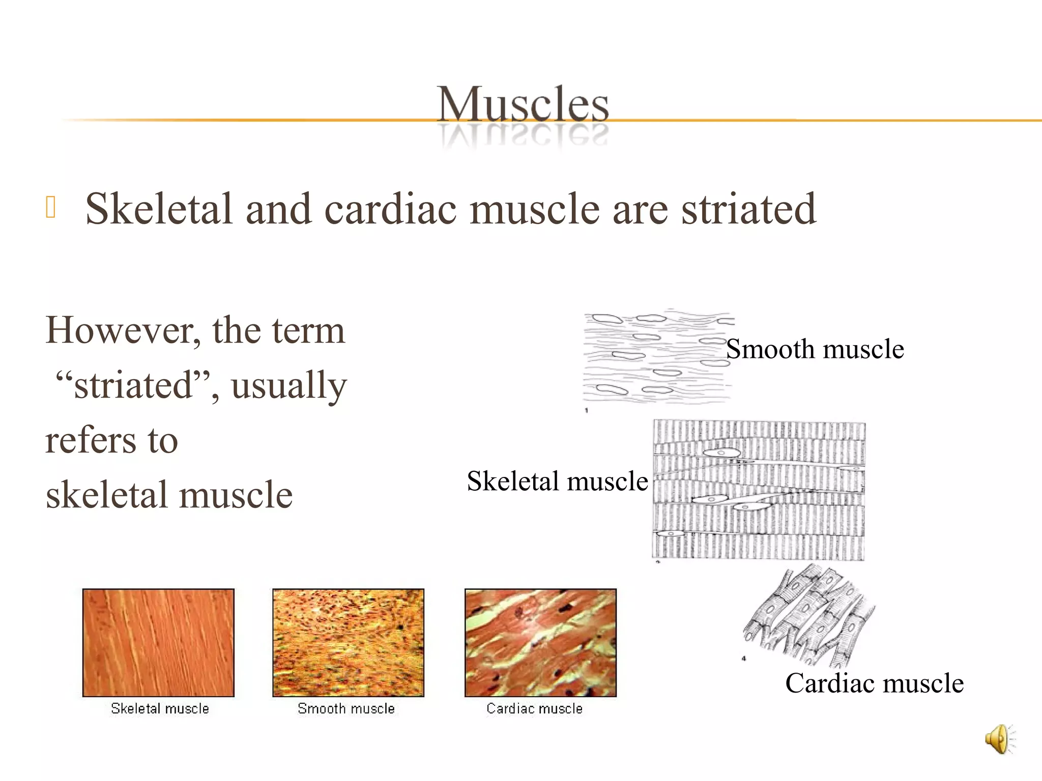
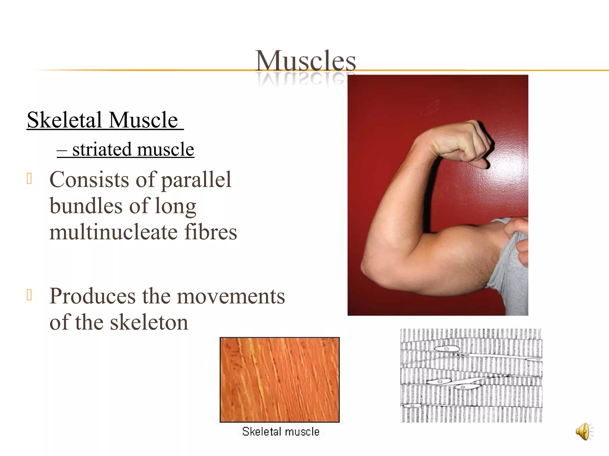
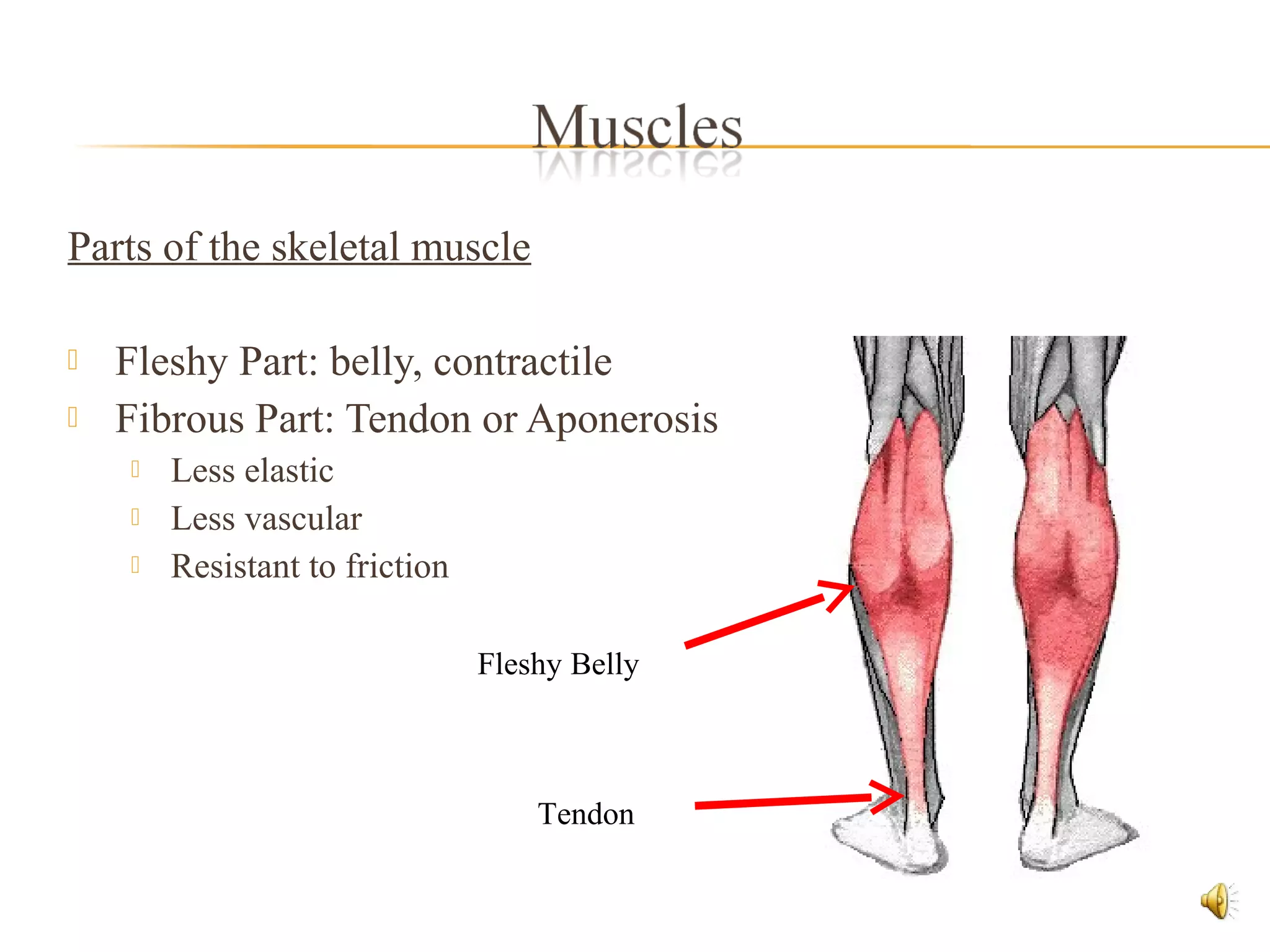
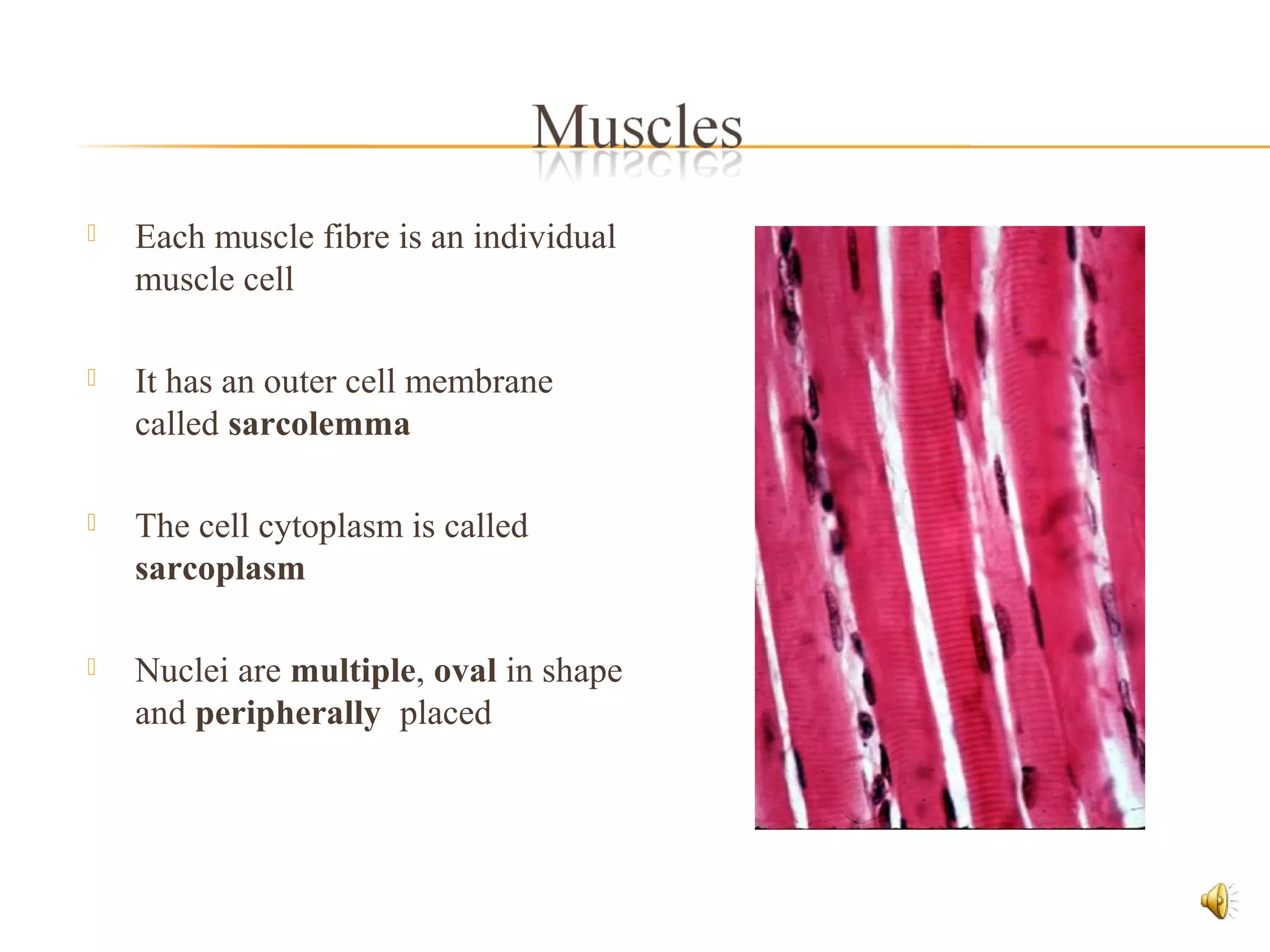
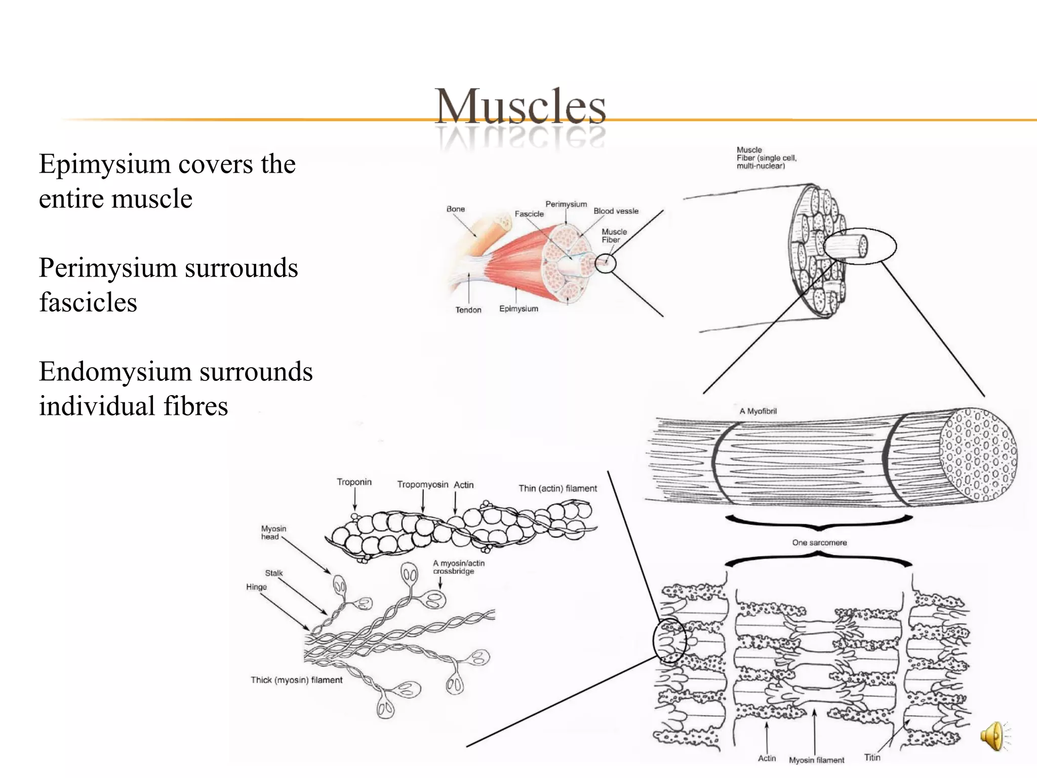
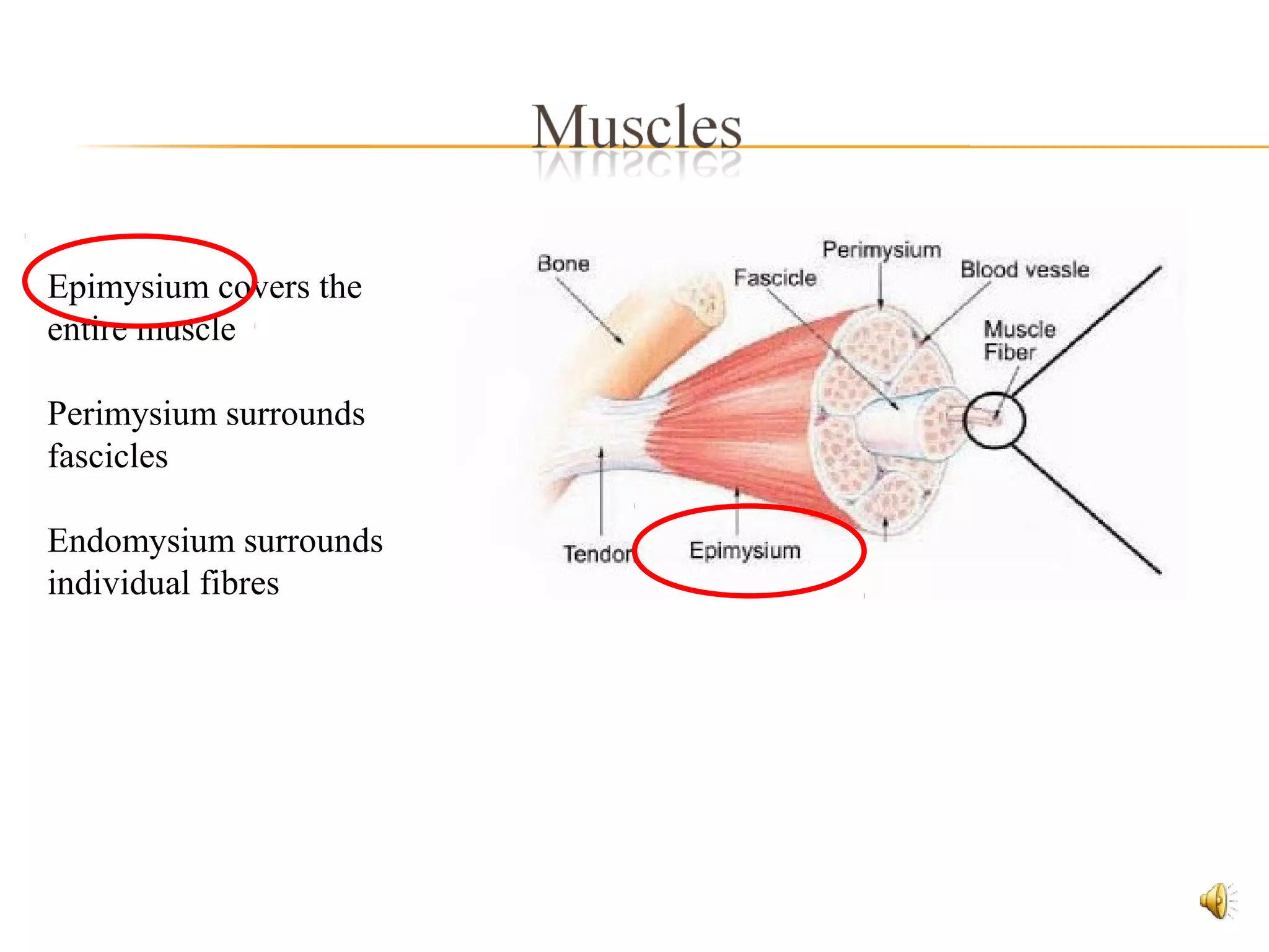
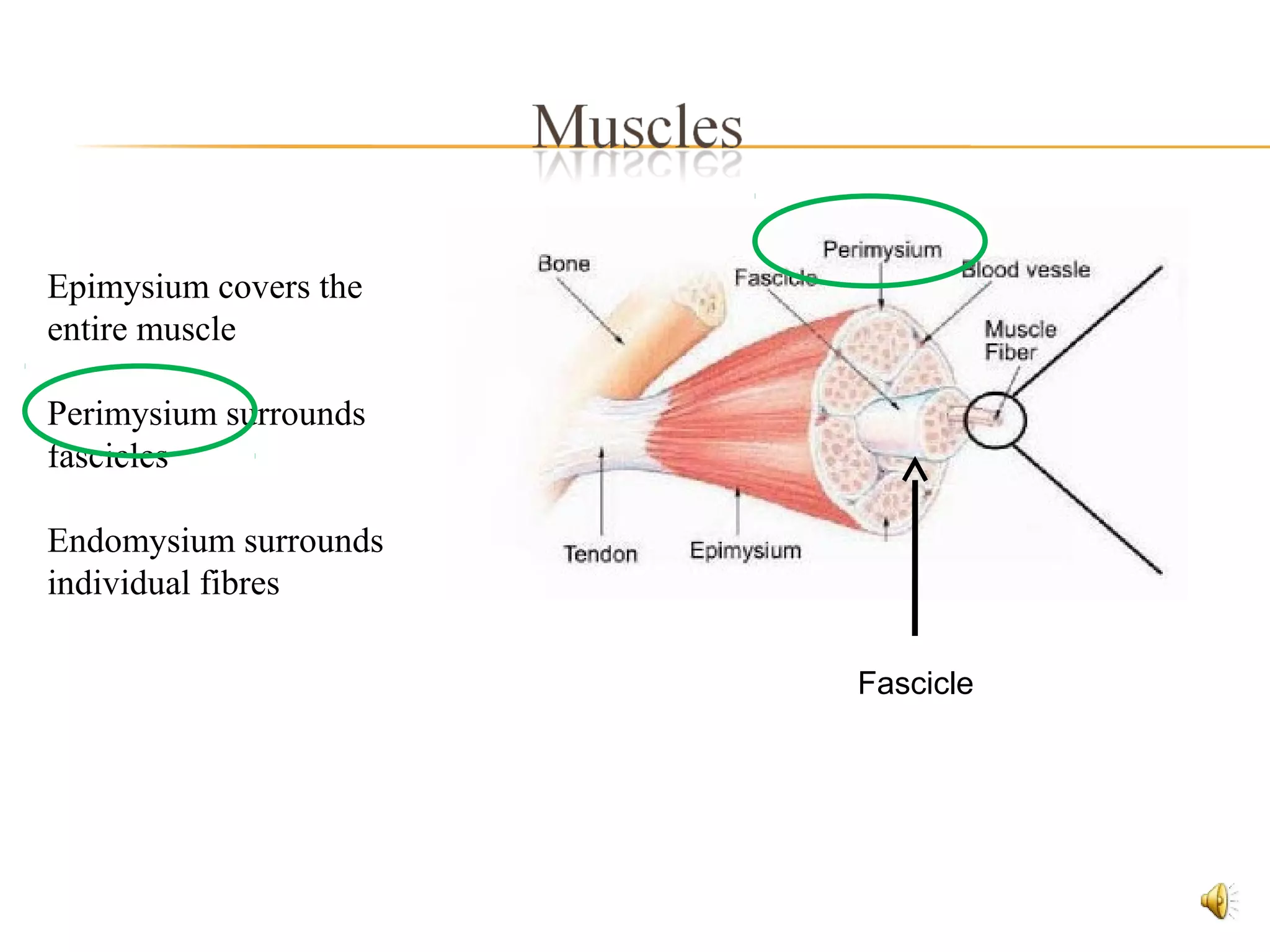
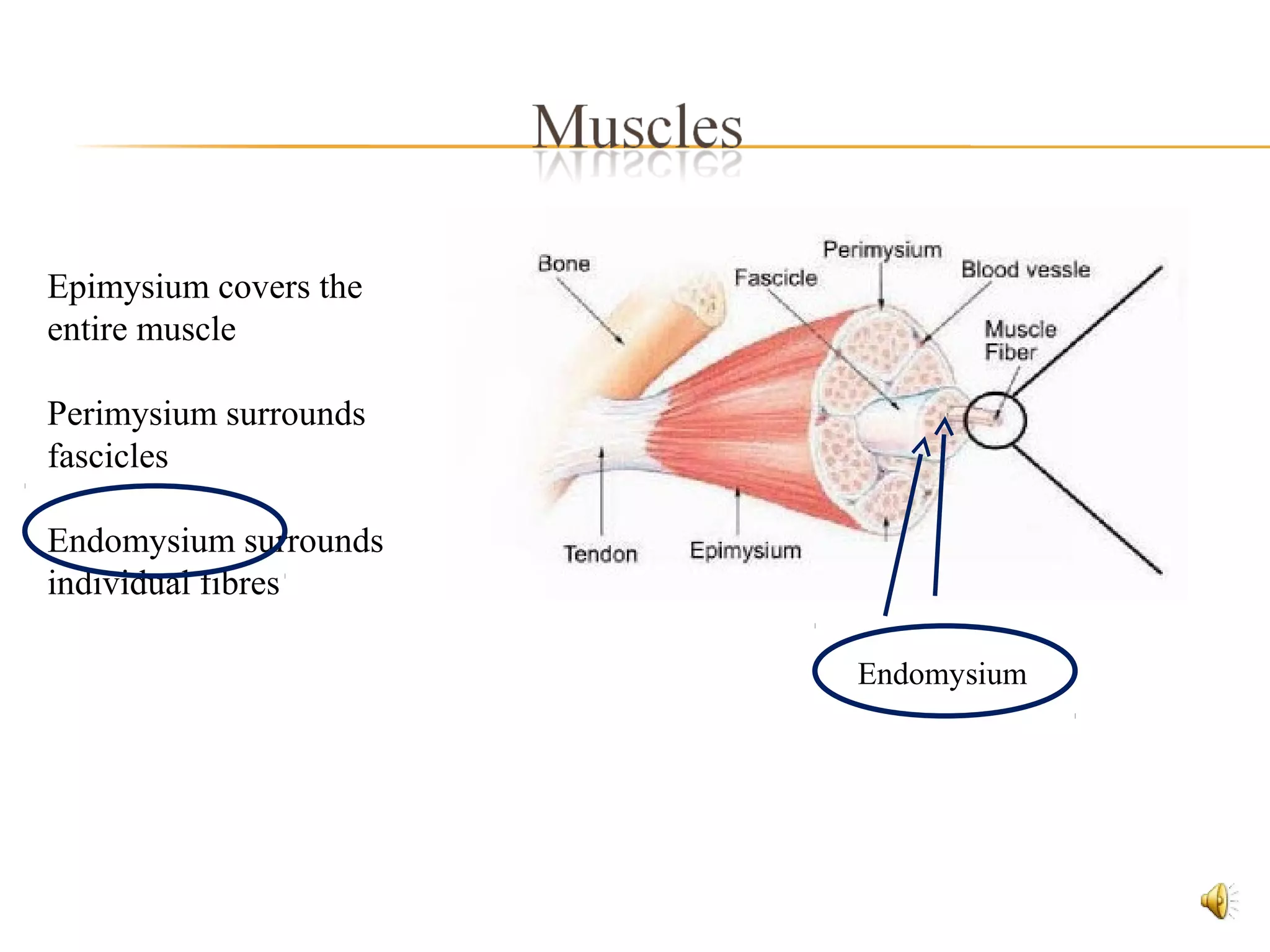
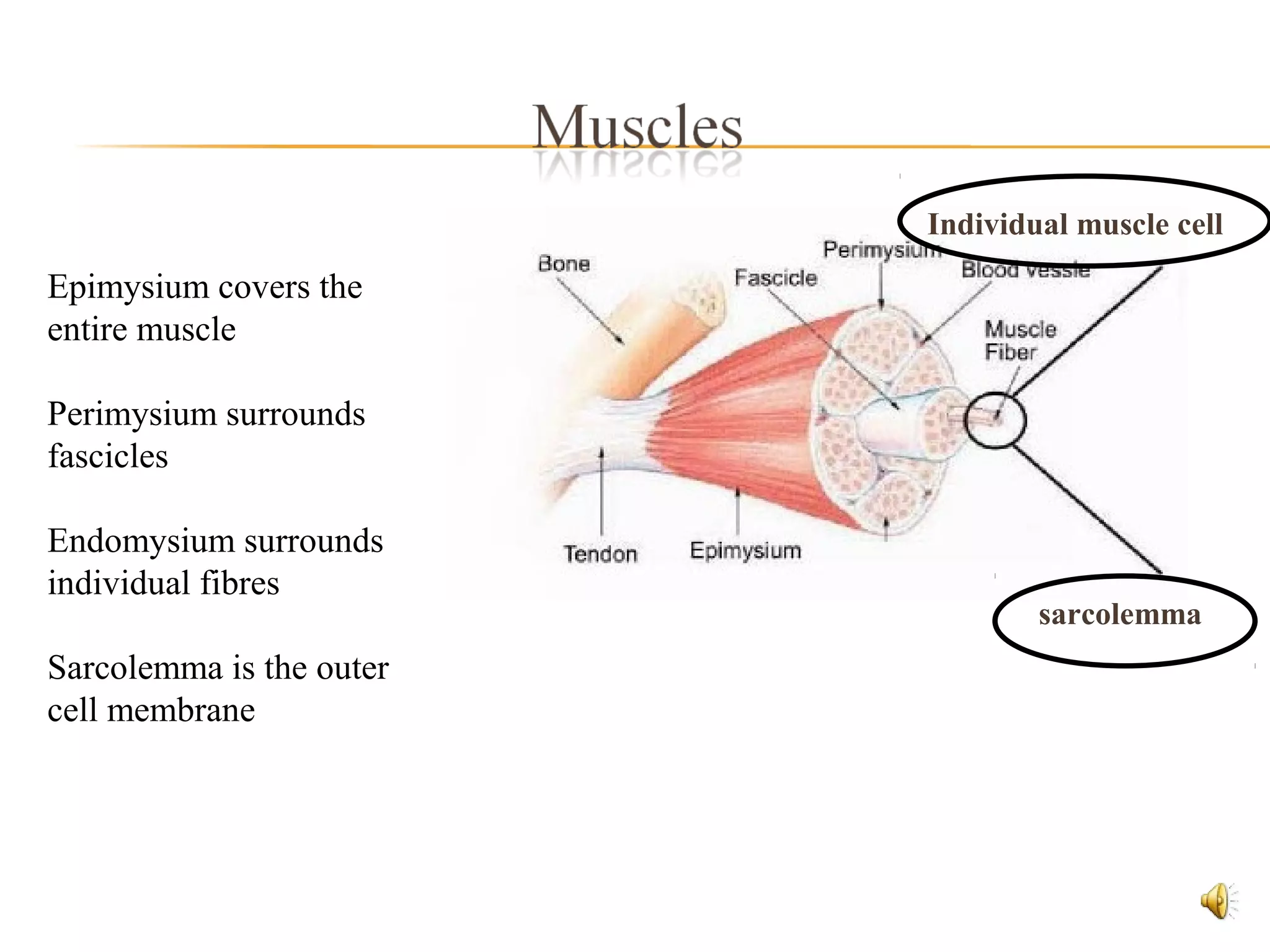
Raymond G Buick FRCS
9 mins
A muscle is a tissue that has the ability to contract
Types of Muscle tissue:
 Skeletal
 Cardiac
 Smooth muscle
 Skeletal and cardiac muscle are striated
However, the term
“striated”, usually
refers to
skeletal muscle
Smooth muscle
Skeletal muscle
Cardiac muscle
Skeletal Muscle
– striated muscle
 Consists of parallel
bundles of long
multinucleate fibres
 Produces the movements
of the skeleton
Parts of the skeletal muscle
 Fleshy Part: belly, contractile
 Fibrous Part: Tendon or Aponerosis
 Less elastic
 Less vascular
 Resistant to friction
Fleshy Belly
Tendon
 Each muscle fibre is an individual
muscle cell
 It has an outer cell membrane
called sarcolemma
 The cell cytoplasm is called
sarcoplasm
 Nuclei are multiple, oval in shape
and peripherally placed
Epimysium covers the
entire muscle
Perimysium surrounds
fascicles
Endomysium surrounds
individual fibres
Epimysium covers the
entire muscle
Perimysium surrounds
fascicles
Endomysium surrounds
individual fibres
Epimysium covers the
entire muscle
Perimysium surrounds
fascicles
Endomysium surrounds
individual fibres
Fascicle
Epimysium covers the
entire muscle
Perimysium surrounds
fascicles
Endomysium surrounds
individual fibres
Endomysium
Epimysium covers the
entire muscle
Perimysium surrounds
fascicles
Endomysium surrounds
individual fibres
Sarcolemma is the outer
cell membrane
sarcolemma
Individual muscle cell
 A tendon (sinew) is a tough band of fibrous connective tissue.
 Usually connects muscle to bone
 Consolodation of collagen fibres in the perimysium and epimysium.
 Maximises efficiency of the muscle in moving a bone
 Strong
 Collagen
Aponeurosis
 Some muscles (especially flattened muscles) are attached to each other.
 aponeurosis (pleural: aponeuroses)
 anterolateral abdominal wall
 a layer of a flat broad tendon
 shiny, whitish-silvery colour,
 histologically similar to tendons
 few blood vessels and nerves
Aponeurosis
 anterolateral abdominal wall
 Lumbar
 Palmer and planter
 scalp
 like tendons
 tightly packed collagen bundles
 arranged in parallel
 respond to stress from a single direction
 attach bones to each other
 stabalise joints
 KNEE JOINT
 foetal remnant
 peritoneal fold
 foetal remnant
 ligamentum arteriosum
 remnant of ductus arteriosus
 peritoneal fold
 ligamentum teres
 layer of connective tissue
 superficial
 deep
 layer of connective tissue
 superficial fascia - subcutaneous connective tissue
 - lies beneath / merges with dermis
 - fibro fatty layer
 -fibroblasts
 - aqueous matrix
 - elastin
 - collagen
 - fat
 - lymphatic tissue
 deep fascia
 beneath the superficial fascia
 ensheaths muscles
 helps attach them to bones
 provides attachment for intermuscular septa which partitian muscle
groups – muscle compartments
 fibrous – sparce fat and fluid
 thin and expansile
 substantial layers, very tough - esp. in neck & limbs
 non-expansile sleeves investing groups of muscles
 very thick bands – retinacula
 deep fascia
 The UP side of fascia
 movement – minimal friction
 venous return in legs
 pus retained within a compartment
 The DOWN side of fascia
 important in disease
 potential spaces between fascia - track through between
facial layers
 Compartment syndrome
 A lubrication device - flattened, fibrous sacs lined with synovial
membranes and containing a film of synovial fluid.
 Reduces friction between:
- tendons and surrounding structures, or
- bone and skin (e.g. prepatellar bursa)
 Occasionally bursae may communicate with the cavity of a
synovial joint, e.g. the suprapatellar bursae of the knee joint, and
the subscapularis bursae of the shoulder joint
Raymond G Buick FRCS

Muscles, Joints and Movement

  • 1.
    Raymond G BuickFRCS 9 mins
  • 3.
    A muscle isa tissue that has the ability to contract Types of Muscle tissue:  Skeletal  Cardiac  Smooth muscle
  • 4.
     Skeletal andcardiac muscle are striated However, the term “striated”, usually refers to skeletal muscle Smooth muscle Skeletal muscle Cardiac muscle
  • 5.
    Skeletal Muscle – striatedmuscle  Consists of parallel bundles of long multinucleate fibres  Produces the movements of the skeleton
  • 6.
    Parts of theskeletal muscle  Fleshy Part: belly, contractile  Fibrous Part: Tendon or Aponerosis  Less elastic  Less vascular  Resistant to friction Fleshy Belly Tendon
  • 7.
     Each musclefibre is an individual muscle cell  It has an outer cell membrane called sarcolemma  The cell cytoplasm is called sarcoplasm  Nuclei are multiple, oval in shape and peripherally placed
  • 8.
    Epimysium covers the entiremuscle Perimysium surrounds fascicles Endomysium surrounds individual fibres
  • 9.
    Epimysium covers the entiremuscle Perimysium surrounds fascicles Endomysium surrounds individual fibres
  • 10.
    Epimysium covers the entiremuscle Perimysium surrounds fascicles Endomysium surrounds individual fibres Fascicle
  • 11.
    Epimysium covers the entiremuscle Perimysium surrounds fascicles Endomysium surrounds individual fibres Endomysium
  • 12.
    Epimysium covers the entiremuscle Perimysium surrounds fascicles Endomysium surrounds individual fibres Sarcolemma is the outer cell membrane sarcolemma Individual muscle cell
  • 14.
     A tendon(sinew) is a tough band of fibrous connective tissue.  Usually connects muscle to bone  Consolodation of collagen fibres in the perimysium and epimysium.  Maximises efficiency of the muscle in moving a bone  Strong  Collagen
  • 15.
    Aponeurosis  Some muscles(especially flattened muscles) are attached to each other.  aponeurosis (pleural: aponeuroses)  anterolateral abdominal wall  a layer of a flat broad tendon  shiny, whitish-silvery colour,  histologically similar to tendons  few blood vessels and nerves
  • 16.
    Aponeurosis  anterolateral abdominalwall  Lumbar  Palmer and planter  scalp
  • 18.
     like tendons tightly packed collagen bundles  arranged in parallel  respond to stress from a single direction  attach bones to each other  stabalise joints
  • 19.
  • 20.
     foetal remnant peritoneal fold
  • 21.
     foetal remnant ligamentum arteriosum  remnant of ductus arteriosus  peritoneal fold  ligamentum teres
  • 23.
     layer ofconnective tissue  superficial  deep
  • 24.
     layer ofconnective tissue  superficial fascia - subcutaneous connective tissue  - lies beneath / merges with dermis  - fibro fatty layer  -fibroblasts  - aqueous matrix  - elastin  - collagen  - fat  - lymphatic tissue
  • 25.
     deep fascia beneath the superficial fascia  ensheaths muscles  helps attach them to bones  provides attachment for intermuscular septa which partitian muscle groups – muscle compartments  fibrous – sparce fat and fluid  thin and expansile  substantial layers, very tough - esp. in neck & limbs  non-expansile sleeves investing groups of muscles  very thick bands – retinacula
  • 26.
     deep fascia The UP side of fascia  movement – minimal friction  venous return in legs  pus retained within a compartment  The DOWN side of fascia  important in disease  potential spaces between fascia - track through between facial layers  Compartment syndrome
  • 28.
     A lubricationdevice - flattened, fibrous sacs lined with synovial membranes and containing a film of synovial fluid.  Reduces friction between: - tendons and surrounding structures, or - bone and skin (e.g. prepatellar bursa)  Occasionally bursae may communicate with the cavity of a synovial joint, e.g. the suprapatellar bursae of the knee joint, and the subscapularis bursae of the shoulder joint
  • 29.