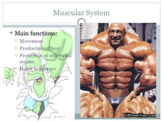
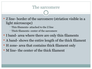
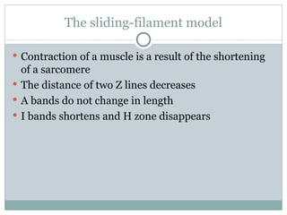
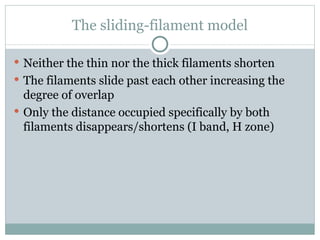
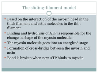
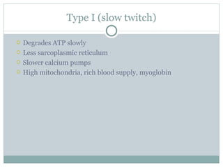
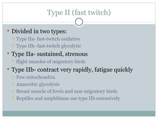
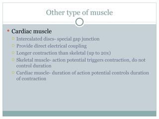
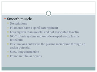
MUSCULAR SYSTEM
Muscular System Main functions: Movement Production of heat Protection of other vital organs Helps in posture
Review of thermoregulation How do ectotherms balance heat loss and heat gain? How do endotherms balance heat loss and heat gain? Importance of daily torpor  Vasoconstriction and vasodilation Countercurrent heat exchanger mechanism
Review of thermoregulation Huddling  Panting Stress-induced proteins Heat-shock proteins
Feedback mechanism in human thermoregulation
Balancing the internal environment Behavioral and physiological adaptations are used to budget heat loss and heat gain E.g. In a desert environment, evaporative cooling is not favored by the body rather conservation of water
Characteristics of skeletal muscles Excitability- receive and respond to stimuli (the all-or-none response) Contractility- shorten and thicken Extensibility- stretch and extend (passively) Elasticity- return to original shape after contraction or extension
Locomotion again.... Again, movement is done only if a muscle is working against some kind of skeleton Action is always to contract Extension is passive Muscles act in pairs (agonist and antagonist) Also synergist (muscles that have the same action)
Origin and insertion A muscle always originates from a specific part of a bone and is inserted also in a specific part of a bone (hahaha, kaya nga tinawag na skeletal muscle) Origin- proximal Insertion- distal
The muscle
Some terms (actually a lot of terms) Skeletal muscle- consists of bundle of muscle fiber Fascicle- a bundle of muscle fiber surrounded by a connective tissue Muscle fiber- a single muscle cell (syncytium) Myofibril – makes up a muscle fiber Divided into two myofilaments (creates the striation) Thin filaments Thick filaments
Still on terms...... Thin filaments- consists of two strands of actin  and one strand of regulatory protein Thick filament- consists of staggered array of myosin Sarcomere- the basic contractile unit of a muscle
The sarcomere Z line- border of the sarcomere (striation visible in a light microscope) Thin filaments- attached to the Z line Thick filaments- center of the sarcomere I band- area where there are only thin filaments A band- shows the entire length of the thick filament H zone- area that contains thick filament only M line- the center of the thick filament
The sliding-filament model Contraction of a muscle is a result of the shortening of a sarcomere The distance of two Z lines decreases A bands do not change in length I bands shortens and H zone disappears
The sliding-filament model Neither the thin nor the thick filaments shorten The filaments slide past each other increasing the degree of overlap Only the distance occupied specifically by both filaments disappears/shortens (I band, H zone)
The sliding-filament model Based on the interaction of the myosin head in the thick filament and actin molecules in the thin filament Binding and hydrolysis of ATP is responsible for the change in shape of the myosin molecule  The myosin molecule goes into an energized stage Formation of cross-bridge between the myosin and actin  Bond is broken when new ATP binds to myosin
Energy for contraction Stored ATP in muscle cell Glycogen that can be degraded and form ATP Phosphagens- supplies a phosphate to ADP to turn it into ATP Vertebrate phosphagen- Creatine phosphate
Control of muscle contraction Contraction is due to stimulation from a motor neuron At rest- tropomyosin blocks binding site in actin molecules Troponin complex- controls position of tropomyosin Calcium ions- binds to troponin Releases the active site of actin High calcium in cytosol- contraction can occur Low calcium in cytosol- contraction cannot occur
The Sarcoplasmic Reticulum Specialized endoplasmic reticulum Controls calcium ion concentration Membrane- actively transports calcium ions Cytosol into the interior of the reticulum
Action potential Stimulus of contraction- action potential from a motor neuron Motor neuron- a type of neuron that is connected to a motor cell (as oppose to sensory) Action potential is due to a change in the net charge of the surrounding muscle cell
Action potential Motor neuron- releases acetylcholine at neuromuscular junction ACh depolarizes the postsynaptic muscle cell creating the action potential The action potential spreads to the T (transverse) tubule T tubule is connected to the sarcoplasmic reticulum Efflux of calcium ions is caused by a change in the permeability of sarcoplasmic reticulum due to the action potential
Overview of muscle contraction Synaptic terminal of motor neuron releases Ach ACh depolarizes postsynaptic muscle cell Action potential is generated in the muscle cell Action potential spreads through T tubules Sarcoplasmic reticulum becomes permeable to calcium ions Calcium ions bind to troponin Troponin releases the active site of actin by moving the tropomyosin
Overview of muscle contraction Myosin head with attached ATP is under low energy state ATP is hydrolyzed creating a high energy state myosin head Cross-bridge is formed Release of energy relaxes the myosin molecule Another ATP molecule detaches the myosin from actin Contraction occurs as the myosin head walks on the thin filament
End of contraction End of contraction occurs if calcium ion concentration in the cytosol falls leading to the blockage of the active site of actin
The all-or-none response Experience and experiments show that whole mucle contraction is graded At the cellular level, contraction is an all-or-none response Frequency of action potential creates the graded contraction Summation of action potential occurs Tetanus- a smooth and sustained contraction
Graded contraction of a muscle  A muscle is innervated by one motor neuron Motor neuron is branched- controls different muscle cell Motor unit- a motor neuron and all muscle fiber it controls Nervous system controls graded contraction by the number of motor unit in action Recruitment of motor neurons- progressive inc in tension in a muscle
Muscle fatigue Caused by depletion of ATP, dissipation of ion gradient for a normal electrical signal, accumulation of lactate Fatigue is prevented by alternating activation of motor units
Fast and slow muscle fibers Based on the duration of a muscle twitch Fast (type II) muscle fibers- short, rapid, powerful contractions Slow (type I) muscle fibers- maintain posture, long contractions, fatigue slowly
Type I (slow twitch) Degrades ATP slowly Less sarcoplasmic reticulum Slower calcium pumps High mitochondria, rich blood supply, myoglobin
Type II (fast twitch) Divided in two types:  Type IIa- fast-twitch oxidative Type IIb- fast-twitch glycolytic  Type IIa- sustained, strenous flight muscles of migratory birds Type IIb- contract very rapidly, fatigue quickly  Few mitochondria Anaerobic glycolysis Breast muscle of fowls and non-migratory birds Reptiles and amphibians use type IIb extensively
Other type of muscle Cardiac muscle Intercalated discs- special gap junction Provide direct electrical coupling Longer contraction than skeletal (up to 20x) Skeletal muscle- action potential triggers contraction, do not control duration Cardiac muscle- duration of action potential controls duration of contraction
Smooth muscle No striations Filaments have a spiral aarangement Less myosin than skeletal and not associated to actin NO T tubule system and well-developed sarcoplasmic reticulum Calcium ions enters via the plasma membrane through an action potential Slow, long contraction Found in tubular organs
Invertebrate muscle Almost similar to vertebrate smooth and skeletal muscle Arthropod skeletal muscle almost identical to vertebrate skeletal muscle Flight muscle of insects- have independent rhythmic contraction faster than action potential Paramyosin- found in clams Regulatory protein that enables muscle fibers to sustain a fixed state of contraction with low energy rate
Lastly  Muscle is derived from mesoderm First muscle is found in Planarians due t the presence of the mesoderm

The Muscular System

  • 1.
  • 2.
    Muscular System Mainfunctions: Movement Production of heat Protection of other vital organs Helps in posture
  • 3.
    Review of thermoregulationHow do ectotherms balance heat loss and heat gain? How do endotherms balance heat loss and heat gain? Importance of daily torpor Vasoconstriction and vasodilation Countercurrent heat exchanger mechanism
  • 4.
    Review of thermoregulationHuddling Panting Stress-induced proteins Heat-shock proteins
  • 5.
    Feedback mechanism inhuman thermoregulation
  • 6.
    Balancing the internalenvironment Behavioral and physiological adaptations are used to budget heat loss and heat gain E.g. In a desert environment, evaporative cooling is not favored by the body rather conservation of water
  • 7.
    Characteristics of skeletalmuscles Excitability- receive and respond to stimuli (the all-or-none response) Contractility- shorten and thicken Extensibility- stretch and extend (passively) Elasticity- return to original shape after contraction or extension
  • 8.
    Locomotion again.... Again,movement is done only if a muscle is working against some kind of skeleton Action is always to contract Extension is passive Muscles act in pairs (agonist and antagonist) Also synergist (muscles that have the same action)
  • 9.
    Origin and insertionA muscle always originates from a specific part of a bone and is inserted also in a specific part of a bone (hahaha, kaya nga tinawag na skeletal muscle) Origin- proximal Insertion- distal
  • 10.
  • 11.
    Some terms (actuallya lot of terms) Skeletal muscle- consists of bundle of muscle fiber Fascicle- a bundle of muscle fiber surrounded by a connective tissue Muscle fiber- a single muscle cell (syncytium) Myofibril – makes up a muscle fiber Divided into two myofilaments (creates the striation) Thin filaments Thick filaments
  • 12.
    Still on terms......Thin filaments- consists of two strands of actin and one strand of regulatory protein Thick filament- consists of staggered array of myosin Sarcomere- the basic contractile unit of a muscle
  • 13.
    The sarcomere Zline- border of the sarcomere (striation visible in a light microscope) Thin filaments- attached to the Z line Thick filaments- center of the sarcomere I band- area where there are only thin filaments A band- shows the entire length of the thick filament H zone- area that contains thick filament only M line- the center of the thick filament
  • 14.
    The sliding-filament modelContraction of a muscle is a result of the shortening of a sarcomere The distance of two Z lines decreases A bands do not change in length I bands shortens and H zone disappears
  • 15.
    The sliding-filament modelNeither the thin nor the thick filaments shorten The filaments slide past each other increasing the degree of overlap Only the distance occupied specifically by both filaments disappears/shortens (I band, H zone)
  • 16.
    The sliding-filament modelBased on the interaction of the myosin head in the thick filament and actin molecules in the thin filament Binding and hydrolysis of ATP is responsible for the change in shape of the myosin molecule The myosin molecule goes into an energized stage Formation of cross-bridge between the myosin and actin Bond is broken when new ATP binds to myosin
  • 17.
    Energy for contractionStored ATP in muscle cell Glycogen that can be degraded and form ATP Phosphagens- supplies a phosphate to ADP to turn it into ATP Vertebrate phosphagen- Creatine phosphate
  • 18.
    Control of musclecontraction Contraction is due to stimulation from a motor neuron At rest- tropomyosin blocks binding site in actin molecules Troponin complex- controls position of tropomyosin Calcium ions- binds to troponin Releases the active site of actin High calcium in cytosol- contraction can occur Low calcium in cytosol- contraction cannot occur
  • 19.
    The Sarcoplasmic ReticulumSpecialized endoplasmic reticulum Controls calcium ion concentration Membrane- actively transports calcium ions Cytosol into the interior of the reticulum
  • 20.
    Action potential Stimulusof contraction- action potential from a motor neuron Motor neuron- a type of neuron that is connected to a motor cell (as oppose to sensory) Action potential is due to a change in the net charge of the surrounding muscle cell
  • 21.
    Action potential Motorneuron- releases acetylcholine at neuromuscular junction ACh depolarizes the postsynaptic muscle cell creating the action potential The action potential spreads to the T (transverse) tubule T tubule is connected to the sarcoplasmic reticulum Efflux of calcium ions is caused by a change in the permeability of sarcoplasmic reticulum due to the action potential
  • 22.
    Overview of musclecontraction Synaptic terminal of motor neuron releases Ach ACh depolarizes postsynaptic muscle cell Action potential is generated in the muscle cell Action potential spreads through T tubules Sarcoplasmic reticulum becomes permeable to calcium ions Calcium ions bind to troponin Troponin releases the active site of actin by moving the tropomyosin
  • 23.
    Overview of musclecontraction Myosin head with attached ATP is under low energy state ATP is hydrolyzed creating a high energy state myosin head Cross-bridge is formed Release of energy relaxes the myosin molecule Another ATP molecule detaches the myosin from actin Contraction occurs as the myosin head walks on the thin filament
  • 24.
    End of contractionEnd of contraction occurs if calcium ion concentration in the cytosol falls leading to the blockage of the active site of actin
  • 25.
    The all-or-none responseExperience and experiments show that whole mucle contraction is graded At the cellular level, contraction is an all-or-none response Frequency of action potential creates the graded contraction Summation of action potential occurs Tetanus- a smooth and sustained contraction
  • 26.
    Graded contraction ofa muscle A muscle is innervated by one motor neuron Motor neuron is branched- controls different muscle cell Motor unit- a motor neuron and all muscle fiber it controls Nervous system controls graded contraction by the number of motor unit in action Recruitment of motor neurons- progressive inc in tension in a muscle
  • 27.
    Muscle fatigue Causedby depletion of ATP, dissipation of ion gradient for a normal electrical signal, accumulation of lactate Fatigue is prevented by alternating activation of motor units
  • 28.
    Fast and slowmuscle fibers Based on the duration of a muscle twitch Fast (type II) muscle fibers- short, rapid, powerful contractions Slow (type I) muscle fibers- maintain posture, long contractions, fatigue slowly
  • 29.
    Type I (slowtwitch) Degrades ATP slowly Less sarcoplasmic reticulum Slower calcium pumps High mitochondria, rich blood supply, myoglobin
  • 30.
    Type II (fasttwitch) Divided in two types: Type IIa- fast-twitch oxidative Type IIb- fast-twitch glycolytic Type IIa- sustained, strenous flight muscles of migratory birds Type IIb- contract very rapidly, fatigue quickly Few mitochondria Anaerobic glycolysis Breast muscle of fowls and non-migratory birds Reptiles and amphibians use type IIb extensively
  • 31.
    Other type ofmuscle Cardiac muscle Intercalated discs- special gap junction Provide direct electrical coupling Longer contraction than skeletal (up to 20x) Skeletal muscle- action potential triggers contraction, do not control duration Cardiac muscle- duration of action potential controls duration of contraction
  • 32.
    Smooth muscle Nostriations Filaments have a spiral aarangement Less myosin than skeletal and not associated to actin NO T tubule system and well-developed sarcoplasmic reticulum Calcium ions enters via the plasma membrane through an action potential Slow, long contraction Found in tubular organs
  • 33.
    Invertebrate muscle Almostsimilar to vertebrate smooth and skeletal muscle Arthropod skeletal muscle almost identical to vertebrate skeletal muscle Flight muscle of insects- have independent rhythmic contraction faster than action potential Paramyosin- found in clams Regulatory protein that enables muscle fibers to sustain a fixed state of contraction with low energy rate
  • 34.
    Lastly Muscleis derived from mesoderm First muscle is found in Planarians due t the presence of the mesoderm

Editor's Notes

  • #14 Thick and thin filaments do not overlap completely
  • #22 Transverse tubule infolding of the plasma memebrane