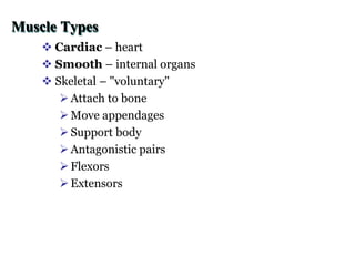
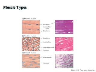
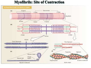
Muscles physiology
Muscles function
Contract!
Generate motion
Generate force
Generate heat
Support
Muscular Functions
 Body movement (Locomotion)
 Maintenance of posture
 Respiration
 Diaphragm and intercostal contractions
 Communication (Verbal and Facial)
 Constriction of organs and vessels
 Peristalsis of intestinal tract
 Vasoconstriction of b.v. and other structures (pupils)
 Heart beat
 Production of body heat (Thermogenesis)
Properties of Muscle
 Excitability: capacity of muscle to respond to a
stimulus
 Contractility: ability of a muscle to shorten and
generate pulling force
 Extensibility: muscle can be stretched back to its
original length
 Elasticity: ability of muscle to recoil to original
resting length after stretched
Muscle Types
 Cardiac – heart
 Smooth – internal organs
 Skeletal – "voluntary"
Attach to bone
Move appendages
Support body
Antagonistic pairs
Flexors
Extensors
Categories of skeletal muscle actions
 Categories Actions
 Extensor - Increases the angle at a joint
 Flexor- Decreases the angle at a joint
 Abductor - Moves limb away from midline of body
 Adductor - Moves limb toward midline of body
 Levator - Moves insertion upward
 Depressor Moves insertion downward
 Rotator - Rotates a bone along its axis
 Sphincter -Constricts an opening
Types of Muscle
 Skeletal
 Attached to bones
 Makes up 40% of body weight
 Responsible for locomotion, facial expressions, posture, respiratory
movements, other types of body movement
 Voluntary in action; controlled by somatic motor neurons
 Smooth
 In the walls of hollow organs, blood vessels, eye, glands, uterus, skin
 Some functions: propel urine, mix food in digestive tract,
dilating/constricting pupils, regulating blood flow,
 In some locations, auto rhythmic
 Controlled involuntarily by endocrine and autonomic nervous systems
 Cardiac
 Heart: major source of movement of blood
 Auto rhythmic
 Controlled involuntarily by endocrine and autonomic nervous systems
Connective Tissue Sheaths
 Connective Tissue of a Muscle
Epimysium. Dense regular c.t. surrounding entire muscle
Separates muscle from surrounding tissues and organs
Connected to the deep fascia
Perimysium. Collagen and elastic fibers surrounding a
group of muscle fibers called a fascicle
Contains b.v and nerves
Endomysium. Loose connective tissue that surrounds
individual muscle fibers
Also contains b.v., nerves, and satellite cells (embryonic
stem cells function in repair of muscle tissue
 Collagen fibers of all 3 layers come together at each end of
muscle to form a tendon or Aponeurosis.
Muscle Types
Figure 12-1: Three types of muscles
 About 40% body mass
 Muscle fibers – cells
 Fascicle – bundle
 Motor unit
 Muscle
 sheath
 Attach to tendons (which attach to bone)
Skeletal Muscle Anatomy
Nerve and Blood Vessel Supply
 Motor neurons
stimulate muscle fibers to contract
Neuron axons branch so that each muscle fiber (muscle
cell) is innervated
Form a neuromuscular junction (= my neural junction)
 Capillary beds surround muscle fibers
Muscles require large amounts of energy
Extensive vascular network delivers necessary oxygen
and nutrients and carries away metabolic waste produced
by muscle fibers
Skeletal Muscle Anatomy
 Multiple nuclei
 Sarcolemma
 T-tubules
 Sarcoplasmic reticulum
 Sarcoplasm
Mitochondria
Glycogen & ions
Myofibrils
Muscle Fiber Structure
Figure 12-3b: ANATOMY SUMMARY: Skeletal Muscle
 Actin –"thin fibers"
 Tropomysin
 Troponin
 Myosin – "thick fibers"
 Titin – elastic anchor
 Nebulin – non-elastic
Myofibrils: Site of Contraction
Sarcomere: Organization of Fibers
Figure 12-6: Titin and nebulin
Skeletal Muscle Contraction: Mechanism
Figure 12-11a: Excitation-contraction coupling
Skeletal Muscle Contraction: Mechanism
Energy for Contraction: ATP & Phosphocreatine
• Aerobic Respiration
• Oxygen
• Glucose
• Fatty acids
• 30-32 ATPs
• Anaerobic Respiration
• Fast but
• 2 ATP/glucose
• Phosphocreatine ATPs
Energy for Contraction: ATP & Phosphocreatine
Figure 12-13: Phosphocreatine
Smooth Muscle
 Fusiform cells
 One nucleus per cell
 Nonstriated
 Involuntary
 Slow, wave-like
contractions
Smooth Muscle
• Cells are not striated
• Fibers smaller than those in skeletal
muscle
• Spindle-shaped; single, central
nucleus
• More actin than myosin
• No sarcomeres
• Not arranged as symmetrically as
in skeletal muscle, thus NO
striations.
• Caveolae: indentations in
sarcolemma;
• May act like T tubules
• Dense bodies instead of Z disks
• Have noncontractile intermediate
filaments
Smooth Muscle
• Grouped into sheets in walls of hollow organs
• Longitudinal layer – muscle fibers run parallel to organ’s long axis
• Circular layer – muscle fibers run around circumference of the organ
• Both layers participate in peristalsis
Smooth Muscle
• Is innervated by autonomic nervous system (ANS)
• Visceral or unitary (single unit) smooth muscle
• Only a few muscle fibers innervated in each group
• Impulse spreads through gap junctions
• Whole sheet contracts as a unit
• Often autorhythmic
• Multiunit:
• Cells or groups of cells act as independent units
• Arrector pili of skin and iris of eye
Smooth Muscle Cell
Myosin of Smooth Muscle
• Different isoform than that found in skeletal
muscle
• Smooth muscle myosin ATPase activity is
much slower, contraction is longer
• Myosin light chain in the myosin head
regulates contraction and relaxation
Smooth Muscle
• Relatively little sarcoplasmic reticulum
• Lacks T-tubules
• Chemically linked to the cell membrane,
rather than mechanically linked
• Ca +2 storage is supplemented by caveolae ,
small vesicles that cluster close to the cell
membrane. Voltage/ligand gated Ca +2
channels
Single-Unit Muscle
Properties of Single-Unit Smooth Muscle
• Gap junctions
• Pacemaker cells
with spontaneous
depolarizations
• Innervation to few
cells
• Tone = level of
contraction without
stimulation
• Increases/decreases
in tension
• Graded Contractions
• No recruitment
• Vary intracellular
calcium
• Stretch Reflex
• Relaxation in
response to sudden
or prolonged stretch
Multi-Unit Muscle
Comparisons Among Skeletal, Smooth, and Cardiac Muscle
Thank you

muscle physiology-1.ppt

  • 1.
  • 2.
  • 3.
    Muscular Functions  Bodymovement (Locomotion)  Maintenance of posture  Respiration  Diaphragm and intercostal contractions  Communication (Verbal and Facial)  Constriction of organs and vessels  Peristalsis of intestinal tract  Vasoconstriction of b.v. and other structures (pupils)  Heart beat  Production of body heat (Thermogenesis)
  • 4.
    Properties of Muscle Excitability: capacity of muscle to respond to a stimulus  Contractility: ability of a muscle to shorten and generate pulling force  Extensibility: muscle can be stretched back to its original length  Elasticity: ability of muscle to recoil to original resting length after stretched
  • 5.
    Muscle Types  Cardiac– heart  Smooth – internal organs  Skeletal – "voluntary" Attach to bone Move appendages Support body Antagonistic pairs Flexors Extensors
  • 6.
    Categories of skeletalmuscle actions  Categories Actions  Extensor - Increases the angle at a joint  Flexor- Decreases the angle at a joint  Abductor - Moves limb away from midline of body  Adductor - Moves limb toward midline of body  Levator - Moves insertion upward  Depressor Moves insertion downward  Rotator - Rotates a bone along its axis  Sphincter -Constricts an opening
  • 7.
    Types of Muscle Skeletal  Attached to bones  Makes up 40% of body weight  Responsible for locomotion, facial expressions, posture, respiratory movements, other types of body movement  Voluntary in action; controlled by somatic motor neurons  Smooth  In the walls of hollow organs, blood vessels, eye, glands, uterus, skin  Some functions: propel urine, mix food in digestive tract, dilating/constricting pupils, regulating blood flow,  In some locations, auto rhythmic  Controlled involuntarily by endocrine and autonomic nervous systems  Cardiac  Heart: major source of movement of blood  Auto rhythmic  Controlled involuntarily by endocrine and autonomic nervous systems
  • 8.
    Connective Tissue Sheaths Connective Tissue of a Muscle Epimysium. Dense regular c.t. surrounding entire muscle Separates muscle from surrounding tissues and organs Connected to the deep fascia Perimysium. Collagen and elastic fibers surrounding a group of muscle fibers called a fascicle Contains b.v and nerves Endomysium. Loose connective tissue that surrounds individual muscle fibers Also contains b.v., nerves, and satellite cells (embryonic stem cells function in repair of muscle tissue  Collagen fibers of all 3 layers come together at each end of muscle to form a tendon or Aponeurosis.
  • 9.
    Muscle Types Figure 12-1:Three types of muscles
  • 10.
     About 40%body mass  Muscle fibers – cells  Fascicle – bundle  Motor unit  Muscle  sheath  Attach to tendons (which attach to bone) Skeletal Muscle Anatomy
  • 11.
    Nerve and BloodVessel Supply  Motor neurons stimulate muscle fibers to contract Neuron axons branch so that each muscle fiber (muscle cell) is innervated Form a neuromuscular junction (= my neural junction)  Capillary beds surround muscle fibers Muscles require large amounts of energy Extensive vascular network delivers necessary oxygen and nutrients and carries away metabolic waste produced by muscle fibers
  • 12.
  • 13.
  • 14.
     Multiple nuclei Sarcolemma  T-tubules  Sarcoplasmic reticulum  Sarcoplasm Mitochondria Glycogen & ions Myofibrils Muscle Fiber Structure
  • 15.
    Muscle Fiber Structure Figure12-3b: ANATOMY SUMMARY: Skeletal Muscle
  • 16.
     Actin –"thinfibers"  Tropomysin  Troponin  Myosin – "thick fibers"  Titin – elastic anchor  Nebulin – non-elastic Myofibrils: Site of Contraction
  • 17.
  • 18.
    Sarcomere: Organization ofFibers Figure 12-6: Titin and nebulin
  • 19.
    Skeletal Muscle Contraction:Mechanism Figure 12-11a: Excitation-contraction coupling
  • 20.
  • 21.
    Energy for Contraction:ATP & Phosphocreatine • Aerobic Respiration • Oxygen • Glucose • Fatty acids • 30-32 ATPs • Anaerobic Respiration • Fast but • 2 ATP/glucose • Phosphocreatine ATPs
  • 22.
    Energy for Contraction:ATP & Phosphocreatine Figure 12-13: Phosphocreatine
  • 23.
    Smooth Muscle  Fusiformcells  One nucleus per cell  Nonstriated  Involuntary  Slow, wave-like contractions
  • 24.
    Smooth Muscle • Cellsare not striated • Fibers smaller than those in skeletal muscle • Spindle-shaped; single, central nucleus • More actin than myosin • No sarcomeres • Not arranged as symmetrically as in skeletal muscle, thus NO striations. • Caveolae: indentations in sarcolemma; • May act like T tubules • Dense bodies instead of Z disks • Have noncontractile intermediate filaments
  • 25.
    Smooth Muscle • Groupedinto sheets in walls of hollow organs • Longitudinal layer – muscle fibers run parallel to organ’s long axis • Circular layer – muscle fibers run around circumference of the organ • Both layers participate in peristalsis
  • 26.
    Smooth Muscle • Isinnervated by autonomic nervous system (ANS) • Visceral or unitary (single unit) smooth muscle • Only a few muscle fibers innervated in each group • Impulse spreads through gap junctions • Whole sheet contracts as a unit • Often autorhythmic • Multiunit: • Cells or groups of cells act as independent units • Arrector pili of skin and iris of eye
  • 27.
  • 28.
    Myosin of SmoothMuscle • Different isoform than that found in skeletal muscle • Smooth muscle myosin ATPase activity is much slower, contraction is longer • Myosin light chain in the myosin head regulates contraction and relaxation
  • 29.
    Smooth Muscle • Relativelylittle sarcoplasmic reticulum • Lacks T-tubules • Chemically linked to the cell membrane, rather than mechanically linked • Ca +2 storage is supplemented by caveolae , small vesicles that cluster close to the cell membrane. Voltage/ligand gated Ca +2 channels
  • 30.
  • 31.
    Properties of Single-UnitSmooth Muscle • Gap junctions • Pacemaker cells with spontaneous depolarizations • Innervation to few cells • Tone = level of contraction without stimulation • Increases/decreases in tension • Graded Contractions • No recruitment • Vary intracellular calcium • Stretch Reflex • Relaxation in response to sudden or prolonged stretch
  • 32.
  • 33.
    Comparisons Among Skeletal,Smooth, and Cardiac Muscle
  • 34.

Editor's Notes

  • #24 Smooth muscle is found in the walls of hollow organs. *Their muscle cells are fusiform in shape. *Smooth muscle cells have just on nucleus per cell. *Smooth muscle is nonstriated. *Smooth muscle is involuntary. *The contractions of smooth muscle are slow and wave-like.